
Research Article
Austin J Dent. 2023; 10(1): 1171.
Comparison of the Chairside Time and Fitness of Michigan Splints Fabricated With Conventional Method and Digital Method: A Self-Controlled Study
Fang Shuobo*
Peking University School of Stomatology, China
*Corresponding author: Fang Shuobo Peking University School of Stomatology, China. Email: [email protected]
Received: March 23, 2023 Accepted: April 27, 2023 Published: May 04, 2023
Abstract
Purpose: To evaluate the chairside time and fitness of Michigan splints fabricated with conventional method and digital method.
Material and Methods: Two Michigan splints were fabricated with conventional method and digital method respectively for 16 participants. In digital workflow, the data of dentitions were obtained by model scanning. Michigan splints were then designed, occlusal surface modified with the aid of fully adjustable virtual articulator, and fabricated by 3D printing technology with light-cured acrylic resin. In conventional workflow, splints were fabricated using waxed up method and fabricated with heat-cured acrylic resin. The chairside time of insertion and occlusal adjustment were recorded. The retention and comfort were evaluated by participants using 10cm Visual Analog Scales (VAS). Highly flowable Polyvinyl Siloxane (PVS) filled the space between tissue side of splints and occlusal surface of the natural dentitions. The fitness of the splints was calculated by scanning the tissue sides of splints with and without PVS replicas respectively. All data were divided into conventional group (control group) and digital group (test group), and self-controlled method was adopted. Differences between two groups were analyzed by paired t-test.
Results: There was no statistical difference in insertion time between two groups (P=0.263). Test group took less chairside time for centric (605±436s vs. 945±427 s, P=0.007), protrusive (97.0±144s vs. 246±211s, P=0.036), and excursive (25.0±101s vs. 114±465s, P=0.026) occlusal adjustment than control group. The space of test group was significantly smaller than control group (350±120μm vs. 470±90.0μm, P<0.001). No statistical difference was detected in the scores of retention, comfort at postural position and comfort at centric occlusion between two groups (P≥0.113).
Conclusion: Michigan splints fabricated with digital method need less chairside time and showed better fitness than conventional method.
Keywords: Michigan splint; Digital workflow; Virtual articulator; 3D printing
Clinical Implications
Compared with conventional method, Michigan splints fabricated with digital method showed higher chairside efficiency and better fitness. However, the therapeutic effect of occlusal splints is still dependent on how well the occlusal adjustment is conducted instead of the method in which an occlusal splint is fabricated.
Introduction
The modern use of biteplanes or occlusal splints to eliminate temporarily occlusal interferences and to allow ideal seating of the condyles was initiated by Posselt in the 1950s [1]. The appliances were recommended for TMJ and muscular disturbances related to occlusal dysfunction. The stabilized splint is the mostly commonly used type of intraoral appliance and has the least potential for adverse effects to the oral structures [2]. The modifications incorporated in the Michigan splint beyond the flat stabilized splint have made it a significantly improved aid in the management of patients with occlusal dysfunction [1]. There are two conventional methods widely used in clinical routines to fabricate splints, including laboratory method and chairside method. The laboratory method is complex, labor-intensive and error-prone [3]. The chairside method is smelly, susceptible to biofilm adhesion and takes amount of chairside time.
With the application of Computer-Aided Design and Computer-Aided Manufacturing (CAD/CAM) technology in the dentistry, several attempts have been launched to introduce digital method to the fabrication of occlusal splints [4-6]. Dedem et al [5] established a fully digital and plasterless fabrication workflow of Michigan splints. By assessing the fit, retention and number of initial occlusal contacts of splints, it was concluded that the Michigan splints fabricated with digital method showed some advantages over conventional method. However, there was lack of occlusal splints fabricated by conventional method as control in their study. Besides, the Michigan splints were milled from PMMA blocks, which took much processing time and inevitably wasted lots of material. 3D printing is another manufacturing process in which an object is formed layer by layer and the advantage is to produce complex shapes using less material than milling processes.
Fitness is crucial for each type of restorations. It is doubtful that a splint can behave as expected since the area between splint and teeth or soft tissue surface is much larger than other restorations, egg. Crown, bridge. This study describes a digital method of using virtual articulator to design occlusal contacts and using 3D printing to fabricate Michigan splints. The chairside time and fitness of splints fabricated with digital method were evaluated by this self-controlled study, and compared with those with conventional method.
Material and Methods
Participants
Healthy participants with complete dentition were recruited from graduate students of Peking University School of Stomatology. Inclusion criteria were Angle class jaw relations, and the vertical distance between upper and maximum opening of more than 40 millimeters. Exclusion criteria were oral infections, acute or chronic pain conditions temporomandibular disorders (diagnosed by one specialist using DC/TMD), various mucocutaneous diseases/disorders, medications affecting the nervous or muscle system. Written informed consents were obtained from all participants. The study was approved by Biomedical ethics committee of Peking University Hospital of Stomatology (PKUSSIRB-AF02).
A self-controlled design was applied in this study and paired t-test was adopted to analyze the differences. The primary outcome was that the difference of centric occlusal adjustment between both groups was up to 120 seconds. The sample size was calculated to be 13 with a significance level of 0.05, a power of 90%, and a SD of 132 seconds. 16 participants were recruited considering a 20% loss rate. Eight females and 8 males were included and the average age was 25 years.
Study Design and Statistical Method
One digital splint and one conventional splint were made for each participant, and the conventional splint was set as control and the digital splint was belonged to test group. The splints fabricated by digital or conventional method were delivered to the participants by one experienced clinician. The study data were input and analyzed in IBM SPSS 18.0 software. A normal test was conducted for variables firstly using Shapiro-Wilk test. Paired t-test and Wilcoxon test were then adopted to compare the differences of two groups according to the outcome of Shapiro-Wilk test. The level of significance was set at 0.05.
Mounting Plaster Model to Mechanical Articulator
Polyether (Impregum, 3M ESPE) impressions were made using metal stock trays (ASA Permalock; ASA Dental) and poured with type dental stone (Pemaco, Pemaco). Mandibular movements were recorded by ultrasonic jaw motion tracking device (ARCUS Digma, KaVo Dental GmbH), and individualized mandibular movement parameters were obtained. Upper jaw models were mounted onto the fully adjustable articulator (PROTARevo 7, KaVo Dental GmbH) with KaVo transfer system. The condyles were located in centric relation with bilateral manual manipulation technique [3]. The vertical dimension provided a thickness that maintained posterior teeth 1-2 mm apart. The therapeutic jaw relations were recorded by combining light-cured resin (Light-cured resin, Huge) and interim crown materials (ProtempTM 4, 3M ESPE). Mandibular movement parameters were set at the articulator joints and lower jaw models were mounted using jaw relation records.
Mechanical Articulator to Virtual Articulator
The occlusal transfer calibration objects (3Shape occlusal transfer calibration object, 3Shape A/S) and the transfer plates (3Shape transfer plate for PROTARevo, 3Shape A/S) were used for transferring the point cloud data and relative position of mounted models from mechanical articulator to virtual articulator. An appliance was designed to make sure that the occlusal transfer calibration object kept in the optimal position in the mechanical articulator, which had already obtained Chinese patent (Patent ZL 2021 1 0042483.1) (Figure 1). After the calibration objects were mounted to mechanical articulator with the appliance, the calibration objects in gypsum were scanned in a lab scanner (D2000, 3Shape A/S). Then the mounted models were scanned in the scanner under the transfer plate pattern and digital models were mounted onto a virtual articulator automatically.
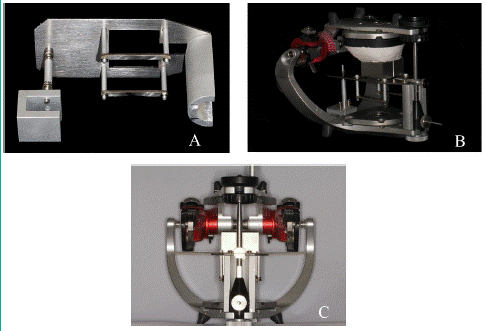
Figure 1: Using custom-made appliance to assist in mounting the 3Shape occlusal transfer calibration object onto the articulator. A: Supporting appliance designed to keep the 3Shape occlusal transfer calibration object in assigned position. B: Supporting appliance with upper part of occlusal transfer calibration object on the articulator. C: Supporting appliance with occlusal transfer calibration object on the articulator from frontal view.
Design and Fabrication of the Digital Splint
The files acquired through scanning were preceded to the design step of Michigan splint. Under the instruction of the Ap pliance Designer software (3Shape Dental System; 3Shape A/S), the first step was to determine the direction of insertion. The undercuts were computed, displayed and properly adjusted. The next step included creating a shell by drawing a spline that determined the rough margin of the splint, and adding a bar for forming occlusal contacts. These two parts were merged after necessary adjustment to form the profile of the splint. Then, the virtual articulator was set personally according to individual condyle movement parameters, which was applied to display and alter occlusal contacts during open-close movement, protrusive and excursive movements. The splints were constructed with stops for all mandibular function cusps or incisors, and control of contact relations in various excursions. Freedom in centric occlusion on a flat plane was adapted, centric occlusion contacts were established in front of centric relation, and with additional freedom of about 1mm from centric occlusion to the cuspid rise in all lateral and protrusive excursions. The splints were as thin as practical, approximately 1-2mm in the molar regions. The cuspid rise should be gradual starting approximately 1mm from centric occlusion and only steep enough to disocclude the balancing side. Incisal guidance beyond what cuspid rise provide was eliminated [1]. Finally, the desired shape was achieved after using the trim and smoothing tool to make adjustment carefully, and design files were exported as Standard Tessellation Language (STL) format (Figure 2).
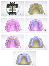
Figure 2: Digital design of Michigan splint. A: Digital models on the fully adjustable virtual articulator. B: Determination of the direction of insertion. C: Determination of the extension range of splints with some points. D: A shell generated on the basis of extension range determined. E: Adjustment of occlusal height and forming occlusal contacts with a bar. F: Combination of the shell and bar and formation of the profile of the splint. G: Occlusal marks displayed during open-close movement, protrusive movement and bilateral movement.
The files were sent to 3D printer. After entering the processing parameters, Michigan splints were fabricated by Shino light-cured 3D printers (3D printer, Shino Tech) with light-cured acrylic resin (Light-cured resin, Shino Tech) using stereolithography technique. Then, the support materials were removed and splints were finished.
Fabrication of the Splint Using Conventional Method
After scanning in 2.5., the stone casts and mechanical articulators were transferred to the laboratory. Michigan splints were fabricated following laboratory workflow of investing a waxed appliance and processing with heat-cured acrylic resin.
Try-in and Adjustment of the Splints
Each splint fabricated by digital or laboratory method was inserted into the dentition by one experienced clinician (Figure 3). After insertion, 100μm red articulating paper was used to examine the number and distribution of static contacts and 100μm blue articulating paper (BK 52 Red, BK 51 Blue, Dr. Jean Bausch GmbH) was chosen to display the protrusive and excursive guidance of each splint (Figure 4). Then occlusal adjustment was conducted by the same clinician. The standard of contacts in centric protrusive and excursive positions was described in 2.5. The time spent on insertion and occlusal adjustment of two types of splints for each participant was recorded. The retention, and comfort at postural position and centric occlusion of each splint were evaluated by a 10cm visual analog scales (VAS, 0=extremely poor, 10cm=extremely good).
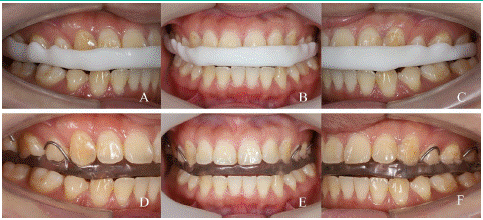
Figure 3: Examples of Michigan splints fabricated by digital and conventional method tried in the mouth on first insertion. A~C: A Michigan splint fabricated by digital method in the mouth from right view, frontal view and left view. D~F: A Michigan splint fabricated by conventional method in the mouth from right view, frontal view and left view.
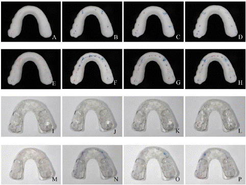
Figure 4: Examples of occlusal contacts of splints before and after adjustment. A: Occlusal contacts before and after occlusal adjustment of splints fabricated by digital and conventional method. A~D: Occlusal contacts of centric occlusion, protrusive occlusion, right lateral occlusion and left lateral occlusion before occlusal adjustment of a splint fabricated by digital method. E~H: Occlusal contacts of centric occlusion, protrusive occlusion, right lateral occlusion and left lateral occlusion after occlusal adjustment of a splint fabricated by digital method. I~L: Occlusal contacts of centric occlusion, protrusive occlusion, right lateral occlusion and left lateral occlusion before occlusal adjustment of a splint fabricated by conventional method. M~P: Occlusal contacts of centric occlusion, protrusive occlusion, right lateral occlusion and left lateral occlusion after occlusal adjustment of a splint fabricated by conventional method.
Evaluation of the Fitness of Splints
After occlusal adjustment, the space between tissue side of splints and occlusal surface of the natural dentitions, which represented fitness of the splint, was calculated. Firstly, the highly flowable Polyvinyl Siloxane (PVS) (Variotime Light Flow, Heraeus Kulzer GmbH) was injected into the tissue side of splint. The splint was then inserted into dentition with sufficient finger pressure to fully seat the prosthesis, which was removed after the material polymerized and the space was replicated by PVS material [7]. The PVS replicas adhered to the tissue side of splints firmly. The tissue sides of splints with and without PVS replicas were scanned respectively. The scanning data were imported into Geomagic Studio 2013 software (3D system) and occlusal areas for comparison were selected by some steps. Step 1: N-point alignment was used to align the scanning data of one splint with and without PVS replicas roughly. Step 2: The area uncovered with PVS was selected in the tissue side of scanning data with PVS replica, the curve of which was then projected onto the scanning data without PVS replica. The corresponding area was generated based on the projected curve. Best fit alignment was adopted to align these two scanning data according to the selected areas. Step 3: Occlusal areas for comparison were selected in one scanning data and corresponding areas were generated like step 2. Occlusal areas for comparison were then saved as Geomagic WRP format and imported into Geomagic Qualify 2013 software (3D system). 3D deviation analysis tool was used to compare the deviation of the two data, which represented the thickness of PVS replicas actually (Figure 5).
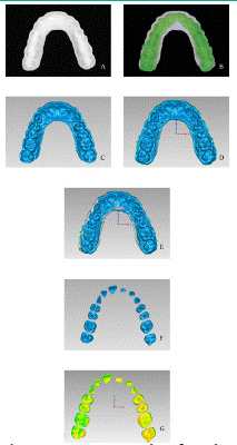
Figure 5: Space between tissue side of occlusal splints and occlusal surface calculated by Polyvinyl Siloxane (PVS) replica and 3D deviation analysis. A: Digital splint without PVS replica. B: PVS replica adhered to the tissue side of occlusal splint fabricated by digital method. C: Scanning data of tissue side of occlusal splint without PVS replica. D: Scanning data of tissue side of occlusal splint with PVS replica. E: Best fit alignment of two scanning data. F, Occlusal areas for comparison selected. G: 3D deviation analysis color map of two scanning data.
Results
The results of Shapiro-Wilk test are summarized in table 1. The occlusal adjustment time spent on centric occlusion and protrusive movement, the space between splint and dentition occlusal surface, and VAS scores of participants obey normal distribution. The results were shown in table 2. The adjustment time spent on insertion and excursive movement doesn’t obey normal distribution. The results were shown in table 3.
![]()
    Variable
    Statistic
    Degree of freedom
    P value


    Time spent on insertion
    0.670
    16
    < 0.001


    Time spent on centric occlusal    equilibration
    0.970
    16
    0.837


    Time spent on excursive occlusal    equilibration
    0.695
    16
    < 0.001


    Time spent on protrusive occlusal    equilibration
    0.943
    16
    0.384


    Space between splint and dentition occlusal    surface
    0.985
    16
    0.991


    VAS scores of retention
    0.937
    16
    0.315


    VAS scores of comfort at postural position
    0.960
    16
    0.659


    VAS scores of comfort at centric occlusion
    0.974
    16
    0.895


    VAS: Visual    Analog Scale
Table 1: Shapiro-Wilk test of differences between test group and control group.
![]()
Variables
Test group
Control group
t value
P value
Mean
SD
Mean
SD
Time spent on centric occlusal equilibration (s)
605
436
945
427
3.122
0.007
Time spent on protrusive occlusal equilibration (s)
97.0
144
246
211
2.304
0.036
350
120
470
90.0
4.442
< 0.001
VAS scores of retention (cm)
8.40
1.50
8.20
1.30
0.267
0.793
VAS scores of comfort at postural position (cm)
8.20
1.10
7.70
0.800
1.386
0.186
VAS scores of comfort at centric occlusion (cm)
7.80
1.30
7.10
1.60
1.682
0.113
SD: Standard Deviation; VAS: Visual Analog Scale.
Table 2: For variables obey normal distribution, differences between test group (digital splints) and control group (conventional splints) were evaluated by a paired t-test.
![]()
    Variables
    Test group

    Control group
    Z value
    P value


    Median
    IQR

    Median
    IQR


    Time spent on insertion (s)
    0
    59.0

    0
    42.0
    1.120
    0.263


    Time spent on excursive occlusal    equilibration (s)
    25.0
    101

    114
    465
    2.229
    0.026


    IQR: Interquartile    Range
Table 3: For variables disobey normal distribution, differences between test group (digital splints) and control group (conventional splints) were evaluated by a Wilcoxon test.
As shown in table 2, digital splint group (test group) took less chairside time for centric and protrusive occlusal adjustment than conventional splint group (control group) (P=0.036). Time spent on centric occlusal adjustment for test group and control group were 605±436s and 945±427s respectively. Time spent on protrusive occlusal adjustment for test group and control group were 97±144s and 246±211s respectively. The space between splint and occlusal surface of dentition of test group was significantly smaller than control group (350±120μm vs. 470±90.0μm, P<0.001). No statistical difference was detected in the scores of VAS in terms of retention, comfort at postural position and comfort at centric occlusion between two groups (P≥0.113).
As is shown in table 3, there was no statistical difference in insertion time between two groups (P=0.263). Test group took less chairside time for excursive occlusal adjustment than control group (25(101)s vs. 114(465)s Median (IQR), P=0.026). Time spent on centric occlusal adjustment was more than eccentric occlusal adjustment for both test group (605±436s vs. 213±312s, P=0.013) and control group (945±427s vs. 539±434s, P=0.022).
Digital technology possesses advantages of easy control and high efficiency. Various CAD/CAM workflows of fabricating stabilized splints have been reported [8-12]. However, when it comes to therapeutic effect of occlusal splints, it is still dependent on how well the occlusal adjustment is conducted instead of the method in which an occlusal splint is fabricated. Therefore, this study focused more on the chairside time and fitness that concerned by most dentists in clinic of splints fabricated by digital method.
This self-controlled study showed that occlusal splints fabricated by digital method took less occlusal adjustment time than conventional method. This phenomenon may be explained: a: There is no surface wear of models that is common in conventional method in virtual environment, and occlusal design can be meticulous and accurate with the aid of convenient visual tools; b: the typical feature resolution of stereolithography technique is up to 50-100 micrometers [13], which avoids the uncontrollable monomer content and shrinkage existing in conventional method. It was also found that time spent on centric occlusal adjustment was more than eccentric occlusal adjustment, which may result from the unique eccentric canine guidance of Michigan splints [1].
The fitness accuracy of splints has rarely been evaluated. The method for quantitative evaluation of fitness in this study was based on PVS replica method described by Parker [14], which was always used in evaluating the fitness of clasp assembly in the field of removable partial denture study [15-17]. Then, many 2D measuring instruments including micrometers [18,19], profile projectors [20,21] and stereo microscopes [7,22] were used to measure the space in these studies. These instruments had been tried in this study, but it was difficult to orient the same anatomy position in two replicas and prone to bring error. Finally, 3D deviation analysis was tried and helped obtain the target data, which could evaluate the deviation both entirely and cloud by cloud with the strong function of the 3D analysis software. Even though the outcome showed significantly difference between two groups, it remained unclear how the difference of 0.12 milometers would influence the occlusal force transmission.
Insertion and retention of splints fabricated by digital method had greater mutant effect than conventional method, because their retention force depended more on undercuts that also influenced insertion seriously, instead of clasps in conventional ones. Undercut parameters setting would influence insertion or retention and result in failure whether the undercuts were too large or too small. As presented in this study, most splints could be inserted directly while a few need some time. It revealed that insertion was not a problem for digital method when the parameters were set appropriately.
VAS scores of retention, comfort at postural position and comfort of occlusion showed no significant difference between two groups in this study. Similar results of VAS scores of retention were reported by previous study [12]. Wang et al. found wearing comfort scores of the digital splint group were significantly higher than conventional splint [12]. This was probably because the buccal and palatal extension of splints in this study was larger than their design and brought greater foreign body sensation. Therefore, there needs more evaluation about the relationship of the extension range, retention and strength of occlusal splints fabricated by 3D printing technology.
Conventional methods for making splints have emerged and been widely used for many years [1,23,24], while digital methods have been established for less than two decades. It is known that various digital workflows of fabricating splints have been proposed in the past decade, which could be described as scanning, design and fabrication roughly [5,6,8-12]. The accuracy of full arch intraoral scanning was still controversial [25,26], which has been used in fabricating splints and showed promising outcome [5,10,11,27]. Model scanning was adopted in this study in order to obtain a high accuracy and apply virtual articulator in designing. Just like mechanical articulators, there are many different types of virtual articulators. As for fully adjustable virtual articulators, models and corresponding mechanical articulators are necessary according to the instruction video on the manufacturer’s website. Exact hinge axis location was obtained by ultrasonic jaw motion tracking device. In the past five years, another type of virtual articulator based on jaw motion data was fused to dental design software [9,27], which may change the workflow greatly in the future and need more studies.
Conclusions
Michigan splints fabricated with digital method need less chairside time and showed better fitness than conventional method.
References
- Ramfjord SP, Ash MM. Reflections on the Michigan occlusal splint. J Oral Rehabil. 1994; 21: 491-500.
- Klasser GD, Greene CS. Oral appliances in the management of temporomandibular disorders. Oral Surgery Oral Medicine Oral Pathology Oral Radiology & Endodontology. 2009; 107: 212-223.
- Okeson JP. Management of Temporomandibular Disorders and Occlusion. 7th ed, 2011; 383-388.
- Beuer F, Schweiger J, Edelhoff D. Digital dentistry: an overview of recent developments for CAD/CAM generated restorations. British Dental Journal. 2008; 204: 505–511.
- Dedem P, Türp JC. Digital Michigan splint-from intraoral scanning to plasterless manufacturing[J]. International Journal of Computerized Dentistry. 2016; 19: 63-76.
- Kim JE, Kwon JH, Kim JH, Shim JS. Recording the trajectory of mouth opening and closing for the fabrication of an occlusal splint. Journal of Prosthetic Dentistry. 2017; 117: 597-600.
- Dunham D, Brudvik JS, Morris WJ, Plummer KD, Cameron SM. A clinical investigation of the fit of removable partial dental prosthesis clasp assemblies. J Prosthet Dent. 2006; 95: 323-326.
- Lauren M, McIntyre F. A new computer-assisted method for design and fabrication of occlusal splints. Am J Orthod Dentofacial Orthop. 2008; 133: S130-S135.
- Kurbad A. Three-dimensional registration of real jaw motion tracking data and its therapeutic consequences. Int J Comput Dent. 2018; 21: 57-70.
- Berntsen C, Kleven M, Heian M, Hjorsjo C. Clinical comparison of conventional and additive manufactured stabilization splints. Acta Biomater Odontol Scand. 2018; 4: 81-89.
- Waldecker M, Leckel M, Rammelsberg P, Bomicke W. Fully digital fabrication of an occlusal device using an intraoral scanner and 3D printing: A dental technique. J Prosthet Dent. 2019; 121: 576-580.
- Wang S, Li Z, Ye H, Zhao W, Liu Y, et al. Preliminary clinical evaluation of traditional and a new digital PEEK occlusal splints for the management of sleep bruxism. J Oral Rehabil. 2020; 47: 1530-1537.
- Ligon SC, Liska R, Stampfl J, Gurr M, Mulhaupt R. Polymers for 3D Printing and Customized Additive Manufacturing. Chem Rev. 2017; 117: 10212-10290.
- Parker MH, Cameron SM, Hughbanks JC, Reid DE. Comparison of occlusal contacts in maximum intercuspation for two impression techniques. Journal of Prosthetic Dentistry. 1997; 78: 255-259.
- Tokue A, Hayakawa T, Ohkubo C. Fatigue resistance and retentive force of cast clasps treated by shot peening. J Prosthodont Res. 2013; 57: 186-194.
- Osada H, Shimpo H, Hayakawa T, Ohkubo C. Influence of thickness and undercut of thermoplastic resin clasps on retentive force. Dent Mater J. 2013; 32: 381-389.
- Torii M, Nakata T, Takahashi K, Kawamura N, Shimpo H, et al. Fitness and retentive force of cobalt-chromium alloy clasps fabricated with repeated laser sintering and milling. J Prosthodont Res. 2018; 62: 342-346.
- Diwan R, Talic Y, Omar N, Sadiq W. The effect of storage time of removable partial denture wax pattern on the accuracy of fit of the cast framework. Journal of Prosthetic Dentistry. 1997; 77: 375-381.
- Viswambaran M, Sundaram RK. Effect of storage time and framework design on the accuracy of maxillary cobalt-chromium cast removable partial dentures. Contemporary Clinical Dentistry. 2015; 6: 471-476.
- Stern M-A, Brudvik J-S, Frank R-P. Clinical evaluation of removable partial denture rest seat adaptation. J Prosthet Dent. 1985; 53: 658-662.
- Nakata T, Shimpo H, Ohkubo C. Clasp fabrication using one-process molding by repeated laser sintering and high-speed milling. J Prosthodont Res. 2017; 61: 276-282.
- Lee JW, Park JM, Park EJ, Heo SJ, Koak JY, et al. Accuracy of a digital removable partial denture fabricated by casting a rapid prototyped pattern: A clinical study. J Prosthet Dent. 2017; 118: 468-474.
- Graham G-S, Rugh J-D. Maxillary splint occlusal guidance patterns and electromyographic activity of the jaw-closing muscles. J Prosthet Dent. 1988; 59: 73-77.
- Pettengill C-A, Growney MR-Jr, Schoff R, Kenworthy CR. A pilot study comparing the efficacy of hard and soft stabilizing appliances in treating patients with temporomandibular disorders. J Prosthet Dent. 1998; 79: 165-168.
- Bohner L, Gamba D-D, Hanisch M, Marcio BS, Neto PT, et al. Accuracy of digital technologies for the scanning of facial, skeletal, and intraoral tissues: A systematic review. J Prosthet Dent. 2019; 121: 246-251.
- Su Ting-shu, Sun Jian. Comparison of repeatability between intraoral digital scanner and extraoral digital scanner: An in-vitro study. J Prosthodont Res. 2015; 59: 236-242.
- Aslanidou K, Kau C-H, Vlachos C, Saleh TA. The fabrication of a customized occlusal splint based on the merging of dynamic jaw tracking records, cone beam computed tomography, and CAD-CAM digital impression. J Orthod Sci. 2017; 6: 104-109.