Case Report
Austin J Dent. 2015;2(1): 1015.
Spontaneous Exfoliation and Osteonecrosis Following Herpes Zoster Infection in an HIV-Seropositive Subject: A Case Report
Santosh Patil1*,Bader K AlZarea2
1Department of Radiology, College of Dentistry, Al Jouf University, Saudi Arabia
2Department of Prosthodontics, College of Dentistry, Al Jouf University, Saudi Arabia
*Corresponding author: Santosh Patil, Department of Radiology, College of Dentistry, Al Jouf University, Sakaka, Saudi Arabia
Received: October 15, 2014; Accepted: February 13, 2015; Published: February 16, 2015
Abstract
Osteonecrosis following herpes zoster infection is a rare but severe complication, and clinicians’ awareness is important for early detection and management of this condition. Herpes zoster in the distribution of the maxillary and mandibular divisions of the trigeminal nerve is characterized by painful vesicular eruptions of the skin and oral mucosa in the distribution of the affected nerves. Oral complications may occur, including post-herpetic neuralgia, devitalization of teeth, abnormal development of permanent teeth, root resorption and periapical lesions. In cases where necrosis of the alveolar bony process occur it may be preceded or accompanied by spontaneous exfoliation of teeth. This article reports a case of 52-year-old man with herpes zoster infection of the trigeminal nerve and related alveolar bone necrosis and teeth loss.
Keywords: Herpes zoster; Maxilla; Osteonecrosis; Trigeminal nerve
Introduction
Herpes zoster is an acute infectious viral disease of extremely painful and incapacitating nature which is characterized by inflammation of dorsal root ganglia or extra medullary cranial nerve ganglia, associated with vesicular eruptions of the skin or mucous membrane in an area supplied by the affected nerve [1]. Herpes zoster or shingles results from reactivation of the varicella-zoster virus and one of its most common chronic is post herpetic neuralgia. Herpes zoster in the distribution of the maxillary and mandibular divisions of the trigeminal nerve is characterized by painful vesicular eruptions of the skin and oral mucosa in the distribution of the affected nerves. The most commonly affected dermatomes are the thoracic (45%), cervical (23%) and trigeminal (15%) [2]. Oral complications may occur, including post-herpetic neuralgia, devitalization of teeth, abnormal development of permanent teeth, root resorption and periapical lesions. In cases where necrosis of the alveolar bony process occur it may be preceded or accompanied by spontaneous exfoliation of teeth. This usually follows the resolution of the acute phase of HZ and is more prevalent in HIV-seropositive than in HIV-seronegative subjects.Very few cases of osteonecrosis and spontaneous teeth exfoliation secondary to herpes zoster are found in the literature. The exact mechanism by which herpes zoster induces these destructive changes in the alveolar bone and teeth cannot be proposed. As Varicella zoster virus is an aneurotropic virus, the possible provoking factors may be the infection of the nerves innervating the periosteum or the chronic inflammatory changes in the form of adverse periodontal disease and delayed healing of the extraction sockets associated with compromised host resistance. Reports of osteonecrosis and spontaneous tooth loss following herpes zoster infection of the fifth cranial are extremely rare. So we here by report a case of spontaneous exfoliation of tooth in an in an HIVseropositive subject following herpes zoster infection [3].
Case Report
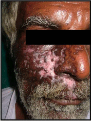
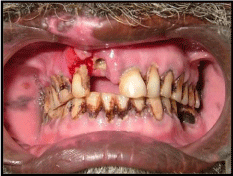
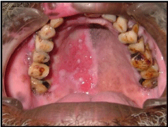
A 58-year-old male patient reported to the Oral medicine Department with the chief complaint of exfoliation of the upper right front teeth which is associated with pain since one week. Patient also gave history of multiple vesicular eruptions containing clear fluid on his right side of face associated with severe pain along the affected area. Vesicles were also observed by the patient in the palatal region. Patient was suffering from persistent fever and rapid weight loss. Extraoral examination revealed healed scars were present on the right side of upper face involving the malar region, the left side of nose, zygoma and upper part of right lip along with pigmentation (Figure 1). Intraoral examination revealed empty tooth socket and exposed alveolar bone with respect to 11 tooth region (Figure 2) and erythematous region over anterolateral part of hard palate was seen (Figure 3). Radiographic examination revealed missing 11and severe alveolar bone destruction. Generalised tender tcervicofacial lymphadenopathy was also observed. Patient was referred to physician to know his immune status and other underlying systemic condition, patient was turned out to be HIV sero positive. The patient was treated with oral acyclovir 800 mg. five times a day for seven days, nutritional supplements and antioxidants. Betadine mouthwash is also prescribed along with. Referral to the prosthodontics department and general physician was made for further treatment.

Figure 1: Extraoral scarring and pigmentation.

Figure 2: Unilateral erosion of the hard palate.

Figure 3: Empty socket.
Discussion
Zoster is a common, predominantly dermal, and neurologic disorder caused by the varicella-zoster virus (VZV), a virus morphologically and antigenically identical to the virus causing
varicella (chickenpox). Difference in clinical manifestations between varicella and zoster apparently depends on the immune status of individual patients; those with no prior immunologic exposure to varicella virus, most commonly children, develop the clinical syndrome of varicella, while those with circulating varicella antibodies develop a localized zoster. Zoster probably results most often from a failure of the immune system to contain latent varicellazoster virus replication. Whether other factors such as radiation, physical trauma, certain medications, other infections, or stress also can trigger zoster has not been determined with certainty. Nor is it entirely clear why circulating varicella antibodies and cellmediated immune mechanisms do not prevent recurrent overt disease, as is common with most other viral illnesses [4]. Zoster most commonly manifests in 1 or more posterior spinal ganglia or cranial sensory ganglia, presumably because viral particles have been preserved within these ganglia in a dormant state since the original episode of varicella. This results in pain and characteristic cutaneous findings along the corresponding sensory dermatomes of the involved ganglia. Less often, involvement of anterior and posterior horn cells, leptomeninges, and peripheral nerves is observed, with consequent muscle weakness or palsy, pleocytosis of spinal fluid, and/or sensory loss. Rarely, myelitis, meningitis, encephalitis, or visceral involvement may occur [5]. Osteonecrosis following herpes zoster infection is a rare but severe complication, and clinicians awareness is important for early detection and management of this condition. The most significant and debilitating complication of herpes zoster is herpetic neuralgia that accompanies and may persist in 10-15% of all zoster patients, particularly those over 60 years of age. Herpes zoster (HZ), a sporadic infection with an estimated lifetime risk of 10-20%, occurs in all age groups, especially among individuals who have had varicella any time in the past, but its prevalence almost doubles in each decade past the age of 50 years [6]. Individuals with immunosuppression are also more prone to it. The most significant and debilitating complication of HZ is herpetic neuralgia that accompanies and may persist in 10-15% of all zoster patients, particularly those over 60 years of age. Cranial and peripheral nerve palsies, encephalitis, pneumonitis, acute retinal necrosis, encephalitis, myelitis, and secondary bacterial infection are other well-known complications of HZ. The described cases had an uncommon complication of spontaneous tooth exfoliation after trigeminal HZ, of which only about 39 cases have been reported before 2012 in the English literature [3]. The precise pathomechanism(s) of these oral complications eventually leading to tooth loss and necrosis of the alveolar bone, periodontal tissue membrane, nerve supply, and adjacent gingival tissue remain largely uncertain. The pre-existing local periodontal disease does not appear to influence the tooth loss [7]. Although the majority of the described cases are aged over 50 years, had severe HZ, were suffering from lymphoproliferative or other malignancies, HIV infection, receiving corticosteroids or chemo-/radiotherapy, the conditions well-known for increased incidence and severity of HZ, few of the reported cases have been young, immunocompetent, and otherwise healthy individuals suggesting that factors like old age, pre-existing systemic illnesses too seem not to precipitate this rare complication. It has been also postulated that it could result from intense vasoconstriction, endarteritis, or vasculitis with ischemia of the blood supply that may be caused by the virus migrating along the trigeminal neurovascular bundle. It is possible that the varicella zoster virus invades the sympathetic nerves that accompanying the blood vessels to the alveolar bone causing vasoconstriction and hampered blood supply or that systemic viral infection perhaps injures odontoblasts and cause degenerative tissue changes/pulp necrosis [6]. Another plausible explanation is that by direct extension of neural inflammation and edema, the adjacent alveolar artery may get compressed in the narrow bony channels and resultant poor tissue perfusion. The postulation of local vasculitis caused by varicella zoster virus leading to infarction and ischemia of the periodontal tissue and alveolar bone has limited evidence despite well-documented vascular damage it causes in the central nervous system [8,9]. It has been reported in a south African study that extensive osteonecrosis and exfoliation of teeth in the area innervated by the nerve affected by HZ was more common in HIV sero positives, and the patient reported here was also HIV positive [10]. Acyclovir has been proved to reduce the duration and severity and the course of the herpes zoster infection in the acute phase significantly, if treatment is initiated within 48 hours of the onset of the symptoms. Acyclovir is considered to be beneficial in treating zoster patients due to its increased rate of phosphorylation in herpes infected cells. The present case is treated on the same backgrounds [11].
Conclusion
The Varicella Zoster Virus (VZV) produces two clinical results: varicella or chickenpox and Herpes Zoster Infection (HZI). Herpes Zoster (HZ) presents as a cutaneous vesicular eruption in the area innervated by the affected sensory nerve, usually associated with severe pain. Oral manifestations of HZ appear when the mandibular or maxillary divisions of the trigeminal nerve are affected. Herpes zoster infection leads to various complications if left untreated. Severe osteonecrosis and exfoliation of teeth in the region innervated by the nerve affected by HZ has been reported after HZ infection. Oral physicians should have a thorough knowledge about this condition, the treatment and prevention of the complications.
References
- Arduino P, Porter S. Herpes Simplex Virus Type 1 infection: overview on relevant clinic pathological features. J Oral Pathol Med. 2008; 37 :107-121.
- Hanvesakul R, Momin A, Gee M, Marrinan MA. Role for video assisted thoracoscopy in stable penetrating chest trauma. Emerg Med J. 2005; 22: 384-386.
- Lambade P, Lambade D, Saha TK, Dolas RS, Pandilwar PK. Maxillary osteonecrosis and spontaneous teeth exfoliation following herpes zoster. Oral Maxillofac Surg. 2012; 16: 369-372.
- Edgerton G. Herpes zoster ophthalmicus: a review of the literature. Arch Ophthalmol. 1945; 34: 40-62; 114–153.
- Burgoon CF Jr, Burgoon JS, Baldridge GD. The natural history of herpes zoster. J Am Med Assoc. 1957; 164: 265-269.
- Van Heerden WF, McEachen SE, Boy SC. Alveolar bone necrosis and tooth exfoliation secondary to herpes zoster in the setting of HIV/AIDS. AIDS. 2005; 19: 2183-2184.
- Wright WE, Davis ML, Geffen DB, Martin SE, Nelson MJ, Straus SE. Alveolar bone necrosis and tooth loss. A rare complication associated with herpes zoster infection of the fifth cranial nerve. Oral Surg Oral Med Oral Pathol. 1983; 56: 39–46.
- Arikawa J, Mizushima J, Higaki Y, Hoshino J, Kawashima M. Mandibular alveolar bone necrosis after trigeminal herpes zoster. Int J Dermatol. 2004; 43: 136–137.
- Chindia ML. HIV-associated fulminating herpes zoster infection with alveolar necrosis and tooth exfoliation: A case report. Dent Update. 1997; 24: 126–128.
- Feller L, Wood NH, Raubenheimer EJ, Meyerov R, Lemmer J. Alveolar bone necrosis and spontaneous tooth exfoliation in an HIV-seropositive subject with herpes zoster. SADJ. 2008; 63: 106-10.
- Strommen GI, Pucino F, Tight RR, Beck CL. Human infection with H. Zoster, etiology, pathophysiology, diagnosis, clinical course and treatment. Pharmacotherapy 1998; 8: 52-68.