
Case Report
Austin J Dent. 2015;2(2): 1020.
Extra Oral Swelling Mimicking a Carcinoma in an Elderly Patient Due to Unusually Impacted Canine- A Case Report
Dalia A Abuelenain¹, Ensanya A Abou Neel1,3,4*and Ayman Al-Dharrab²
¹1Department of Oral Medicine and Radiology, A B Shetty Memorial Institute of Dental Sciences, Nitte University,India
²2Department of Oral Medicine and Radiology, Sree Aanjenaya Institute of Dental Sciences, Kerala University of Health Sciences, India
³Department of Oral Medicine and Radiology, Century International Institute of Dental Sciences and Research Centre, India-575018
*Corresponding author: Renita Lorina Castelino,Department of Oral Medicine and Radiology, A B Shetty Memorial Institute of Dental Sciences, Nitte University,Mangalore- 575018, India
Received: April 21, 2015; Accepted: June 02, 2015; Published: June 04, 2015
Abstract
Inverted teeth have been reported in both maxilla and mandible and most of them are inverted impacted third molars and premolars. In the oral cavity maxillary canines are called as “corner stones” and are most important teeth in terms of aesthetics and function. Impacted teeth are those teeth with a delayed eruption time or that are not expected to erupt completely based on clinical and radiographic assessment. The permanent maxillary canine impaction has been reported in about 1% to 5% of the population. They are known to be second most frequently impacted teeth after the third molars. We hereby report a case of a canine space infection causing the involvement of the skin mimicking carcinoma of skin on the right side of the face due to an impacted canine in an elderly female patient.
Keywords: Inverted; Canine; Carcinoma
Introduction
Impaction of tooth is retardation or halt in the normal process of eruption. A canine is considered as being impacted if it is interrupted after complete root development or if the contra lateral tooth is erupted for at least 6 months with complete root formation [1,2]. Maxillary canine impactions occur twice as often in females than in males in the ratio 2.3:1 [3]. Most of the canine impactions are palatal (85%) and it is also reported that only 8% of canine impactions are bilateral in nature [4]. The case presented here is a case of an elderly woman who had canine space infection involving the skin mimicking a skin carcinoma due to the impacted canine.
Case Report
A 65 year old female patient reported to the Department Of Oral Medicine And Radiology with the complaint of swelling in the right side of the face since 3 months (Figure 1). The swelling was gradually increasing in size. There were no constitutional symptoms reported by the patient. The family, drug and medical history were non-contributory. On further questioning, the patient reported of consulting a physician who provisionally diagnosed the swelling as carcinoma of skin based on the history and advised biopsy of the same. The biopsy report ruled out malignancy and stated the underlying connective tissue showed dense chronic inflammatory infiltrate consisting of lymphocytes and plasma cells suggestive of chronic infection (Figure2). The patient was then referred to the dental department for further evaluation. On extra oral examination a diffuse swelling was noted on the right middle one third of the face measuring approximately 3x3 cm in size. The overlying skin appeared hypo pigmented over a large area. The peeling of skin surrounded by pinkish area was noted on the superior part of the swelling (Figure 1). There was increase in the temperature on the right side when compared to the left side. The swelling was firm in consistency and slightly tender on palpation and the overlying skin was not pinchable. On intra oral examination, the patient was partially edentulous in the maxillary and mandibular arch and had generalised periodontitis (Figure 3). There was no swelling or abnormality noted intra orally. Based on the history and clinical features a provisional diagnosis of chronic skin infection was made. As part of routine examination, radiographic investigations were made. An Orthopantomogram (OPG) was made which revealed an interesting finding of inverted bilateral impacted canines on the maxillary arch between the maxillary antrum and the lateral wall of the nasal cavity. The other findings were generalised periodontitis and multiple missing maxillary and mandibular teeth. It was also noted that root formation of inverted maxillary canines was almost complete indicating that they were impacted. The root tip of the right impacted inverted canine appeared to be beyond the alveolar crest and probably was just covered by the overlying mucosa and a breach in the mucosa through the overlying mucosa resulted in the swelling and infection. The root tip of the left inverted impacted canine was seen near the root tip of the premolar tooth (Figure 4). A lateral cephalogram was made which showed the inverted canines near the nasal cavity (Figure 5). The patient was advised surgical removal of the impacted teeth along with total extraction of the remaining teeth followed by prosthetic rehabilitation. Following the surgical removal of the impacted teeth the swelling regressed in size.
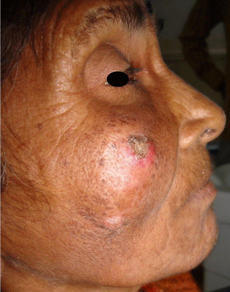
Figure 1: Extra oral photograph of the patient.
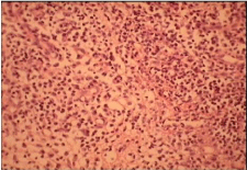
Figure 2: Histopathology of the lesion.
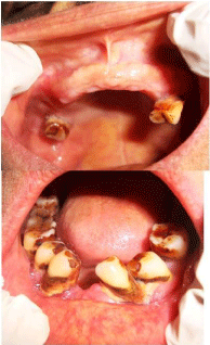
Figure 3: Intra oral picture of the maxillary and the mandibular arch.
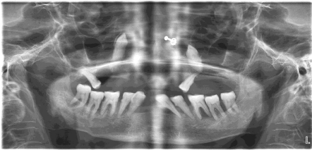
Figure 4: OPG showing bilateral inverted impacted canines.
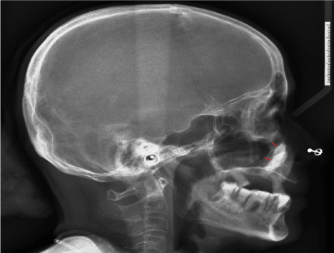
    Figure 5:  Lateral cephalogram showing the unusual pattern of the impacted
inverted canines marked by red lines.
Discussion
Eruption is a continuous movement of a tooth from its developmental location to its functional position. Localized disturbances include primary impaction, ankylosis, malpositioning of teeth etc [5]. Inversion has been defined as ‘the malposition of a tooth in which the tooth has reversed and is positioned upside down [6]. Inverted teeth have been reported in both maxilla and mandible, and most of them are inverted impacted third molars and premolars [7]. Although inverted impacted teeth may remain in position for many years without clinical manifestations and may be detected in radiographic examinations incidentally, many complications including delayed or ectopic eruption, crowding, diastema, eruption into the nasal floor, resorption of the adjacent root and development of a dentigerous or primordial cysts may arise [8,9]. In the case reported here the impacted canine tooth caused space infection which was diagnosed provisionally as skin carcinoma leading to misdiagnosis. It was only after the radiographic examination the exact cause of the swelling could be made out stressing on the fact that routine radiographic investigations do help in early detection and diagnosis. As the patient was old she could not recollect about the missing tooth in the oral cavity. The cause for impaction is unknown but it is now being predicted that a deficiency in the cell signalling process of one tooth or more adjacent teeth at an early stage, could cause the tooth buds to move in the wrong direction leading to transposition of the tooth germ [10]. The most common causes for canine impactions are usually localized and they occur as a result of any one or combination of the following factors: tooth size/arch length discrepancy, prolonged retention or early loss of the primary canine, abnormal position of the tooth bud and the long path of eruption, presence of an alveolar cleft, ankylosis, follicular disturbance and cyst or neoplasm formation, dilacerations of the root or trauma, and idiopathic factors including primary failure of eruption [11]. Most of the canine impactions are palatal (85%) and it is also reported that only 8% of canine impactions are bilateral in nature as seen in our case. The case presented here is unusual because the impacted tooth caused space infection of the facial region without obvious intra oral findings and also being misdiagnosed as skin carcinoma resulting in mental trauma to the patient.
Conclusion
Impacted teeth can cause various problems and hence routine radiographic examination must be carried out in patients with congenitally missing teeth to prevent further problems like malocclusion, space infections, cysts, tumours etc. in the long course of time.
References
- Lindauer SJ, Rubenstein LK, Hang WM, Andersen WC, Isaacson RJ. Canine impaction identified early with panoramic radiographs. J Am Dent Assoc. 1992; 123: 91-92, 95-97.
- Goel A, Loomba A, Goel P, Sharma N. Interdisciplinary approach to palatally impacted canine. Natl J Maxillofac Surg. 2010; 1: 53-57.
- Aydin U, Yilmaz HH, Yildirim D. Incidence of canine impaction and transmigration in a patient population. Dentomaxillofac Radiol. 2004; 33: 164-169.
- Millet D, Welbury R. Clinical problem solving in orthodontics and paediatric dentistry. 1st ed. New York. Elsevier; 2005; 194.
- Jacobs R, Willems G. Inverted eruption of a supplemental lower premolar: report of an unusual case. Int J Paediatr Dent. 2003; 13: 46-50.
- Ulusoy AT, Akkocaoglu M, Akan S, Kocadereli I, Cehreli ZC. Reimplantation of an inverted maxillary premolar: case report of a multidisciplinary treatment approach. J Clin Pediatr Dent. 2009; 33: 279-282.
- Mori S, Kitamura K, Ohmori T. Inverted tooth eruption. Report of a case. Oral Surg Oral Med Oral Pathol. 1979; 47: 389-390.
- Avsever H, Gunduz K, Orhan K, Aksoy S. An inverted eruption of mesiodens: Report of a Rare Case. MÜSBED. 2012; 2: 37-39.
- Lakshman AR, Kanneppady SK, Balan P. A Rare Case Report of Inverted Eruption of Supernumerary Tooth in the Anterior Maxilla. MÜSBED. 2013; 3: 164-166.
- Nimbeni B, Golai S, Kumar H, Rai S. Unusual case of bilateral impacted and inverted maxillary canines. IJSS Case Reports & Reviews. 2015; 1: 11.
- Sajnani AK. Permanent maxillary canines - review of eruption pattern and local etiological factors leading to impaction. J Investig Clin Dent. 2015; 6: 1-7.