
Research Article
Austin J Dent. 2015;2(3): 1023.
Mandibular Reconstruction and Implantology: Anatomical Study and CT Scan of Dried Human Fibulas
Almeida FCS¹*, Moreira MS², Farias BUL³, Dias RB4, Crossato EM¹ and Silva DP4
¹Department of Community Dentistry, Universidade de São Paulo, Brazil
²Postgraduate Course in Dentistry, Universidade Ibirapuera, Brazil
³Department of Pediatric and Orthodontics, University of São Paulo, Brazil
4Department of Maxillofacial Surgery, University of Sao Paulo, Brazil
*Corresponding author: Almeida FCS, Department of Community Dentistry, University of Sao Paulo, Av. Prof Lineu Prestes, 2227, Cidade Universitária, São Paulo, SP, Brazil
Received: April 08, 2015; Accepted: June 22, 2015; Published: June 24, 2015
Abstract
The aim of this study was to evaluate the potential presented by human dried fibula on regular implants installation (7 mm or more) and to search for a standard anatomical position of more appropriate areas. Thirty human dried fibulas, in five distinct areas of each bone, were evaluated in a tomographic study and the data were processed statistically. The results revealed that the thirty bones examined showed significant differences between the maximum and minimum values measured, without any anatomical standard for these differences and that only three of these bones showed values less than the standard 7 mm, but these same bones presented other dimensions compatible with the installation of implants. The differences between maximum and minimum values ranged from 2.50 mm up to 11.50 mm (p=0 000). The analyzed data showed that 29 of the 30 bones presented viable areas for the installation of regular implants. The indication for CT scan of the patient’s fibulae can be a valuable test, to the installation of larger implants, thus increasing the survival of the implants and the success of oral rehabilitation.
Keywords: Free flap fibula; Computed tomography; Maxillofacial rehabilitation
Introduction
The fibula has been the bone of choice in mandibular reconstruction, also starting to be used and applied with great efficiency in maxillary reconstructions [1]. This bone has characteristics that facilitate its plasticity by the surgeon, do not cause much morbidity, allow multiple osteotomies and promote good modeling in mandibular reconstruction. This is a bone that has good quality for being bicortical, offers sufficient quantity for bone reconstruction, besides being a good bed for receiving dental implants [2-4].
On the other hand, the prosthetic rehabilitation of a jaw segment reconstructed by fibula without the aid of implants is a very difficult task. Considering that sometimes a lack of vestibule and the presence of excess skin, replacing the keratinized mucosa from the oral cavity, are observed in the reconstructed section. Also, in these cases, an occlusal discrepancy (anteroposterior) relationship between the maxilla and mandible, and large vertical differences between the fibula and the remaining bone are still common [5].
Additionally, the literature alerts us to the fact that the vast majority of patients reconstructed by fibula does not use functional prostheses and does not receive dental rehabilitation; and, in some cases, these numbers may exceed the 80% So, this is a challenge to be overcome, as the quest for quality of life should be the ultimate goal of any treatment. Implants can also increase the rate of use of prostheses by these patients, since they assist in the retention and stability of the parts. Assuming advantages in the use of osseointegrated implants in the prosthetic rehabilitation of jaws reconstructed with the fibula, anatomic and radiographic knowledge of this bone is required so that the maxillo-facial professional can be prepared and secure when discussing the rehabilitation, the means of retention and the stability of the prostheses.
Materials and Methods
Thirty dried human fibulae were selected at random from the Department of Anatomy, Institute of Biomedical Sciences (ICB) of the University of Sao Paulo. Inclusion criteria for selection were that the bones had to have good anatomic integrity and possess the essential proximal and distal ends in great condition. The pieces were measured on the long axis by the same observer. Then, the center of each bone was marked and, to the right and left of this center, two new points were marked at a distance of 3 cm from each other, thus forming two areas on the right and two on the left of the center. These areas served as markers for the tomographic images taken images and were classified in sections 1, 2, 3, 4, 5 (Figure 1); and, each section was evaluated in three distinct areas A, B, C (Figures 2A and 2B). After marking all the bones, they underwent CT examination performed using the General Electric Prospeed helical unit (WW 2500, WL 1000) (Figure 3). The fibulae were examined in groups of four parts, making five CT slices of 1 mm collimation, with 30 mm spacing in the part during the anatomical analysis. In each section, the presence of three cortical bones per piece was observed. The cortical thickness was measured by the same observer, resulting in a (N) total of 15 measurements for each fibula (Table 1). The data were entered and analyzed using the statistical package Stata 10. Descriptive measurements were carried out (minimum and maximum values, standard deviation, differences between minimum and maximum values). To investigate the differences between the heights of fibulae, the t-test for paired data was performed. Based on the supply of regular implants, on the international market, a minimum value of 7.00 mm thickness was established as the minimum acceptable for the installation of implants. The significance level was 95%.
![]()
    Fibulae
    N
    Median
    Min
    Max
    SD
    Diff
    p


    1
    15.00
    12.10
    10.00
    14.00
    1.07
    4.00
    0.00


    2
    15.00
    11.60
    8.00
    14.50
    2.05
    6.50
    0.00


    3
    15.00
    10.50
    9.00
    13.50
    1.28
    4.50
    0.00


    4
    15.00
    11.37
    8.00
    14.00
    1.74
    6.00
    0.00


    5
    15.00
    6.70
    4.00
    11.50
    2.27
    7.50
    0.00


    6
    15.00
    8.03
    5.00
    10.00
    1.62
    5.00
    0.00


    7
    15.00
    7.57
    5.00
    10.00
    1.47
    5.00
    0.00


    8
    15.00
    7.57
    6.00
    8.50
    0.59
    2.50
    0.00


    9
    15.00
    9.43
    6.50
    14.00
    2.44
    7.50
    0.00


    10
    15.00
    11.87
    10.00
    15.50
    1.95
    5.50
    0.00


    11
    15.00
    13.60
    10.00
    17.50
    2.32
    7.50
    0.00


    12
    15.00
    9.13
    6.00
    14.00
    2.26
    8.00
    0.00


    13
    15.00
    9.80
    8.00
    12.50
    1.41
    4.50
    0.00


    14
    15.00
    12.30
    9.00
    15.50
    1.88
    6.50
    0.00


    15
    15.00
    9.93
    7.00
    14.00
    1.90
    7.00
    0.00


    16
    15.00
    11.03
    8.00
    14.50
    2.08
    6.50
    0.00


    17
    15.00
    8.13
    5.00
    12.50
    2.06
    7.50
    0.00


    18
    15.00
    12.80
    8.50
    17.00
    2.58
    8.50
    0.00


    19
    15.00
    10.77
    8.00
    13.00
    1.60
    5.00
    0.00


    20
    15.00
    8.63
    6.50
    10.50
    1.43
    4.00
    0.00


    21
    15.00
    9.95
    7.00
    12.00
    1.22
    5.00
    0.00


    22
    15.00
    9.80
    6.00
    14.80
    2.54
    8.80
    0.00


    23
    15.00
    10.41
    8.60
    12.50
    1.13
    3.90
    0.00


    24
    15.00
    10.39
    8.70
    12.00
    0.94
    3.30
    0.00


    25
    15.00
    10.81
    9.80
    14.90
    1.31
    5.10
    0.00


    26
    15.00
    9.71
    7.20
    13.30
    1.56
    6.10
    0.00


    27
    15.00
    8.38
    7.00
    11.80
    1.57
    4.80
    0.00


    28
    15.00
    12.13
    9.50
    15.80
    2.25
    6.30
    0.00


    29
    15.00
    11.75
    9.80
    16.00
    2.39
    6.20
    0.00


    30
    15.00
    9.28
    4.50
    16.00
    4.01
    11.50
    0.00
Table 1: Descriptive measurements of the height of the fibula.
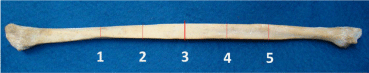
Figure 1: Example of Fibula with five sections analyzed.
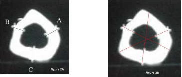
    Figure 2A and 2B:  Example of the three areas analyzed for each selected
section one of the five.
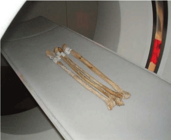
Figure 3: Dry bones underwent CT examination.
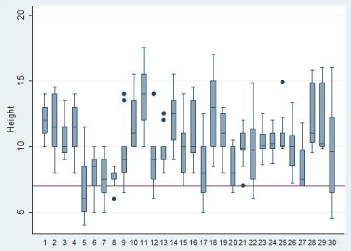
    Figure 4:  Graphic of the maximum and minimum measurements in relation to
the established standard of 7mm.
Results
Data analysis aimed at assessing the fifteen Minimum measurements (Min) and fifteen Maximum measurements (Max) of the thirty fibulae studied. When analyzing the data of fibula 1 (one), for example, it was found that the average of the fifteen measurements was 12.10 mm with the minimum measurement 10.00 mm and the maximum 14.00 mm. The difference between the maximum and minimum values (Max - Min) was also calculated which, in the case of fibula 1 (one), was 4.00 mm; the standard deviation was 1.07; and, p = 0.00. For all other 29 bones analyzed, important differences were also observed between the measurements obtained, as described for fibula 1. Descriptive analysis of the data obtained from the CT scans revealed that, in the sample examined, only fibula five [5] had the mean of the measurements less than the standard of 7.00 mm; although, three other bones (7, 17 and 30) presented the minimum measurement below the standard, the first two were 5 mm and the last was 4.50 mm. But, when all fifteen measurements are taken into account, the average value is greater than the standard; on the other hand, the maximum measurement observed was 17.50 mm. Other data relate to differences between the minimum and maximum measurements (Diff), which ranged from 2.50 mm in fibula eight (8) to 11.50 mm in fibula thirty (30). To check the real difference among the measurements obtained, the Student’s t-test was applied, which confirmed the statistically significant difference among the heights of all fibulae examined (Figure 4).
Discussion
Mandibulectomies are due to the resection of benign and malignant tumors of the oral cavity, maxillofacial trauma, and infections; and, they can cause functional deficits which include difficulties in mastication and swallowing, as well as changes in social relations, and immediate reconstruction is desirable in order to reduce the impact of the resection on the patient’s life [6].
There is no doubt about the advantages of the fibula, compared with other donors, in mandibular reconstruction [3]. These advantages include the possibility of several osteotomies with low morbidity rate, compared to the iliac bone, for example; the possibility of combining skin flaps for the reconstruction of soft tissue defects; and, the possibility of osseointegration of implants (being a bone with two dense cortical). However, despite the many advantages, the bone “height” limits the rehabilitation with implants, and the discrepancy in “height” between the fibula and the remaining jaw, especially in the dentates, will always exist. However much higher the height of the fibula, the smaller will be the vertical discrepancy between the flap and the native mandible [7].
Papers published around the world try to minimize the vertical gap in partial dentates reconstructed by fibula, as this seems to be one of the major obstacles in this type of reconstruction. However, these methods are not applied to all patients, and are usually reported in small samples with short follow up [2,3]. Searching for less invasive alternatives that reduce risks to the patient, seems to be a desirable alternative.
Another important aspect, that must be considered and that should guide the rehabilitative procedure, concerns the comprehensive rehabilitation in which the center is the patients, their needs, desires, expectations and the return to normal activities prior to which the illness crippled them. These include work, study, family and emotional relationships, and are good indicators of the success of the comprehensive rehabilitation of the patient [3,8]. The literature warns that only 45% of patients rehabilitated by fibula returned to an unrestricted diet, 45% soft diet, 5% use only liquid diet, and 5% enteral feeding via nasogastric tube [9]. Recent studies corroborate the findings of Cordeiro, as they find that 58% of men and 25% of women who received fibula report difficulty in speaking. On the other hand, 62% of women and 34% of men have esthetic complaints about the region considered. However, a third of these patients did not receive prosthetic rehabilitation and 43% of them received implant prostheses [10].
Other studies show that most reconstructed fibulae do not receive osseointegrated implants; however, there is no consensus among the authors about what motivates the non-use of implants to rehabilitate these patients, since we know that these improve the retention and stability of the prostheses. The high costs, the adjuvant treatments (chemotherapy and / or radiotherapy), the fatigue of the patient, the low survival rate of patients, may be hypothesized as justifying this picture, although we cannot reject the hypothesis that the lack of knowledge about the real potential of fibula rehabilitation, the advantages of installing implants under the best possible conditions and, essentially, the patient’s desire to be fully rehabilitated, can change this situation [7].
Our data show that the fibula is not a uniform bone with regard to its height and may vary, as in bone 30, from 16.00 mm at its maximum to 4.50 mm at its minimum. Fibula 11, in turn, which was 17.50 mm at maximum and 10 mm at minimum, is a good example for our discussion. Although it had viable bone in all areas analyzed, from the standpoint of regular placement of implants it revealed areas with more bone height than others, allowing far greater implant placement with consequent decrease in the vertical gap and possibly increased longevity of these implants. Another example is fibula 18 because, while this bone showed areas bordering the placement of regular 8.5 mm implants, there were areas with significant 17.00 mm bone height; thus, suggesting that the surgeon’s election of regions from within the same fibula, more favorable to rehabilitation by implants, could reverse the numbers indicating that the majority of patients reconstructed by fibula receive neither dental reconstruction nor implant placement (Table 1).
Despite 29 of the 30 bones examined showing sufficient height for placement of regular implants in some of the areas studied, it is important to note the discrepancy between the measurements and the lack of a pattern among them, indicating that the bones do not have heights with uniform distribution.
A recent study reveals that the CT scan is a important tool to analysis of the anatomy of the fibula for the optimal site of installation of osseointegrated implants in maxillofacial reconstruction [11]. Therefore, our data to suggest that the tomographic study of both fibulae of the patient may be interesting, so that the surgeon could have more options to decide which bones and regions are larger, to minimize some of the problems reported in the literature; especially, the great gap produced by the difference between the height of the transplanted fibula and the native mandible, because small implants, theoretically, would not handle the masticatory load and could be lost in a short time. Also, during the surgical rehabilitation planning for the patients are treatments for infection, trauma, or benign and malignant tumors, and we have to consider survival as a decisive factor in searching for the most favorable conditions possible for a rehabilitation, that will restore the lost form of the face and jaw line, the oral functions (chewing, speaking and swallowing), and return the patient’s desire to smile, preferably with a suitable prosthesis with good retention and stability that can restore the integrity of the conditions lost by the preoperative patient.
Conclusion
CT scan of the human fibulae revealed that 29 of the 30 bones examined had viable areas for the placement of regular implants;
There was great variation between the maximum and minimum measurements analyzed in each bone;
There was no pattern among the measurements obtained, with respect to the examined areas;
The indication for CT scan of the patient’s fibulae can be a valuable test, in the search for areas with more bone height to allow the installation of larger implants, thus increasing the survival of the implants and the success of oral rehabilitation.
Acknowledgment
The authors thank the Departments of Radiology, School of Dentistry, Hospital das Clinicas of the School of Medicine of USP for their collaboration, and the Institute of Biomedical Sciences for providing the fibulas.
References
- Mukohyama H, Haraguchi M, Sumita YI, Iida H, Hata Y, Kishimoto S, et al. Rehabilitation of a bilateral maxillectomy patient with a free fibula osteocutaneous flap. J Oral Rehabil. 2005; 32: 541-544.
- Wang KH, Inman JC, Hayden RE. Modern concepts in mandibular reconstruction in oral and oropharyngeal cancer. Curr Opin Otolaryngol Head Neck Surg. 2011; 19: 119-124.
- Anne-Gaëlle B, Samuel S, Julie B, Renaud L, Pierre B. Dental implant placement after mandibular reconstruction by microvascular free fibula flap: current knowledge and remaining questions. Oral Oncol. 2011; 47: 1099-1104.
- Fernandes R. Fibula free flap in mandibular reconstruction. Atlas Oral Maxillofac Surg Clin North Am. 2006; 14: 143-150.
- Reychler H, Iriarte Ortabe J. Mandibular reconstruction with the free fibula osteocutaneous flap. Int J Oral Maxillofac Surg. 1994; 23: 209-213.
- Kadota C, Sumita YI, Wang Y, Otomaru T, Mukohyama H, Fueki K, et al. Comparison of food mixing ability among mandibulectomy patients. J Oral Rehabil. 2008; 35: 408-414.
- Garrett N, Roumanas ED, Blackwell KE, Freymiller E, Abemayor E, Wong WK, et al. Efficacy of conventional and implant-supported mandibular resection prostheses: study overview and treatment outcomes. J Prosthet Dent. 2006; 96: 13-24.
- Vu DD, Schmidt BL. Quality of life evaluation for patients receiving vascularized versus nonvascularized bone graft reconstruction of segmental mandibular defects. J Oral Maxillofac Surg. 2008; 66: 1856-1863.
- Cordeiro PG, Disa JJ, Hidalgo DA, Hu QY. Reconstruction of the mandible with osseous free flaps: a 10-year experience with 150 consecutive patients. Plast Reconstr Surg. 1999; 104: 1314-1320.
- Hölzle F, Kesting MR, Hölzle G, Watola A, Loeffelbein DJ, Ervens J, et al. Clinical outcome and patient satisfaction after mandibular reconstruction with free fibula flaps. Int J Oral Maxillofac Surg. 2007; 36: 802-806.
- Ide Y, Matsunaga S, Harris J, O’ Connell D, Seikaly H, Wolfaardt J. Anatomical examination of the fibula: digital imaging study for osseointegrated implant installation. J Otolaryngol Head Neck Surg. 2015; 44: 1.