
Case Report
Austin J Dent. 2016; 3(1): 1029.
Odontoma Associated with Over Retained Primary Teeth that Caused Ectopic Eruption of Canine: A Case Report
Jose D*
Department of Pedodontics, Kannur Dental College, India
*Corresponding author: Deepak Jose, Department of Pedodontics, Kannur Dental College, Anjarakandy, kannur, Kerala, India
Received: January 05, 2016; Accepted: April 04, 2016; Published: April 06, 2016
Abstract
Odontomas are the most common type of odontogenic tumours and usually they are asymptomatic. However, odontomas are commonly causes ectopic position and/or impacted permanent teeth. There are few reports on odontomas which are associated with over retained deciduous tooth. Odontoma are commonly unerupted and surgical removal of odontoma is regular treatment of line. The purpose of the present case report is to describe a case of an odontoma which was associated with an over retained deciduous tooth. The over retained deciduous tooth was removed and the odontomas was surgically removed and over twenty seven denticles were obtained from the lesion.
Keywords: Odontoma; Ectopic canine; Primary teeth; Dental anomaly
Introduction
Odontomas are developmental anomalies caused by the growth of completely differentiated epithelial and mesenchymal cells which give rise to ameloblasts and odontoblasts [1]. These are hamartomas rather than true neoplasms [2] and they may contain various dental tissues, i.e., enamel, dentin, cementum some times pulpal tissue [3]. Most of the odontomas are asymptomatic in nature and they are found during routine radiographic examination accidentally. They are the most common odontogenic tumour and among all odontogenis tumors, odontomas account for 50% [1]. Odontomas can be classified as complex and compound odontomas. Compound odontomas are commonly found in the maxillary anterior region and they are similar to normal teeth whereas compound odontomas are seen in posterior mandible followed by anterior maxillary region and they are mass like irregular structures. Radiographically compound odontomas are seen as a mass of multiple calcified structures which resemble normal teeth surrounded by a narrow radiolucent zone. Complex odontomas are more or less amorphous mass of calcified material which is also surrounded by a narrow radiolucent zone [3].
It has been suggested that trauma and infection at the place of the are common reasons for its appearance, however, the aetiology of odontomas are not clearly documented Odontomas are usually asymptomatic, and they may be detected by chance on a routine radiograph (panoramic and/or intra-oral X-rays), or when they are large enough to cause a swelling of the jaw. Retained deciduous tooth or an impacted tooth, are clinical signs suggestive of an odontoma. The treatment of these lesions includes surgical removal followed by orthodontic correction if needed and follow up [4]. Odontomas could be diagnosed at any age and mostly commonly found in first two decades of life. Though diagnosis can be confirmed by radiographs, it is recommended to carry out a histological examination to confirm the diagnosis [5]. The purpose of the present case is to report a case of a compound odontoma with twenty seven denticles caused over retained of deciduous teeth and ectopic position of maxillary canine.
Case Presentation
A 14 year old male patient presented to the Department of Pedodontics and preventive dentistry with a chief complaint of irregularly aligned maxillary anterior teeth. Upon examination, the primary maxillary right lateral incisor was over retained, the permanent maxillary right lateral incisor had erupted distal to it and buccaly erupted canine were observed (Figure 1). An intra-oral periapical radiograph revealed the presence of compound odontome in relation to the deciduous lateral incisor perioapically. A maxillary anterior occlusal radiograph and a panoramic radiograph (Kuer’s Technique or Verticle tube shift technique) were taken for further to locate the odontome (Figure 2 a and b). Based on clinical and radiographic examinations the diagnosis of compound odontome was confirmed. Parents and patient were informed regarding the presence of the odontome. The options that were included that extraction of lateral incisor, surgical removal of odontome and fixed orthodontic treatment for the alignment of the teeth in maxillary arch. The deciduous lateral incisor and surgery removal of odontoma were performed under local anesthesia. The mineralized structures showing a tooth-like appearance were found within the lesion (Figure 3). Overall Twenty seven denticles were removed from the surgical site. The patient was referred to the department of Orthodontics for further evaluation and treatment.
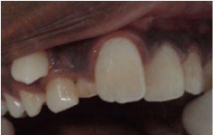
    Figure 1:  Intraoral view of maxilla showing retained teeth 53 and 52 and
buccally erupted 13.
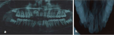
    Figure 2:  Panoramic radiograph (a) and maxillary anterior occulasal
radiograph and (b) showing radio opaque mass and impacted denticles in
tooth 13 region.
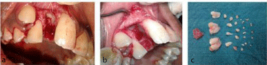
    Figure 3:  (a) Showing the odontoma after surgical exposure (b) surgical site
after removal of the lesion and (c) denticles removed along with the cystic
lining.
Discussion
Odontomas are most common type of odontogenic tumours however, most of the authors preferred to refer to it as hamartoma not a true tumour [3]. It is been reported that one third of compound odontomas and one half complex odontomas prevented eruption of teeth [6]. Odontomas are common among Caucasian population [7]. They are rarely associated with primary teeth and often cause impaction of the associated primary teeth [8,9]. In this case it was associated with an over retained deciduous tooth and the successor tooth erupted distal to it. Odontomas of all types composite approximately 22% of odontogenic tumours of jaws [6]. There is no gender predilection and odontoma can occur at any age but most commonly occurs in the second decade of life (Mallineni). Two third of all odontomas combined occur in the maxillary arch and 33% in the mandible. The compound odontoma has predilection towards the anterior maxilla is reported to be high (61%) whereas only 34% of complex odontoma occurred here. In general complex odontoma had a predilection for the posterior jaws 59% and lastly the premolar area (7%). Fascinatingly both types of odontoma reported more often on the right side of the jaw [10-14]. Odontomas show no gender predilection and are usually diagnosed in the second decade of life [14]. They are commonly found in the anterior maxillary region. Delayed eruption, persistence of the primary tooth, and the presence of a tumour are the most common clinical finding [14-16]. There are few reports of odontomas which has erupted through the soft tissue [12]. There are few reports of relapse of this condition.
Literature suggest that radiographic investigation should be carried out in any pediatric patients who shows clinical evidence of delayed eruption of permanent tooth or temporary tooth displacement with or without a history of dental trauma [3]. Especially for the location of impacted tooth or any odontogenic structure vertical tube shift technique id most useful [17]. It has been suggested clinical and radiographic investigations are useful in diagnosis of odontomas, most recently it has been suggested radiographic, histological and micro radiography have been suggested for the diagnosis [18]. In the present case the odontoma was diagnosed based the both clinical and radiographic evaluation. Clinically there was evidence of retained primary tooth 52 with buccally displaced canine, vertical tube shift technique (panoramic radiograph and upper maxillary occulsal radiograph) [19] used for the localization of odontoma.
The odontoma presents as a well-de ned radiopacity situated in bone, but with a density that is greater than bone and equal to or greater than that of a tooth. It contains foci of variable density. Odontomas have been associated with trauma during primary dentition, as well as with inflammatory and infectious processes, hereditary anomalies (Gardner syndrome, Hermann’s syndrome), odontoblastic hyperactivity and alterations in the genetic components responsible for controlling dental development [11]. However, the etiology of this malformation is not clearly understood, there is some reported evidence to show the genetic involvement for both complex and compound odontomas. It has been suggested that trauma and infection at the place of the common reasons. None of these etiological factors were evident at our case. In the present case the presence of retained primary tooth and displacement of both lateral incisor and canine have lead for opting the radiographic evaluation. Through clinical and radiographic investigations in case of retained primary teeth along with ectopically placed permanent teeth would reveal underlying pathology.
Conclusion
Early diagnosis of odontomas allows adoption of a less complex and less expensive treatment and ensures better prognosis. Retained deciduous teeth and ectopic position of permanent teeth may suggest underlying odontogenic pathology. Through clinical and radiographic investigations and appropriate multi-disciplinary planning is essential to overcome potential complications.
References
- Santos JN, Pinto LP, de Figueredo CR, de souza LB. Odontogenic tumors: analysis of 127 cases. Pesqui Odontol Bras. 2001; 15: 308-313.
- Salgado H, Mesquita P. Compound odontoma- case report. Rev port estomatol med dent Cir Maxilofac. 2013; 54: 161-165.
- Oliveira B, Campos V, Marçal S. Compound odontoma- diagnosis and treatment: 3 case reports. Pediatr Dent. 2001; 23: 151-157.
- An S, An C, Choi K. Odontoma: a retrospective study of 73 cases. Imaging Sci Dent. 2012; 42: 77-81.
- Iatrous I, Vardas E, Theologie-Lygidakis N, Leventis M. A retrospective analysis of the characteristics, treatment and follow-up of 26 odontomas in Greek children. J Oral Sci. 2010; 52: 439-447.
- Katz RW. An analysis of compound and complex odontomas. ASDC J Dent Child. 1989; 56: 445-449.
- Yeung K, Cheung R, Tsang M. Compound odontoma associated with an unerupted and dilacerated maxillary primary central incisor in a young patient. Int J Paediatr Dent. 2003; 13: 208-212.
- Stajcic ZZ. Odontoma associated with a primary tooth. J Pedod. 1988; 12: 415-420.
- Otsuka Y, Mitomi T, Tomizawa M, Noda T. A review of clinical features in 13 cases of impacted primary teeth. Int J Paediatr Dent. 2001; 11: 57-63.
- Mallineni SK. Supernumerary teeth: Review of the literature with recent updates. Conference Papers in Science. 2014.
- Shafer, Hue, Levy. Cysts and Tumors of the jaws. In: a Textbook of Oral Pathology, 5th edn. Saunders WB Company. 258-317.
- Cawson RA, Odell EW. Odontogenic tumors and Tumor like lesion of the jaws. In: Essentials of Oral Pathology and Oral Medicine. 6th edn. Churchill Livingstoe. 1998; 117-131.
- Hidalgo-Sánchez O, Leco-Berrocal MI, Martínez- González JM. Meta-analysis of the Epidemiology and clinical manifestation of odontomas. Med Oral P Patol Oral Cir Bucal. 2008; 13: E730-734.
- Ferrer Ramírez MJ, Silvestre Donat FJ, Estelles Ferriol E, Grau García Moreno D, López Martínez R. Recurrent infection of a complex odontoma following eruption in the mouth. Med Oral. 2001; 6: 269-275.
- Vengal M, Arora H, Ghosh S, Pai KM. Large erupting complex odontoma: A case report. J Can Dent Assoc. 2007; 73: 169-173.
- Fernández López RG, López Buendía MC, Ruiz González E. Plasma rico en factors de crecimiento en cirugía bucal. Presentación de caso clínico. Rev Odont Mex. 2005; 9: 141-146.
- Mallineni SK, Nuvvula S. Management of supernumerary teeth in children: A narrative overview of published literature. J Cranio Max Dis. 2015; 4: 62-68.
- Satish V, Prabha devi MC, Sharma R. Odontomas: A brief overview. Int J Clin Pediatr Dent. 2011; 4: 177-185.
- Keur JJ. Radiographic localization techniques. Aus Dent J. 1986; 31: 86-90.