
Case Report
A Case Report. Austin J Dent. 2017; 4(1): 1062.
Bilateral Speckled Leukoplakia: A Case Report
Nair SN*, Holla V, Kini R and Rao PK
Department of Oral Medicine and Radiology, A J Institute of Dental Sciences, India
*Corresponding author: Sreelakshmi N Nair, Department of Oral Medicine and Radiology A J Institute of Dental Sciences, PIN– 575004, India
Received: December 09, 2016; Accepted: January 12, 2017; Published: January 17, 2017
Abstract
Lesions are named erythroleukoplakia, leukoerythroplakia or speckled leukoplakia when both red and white areas are associated with a lesion. Speckled leukoplakia even though is rare, has the highest malignant potential among the various types of leukoplakia, so timely diagnosis is required for the early detection and proper management of the lesion. The etiology of erythroleukoplakia may be related to smoking, alcohol¬ism, microorganisms, and other agents so the dental practitioner performing the oral examination should be aware of the habits of the patients. Because of its high malignant potential, periodic monitoring of these patients and cessation of the associated risk factors are essential measures in such cases.
Keywords: Buccal mucosa; Toluidine blue; Speckled
Case Presentation
A 55 year old medically fit male patient came to the Dental Outpatient department with a chief complaint of whitish area in relation to left and right inside of the cheek since 3 years. There were no abnormalities detected extraorally. On intraoral examination, Erythematous patch with white specks were seen on left (Figure 1A) and right buccal mucosa (Figure 1B) measuring around 5 cmx3 cm in diameter, extending anteriorly from commisure of the lip and extending 5 cm posteriorly till second molar region. Superiorly, 3 cm above vestibule to 0.5 cm above vestibule inferiorly. Lesions were irregular in shape, surrounding areas appeared normal. On palpation the lesions were nontender, hard in consistency, non scrapable and did not bleed. So considering the patient history and clinical findings, speckled leukoplakia irt left and right bucccal mucosa. Investigations such as Toluidine blue stain and incision biopsy were carried out. Toluidine blue stain did not show retentive areas on both the buccal mucosa. Incision biopsy specimen showed epithelium and connective tissue, epithelium was of various thicknesses and was hyperkeratinized with mild dysplatic changes. Based on history, clinical examinations and investigations, a final diagnosis of speckled leukoplakia in relation to left and right bucccal mucosa was given.
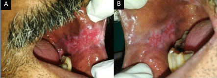
    Figure 1:  Left buccal mucosa showing erythematous patch with white
specks. Right buccal mucosa showing erythematous patch with white specks.
Discussion
WHO defines leukoplakia as a whitish patch or plaque that cannot be characterized, clinically or pathologically, as any other disease and which is not associated with any other physical or chemical causative agent except the use of tobacco [1].The World Health Organization (WHO) employs the term Speckled Leukoplakia (SL) to describe the presence of both white and red patches on the oral mucosa [2]. The two main clinical types of leukoplakia are homogeneous and nonhomogeneous leukoplakia. Speckled leukoplakia falls under the category of non-homogenous leukoplakia.
Etiological factors involved are alcohol use and smoking, diets lacking antioxidants (such as vitamins C, E, and beta-carotenes), occupational exposure to carcinogens, viral infections, and genetic and hereditary factors. Smoking of tobacco was found to be the strongest independent risk factor. Other forms of tobacco, hyperacidity, lipstick, and ill-fitting dentures were found to be a causative factor, which shows that socioeconomic status and lifestyle are involved in causing premalignant lesions [3].
The various treatment modalities include cessation of tobacco and alcohol use, topical application of retinoids, systemic treatment with Vitamin A, beta-carotene, lycopene, isotretinoin. Surgical management includes conventional surgical excision, laser excision, photodynamic therapy, cryotherapy and electrocautery. Speckled leukoplakia carries a higher risk of developing into malignancy than the other types. So early diagnosis by biopsy has to be done to avoid the dangerous malignant transformation [4].
Conclusion
The early recognition oral speckled leukoplakia is mandatory. Since the malignant potential of speckled leukoplakia is high, observation of the lesion alone without biopsy must be discouraged. Biopsy must be carried out to arrive at a proper diagnosis and to do immediate treatment planning.
References
- Pindborg JJ, Reichart P, Smith CJ, Van der Waal I. World Health Organization: histological typing of cancer and precancer of the oral mucosa. Berlin: Springer-Verlag. 1997.
- Eversole LR. Dysplasia of the Upper Aerodigestive Tract Squamous Epithelium. Head and Neck Pathol. 2009; 3: 63-68.
- Reibel J. Prognosis of oral pre-malignant lesions: significance of clinical, histopathological, and molecular biological characteristics. Crit Rev Oral Biol Med. 2003; 14: 47-62.
- Parlatescu Ioanina, Tovaru Serban, Mihai Lelia. Treatment approach of oral leukoplakia. Review of literature. 2013; 8: 39-43.