Clinical Image
Austin J Dent. 2017; 4(2): 1068.
Stafne’s Bone Cavity
Cameron Y. S. Lee*
Department of Oral Maxillofacial and Reconstructive Surgery, Temple University, USA
*Corresponding author: Cameron Y. S. Lee, Department of Oral Maxillofacial and Reconstructive Surgery, Temple University, Kornberg School of Dentistry, Philadelphia, PA, USA
Received: February 12, 2017; Accepted: February 27, 2017; Published: March 01, 2017
Clinical Image
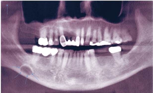
A 51-year old Asian male presented to the office for evaluation of a radiolucency of the right posterior mandible. The radiographic lesion was observed during a dental implant consultation with his dentist to replace several missing teeth in the left posterior mandible. Panoramic radiograph revealed a well-defined unilocular radiolucency in the right posterior mandible between the inferior alveolar canal and the inferior border of the mandible (Figure 1). Cone beam CT scan was completed to obtain a definitive diagnosis which was consistent with Stafne’s Bone Cavity (SBC).

    Figure 1: Panoramic radiographic showing elliptical shaped radiolucent
lesion (arrows) in molar region of right posterior mandible between inferior
alveolar canal and inferior border of mandible.
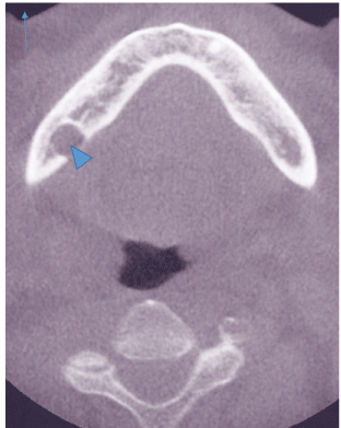
SBC, also known as Stafne’s cyst and lingual salivary gland inclusion defect is a lingual (medial) cortical defect of the molar region between the inferior alveolar canal and inferior border of the mandible (Figure 2,3). It is often observed during a dental examination. It represents remodeling of the bone on the medial aspect of the mandible from pressure of the submandibular salivary gland. No treatment is indicated for this benign pathologic entity.

    Figure 2: Lingual cortical concavity of the mandible (arrow head) observed
with sagittal views of cone beam CT scan consistent with Stafne’s bone
concavity.
