
Clinical Image
Austin J Emergency & Crit Care Med. 2014;1(1): 2.
Concealed Penetrating Parietal Lobe Injury Due to Stab Wound: Case Report
Enver Sösüncü1, Hakan AK2*, Ismail Gülsen1, Yurdanur Akyüz3 and Mehmet Arslan1
1Department of Neurosurgery, Yüzüncü Yil University, Turkey
2Department of Neurosurgery, Bozok University, Turkey
3Department of Radiology, Bozok University, Turkey
*Corresponding author: Hakan Ak, Department of Neurosurgery, Bozok University, School of Medicine, Yozgat, Turkey
Received: July 11, 2014; Accepted: August 22, 2014; Published: August 26, 2014
Abstract
Penetrating brain injury may occur as a result of the bullets shrapnel parts of gunshot wounds, wood, knife, and glass. Thick and hard structure of the skull protects the brain from external forces. However, relatively thin structures roof of orbital cavity, temporal bone, and cribriform plate make the brain susceptible to penetrating injury. These injuries mostly occur in maxillofacial region. Herein, we present a penetrating parietal lobe injury due to stab wound.
Keywords: Penetrating brain injury; Stab wound; Knife; Parietal lobe
Introduction
Thick and hard structure of skull protects the brain from external forces, however, in rare instances penetrating brain injury may occur depending on the severity of trauma [1]. This type of injury may occur in every period of life. Glass, knife, wood, metal splinters and bullet injuries are the some examples in the etiology of these injuries [1-4]. Penetrating brain injury may lead to serious vascular and neurologic deficits such as intracerebral hematoma, cerebral contusion, intraventricular hemorrhage, pneumocephalus, brain stem injury, and fistula of carotid-cavernous sinus [5,6].
Most of the penetrating head injuries by stab wound are visible. However the concealed penetrating head Injuries are rare and the diagnosis is easily missed. Herein, we report a parietal lobe injury due to stab wound after assaulting in a 29 year old man whose diagnosis was made fortuitously and who recovered without any neurological deficit.
Case Presentation
A 29 year-old man attended to the emergency room due to assaulting. In the physical examination there were multiple scalp injuries due to stabbing. There was not any foreign body on the scalp (Figure 1). Neurological examination was normal. In spite of normal neurological examination, brain computed tomography (CT) was received and it revealed a foreign body looks like tip of knife blade on the right parietal region. It was extending about 2,5cm to inside of brain parenchyma it was showing close neighborhood with sagittal sinus (Figure 2 & 3). However, we didn’t perform angiography in spite of close neighborhood. Patient was hospitalized and underwent operation under general anesthesia. Craniectomy with drilling around knife was performed. Dural tear was extended. After that knife was removed with gentle manipulation (Figure 4 & 5). We didn’t see any complication in the postoperative period like hemorrhage or neurologic deficit. Patient was discharged without any deficit.
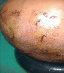
Figure 1: Physical examination of the patient revealed only scalp incisions.
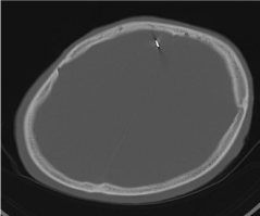
Figure 2: Preoperative CT image showing knife tip in the brain parenchyma with close relationship with sagittal sinus.
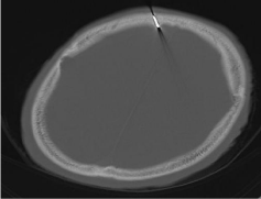
Figure 3: Preoperative CT image showing knife tip in the brain parenchyma with close relationship with sagittal sinus.
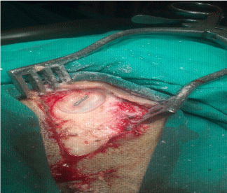
Figure 4: An intraoperative image showing craniectomy borders and knife.
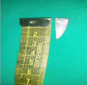
Figure 5: Image showing removed knife from the brain.
Discussion
Penetrating brain injury usually occurs as a result of gunshot wounds. However, penetrating injury due to glass, wood, and knife have been reported. These injuries usually occur accidentally. Besides this, it may also occur as a result of suicide attempt, crime, and in severe psychological disorders [1,7,8].
Thick and hard structure of skull protects the brain from external forces. However, roof of orbital cavity, temporal bone, and cribriform plate are susceptible to penetrating trauma due to their relatively thin structure. When literature search is performed, penetrating brain injury usually occur on orbital, frontal sinus, and nasal region [1,7-9]. Miller et al. reviewed the 42 case reports from the literature about peri-orbital puncture wounds by sharp wooden objects. They reported permanent neurologic sequelae in 74% of the cases. Intracranial suppuration was defined as the major complication, with brain abscess which occurred in about half of the all cases. They also reported high rates of mortality [9]. DiRoio et al. defined penetrating intracranial injury by transorbital way [10]. Nagakawa et al. ad Yilmaz et al. reported penetrating brain injury due to glass at the temporal and parietal regions, respectively [1,4].
Literature contains only a few cases about penetrating brain injury due to stabbing. In these injuries, neurological sequelae may be seen due to damage of parenchyma and vascular structures with direct effect of trauma. Also, thrombosis, intracerebral hematoma, meningitis, sepsis may be seen [1,11]. Fortunately, none of these complications was observed in our case.
The diagnosis of this type of brain injury starts with physical and neurological examination and most of the penetrating head injuries by stab wound are visible. However, in some cases, like ours, only physical and neurological examination may not be enough and the diagnosis may be easily missed. Sometimes, as in our case, only physical and neurological examination may not be enough. Missed cases may lead to some undesirable medical and legal results such as especially malpractice litigation. Because of this reason, we recommend to receive CT in these cases routinely.
In conclusion, concealed penetrating brain injury in parietal lobe due to stab wound is a rare pathology. Brain CT should be received in every case attended due to stabbing even though the normal physical and neurological examinations.
References
- Yilmaz N, Kiymaz N, Mumcu Ç, Yilmaz C, Etlik O. Cam parçasina bagli görülen nadir penetran kafa travmasi. Van Tip Dergisi. 2004; 11: 152-154.
- Gajdos M, Stancák M, Lacko F. [A brain injury caused by a fragment from a soda water carbon dioxide cartridge]. Acta Chir Orthop Traumatol Cech. 1993; 60: 247-249.
- Hansen JE, Gudeman SK, Holgate RC, Saunders RA. Penetrating intracranial wood wounds: clinical limitations of computerized tomography. J Neurosurg. 1988; 68: 752-756.
- Nakagawa A, Su CC, Yamashita Y, Endo T, Shirane R. [A temporal head injury involving intracranial penetration by glass]. No Shinkei Geka. 2002; 30: 529-533.
- Iwakura M, Kawaguchi T, Hosoda K, Shibata Y, Komatsu H, Yanagisawa A, et al. Knife blade penetrating stab wound to the brain--case report--. Neurol Med Chir (Tokyo). 2005; 45: 172-175.
- Fujimoto S, Onuma T, Amagasa M, Okudaira Y. [Three cases of an intracranial wooden foreign body]. No Shinkei Geka. 1987; 15: 751-756.
- Greene KA, Dickman CA, Smith KA, Kinder EJ, Zabramski JM. Self-inflicted orbital and intracranial injury with a retained foreign body, associated with psychotic depression: case report and review. Surg Neurol. 1993; 40: 499-503.
- Arunkumar MJ, Selvapandian S, Rajshekhar V. Penetrating intracranial wooden object: case report and review of CT morphology, complications, and management. Surg Neurol. 1999; 51: 617-620.
- Miller CF, Brodkey JS, Colombi BJ. The danger of intracranial wood. Surg Neurol. 1977; 7: 95-103.
- Di Roio C, Jourdan C, Mottolese C, Convert J, Artru F. Craniocerebral injury resulting from transorbital stick penetration in children. Childs Nerv Syst. 2000; 16: 503-506.
- Gulati A, Srinivasan B, Hunter R, Flood TR. Penetrating knife injury to the frontal lobe--a case report. Ann R Coll Surg Engl. 2010; 92: W41-42.