
Special Article – Emergency Medicine and Clinical Case Reports
Austin J Emergency & Crit Care Med. 2016; 3(1): 1045.
A Patient with Extensive Pneumocephalus
Heidari SF*
Department of Emergency Medicine, Emam Khomeini Hospital, Medical Faculty, Mazandaran University of Medical Sciences, Sari, Iran
*Corresponding author: Heidari SF, Department of Emergency Medicine, Emam Khomeini Hospital, Medical Faculty, Mazandaran University of Medical Sciences, Sari, Iran
Received: September 23, 2016; Accepted: September 26, 2016; Published: September 28, 2016
Clinical Image
A 63-year-old man presented to the emergency department by EMS because of head trauma. The incidence of pedestrian with car was mechanism of trauma. Patient had nausea, vomiting and rhinorrheagia. On physical examination, he was found to have confusion, PTA and GCS of 14/15. Vital sign was stable. There was bilateral periorbital ecchymosis and one laceration with size of 2cm in left eyebrow. A remnant examination was unremarkable. Axial images of the brain on a Computed Tomographic scan (CT scan) showed an extensive pneumocephalus in frontal on both sides without midline shift (Figure 1). Also, there was evidence of fracture in left maxillary sinus and skull base fracture. The patient was transferred to the ICU and treated conservatively with head elevation 30 degrees and oxygen therapy. On evaluation of 5 days later, he was alert and asymptomatic. Control brains CT scan have minimal pneumocephalus so that numerous amount of pneumocephale was absorbed. Then, the patient was discharged from hospital.
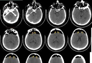
Figure 1: Brain CT scan of a patient with extensive pneumocephalus.