
Case Report
Austin Emerg Med. 2016; 2(3): 1019.
Is Computerized Tomography Sufficient for the Diagnosis of Brain Abscess?
Karagöz A¹* and Ünlüer EE²
¹Department of Emergency Medicine, Karşıyaka State Hospital, Karşıyaka, Turkey
²Department of Emergency Medicine, Katip Çelebi University Atatürk Research and Training Hospital, Karabaglar, Turkey
*Corresponding author: Arif Karagöz, Department of Emergency Medicine, Karşıyaka State Hospital, Karşıyaka, Turkey
Received: February 16, 2016; Accepted: March 05, 2016; Published: March 08, 2016
Abstract
Brain Abscess (BA) is a focal suppurative process in the brain parenchyma. It is seen rarely in the Western population, with approximately 1500-2500 cases per year. They arise after the direct extension of infection from neighboring structures, hematogenous seeding of infectious agents, or trauma and neurosurgical complications. Symptoms of BA are non-specific and laboratory tests add little to the diagnosis. They develop in four stages and imaging characteristics of BAs differ according to the stage. The most important prognostic factor for BAs is the initial neurologic grade and diagnostic delay is a key contributing factor to the severity and outcome of BAs. This fact makes the timely diagnosis of BA in the Emergency Department (ED) very important. Here, we describe the case of a 20-year-old male who presented to ED with confusion, drowsiness and hypoacusis. While cranial computerized tomography reveals only localized brain edema, magnetic resonance imaging revealed the abscess with a fluid-fluid level and the patient was diagnosed as having a BA.
The changing imaging characteristics of BAs make their diagnosis difficult. Timely diagnosis of BA is very important in ED. In selected patients, further imaging modalities such as magnetic resonance help to make the diagnosis properly.
Keywords: Brain abscess, Emergency, Computerized tomography
Abbreviations
BA: Brain Abscess; ED: Emergency Department; CCT: Cranial Computed Tomography; CMRI: Cranial Magnetic Resonance Imaging; DW-MRI: Diffusion-Weighted Magnetic Resonance Imaging.
Introduction
Brain Abscess (BA) is a focal suppurative process in the brain parenchyma and patterns in the diagnostic imaging considerably change depending on the onset of the disease process. Here, we describe the case of a 20-year-old male who presented to the Emergency Department (ED) with brain edema in Cranial Computed Tomography (CCT), but diagnosed as BA containing a fluid-fluid level in Cranial Magnetic Resonance Imaging (CMRI) and Diffusion- Weighted Magnetic Resonance Imaging (DW-MRI) due to early presentation of the patient.
Case Report
A 20-year-old male patient presented to ED with confusion, drowsiness and hypoacusis. On admission, his Glasgow coma scale was 12 (E3M5V4). On his right extremities, muscle strengths were 4/5. His blood pressure was 120/80 mmHg, heart rate 108 and he had a fever of 38.5°C. His history revealed that, a week ago, he had seen an otolaryngologist because of the pain in his left ear and was prescribed amoxicillin clavulanate. He had had a fever for four days.
In laboratory tests, the leucocyte count was 13000/mm3 (4500- 10000) with 86% neutrophils, biochemistry was normal and C reactive protein level was 36 mg/L (<4.9 mg/L). A non-contrast CCT revealed a low attenuated and indefinite bordered area causing sulcal effacement in the left temporal lobe (Figure 1). Then, CMRI and DWMRI were ordered. On the CMRI and DW-MRI, a 14mm diameter cystic lesion with a fluid-fluid level and rounded with cerebral edema causing sulcal effacement was diagnosed (Figures 2,3). Both CCT and CMRI showed inflammatory process in the left mastoid sinus (Figure 4). Ceftriaxone and metronidazole were administered intravenously as empirical antibiotherapy and neurosurgery and otolaryngology consultation were ordered. He was admitted to the surgical intensive care unit and a mastoidectomy was planned.
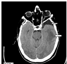
    Figure 1:  Non-contrast cranial computerized tomography revealed a low
attenuated and indefinite bordered area causing sulcal effacement in the left
temporal lobe (large arrow). There is an incidental arachnoid cyst in front of
the left temporal lobe (small arrow).

    Figure 2:  T2-weighted cranial magnetic resonance showing abscess cavity
with fluid-fluid level. The vasogenic edema surrounding the abscess cavity is
characterized by surrounding hyperintensity on the T2-weighted sequence.
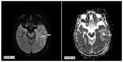
    Figure 3:  Figure 3A: On diffusion-weighted sequence, the facilitatedand
restricted-diffusion are together and they create a clear level. Brain
edema surrounds the abscess cavity. Figure 3B: On the apparent diffusion
coefficient, induced- and reduced- apparent diffusion coefficiency values are
together and they create a clear level. Brain edema surrounds the abscess
cavity.
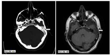
    Figure 4:  Figure 4A: Cranial computerized tomography and Figure 4B:
Magnetic resonance imaging showing inflammation in left mastoid sinus.
Discussion
The incidence of BAs in the Western population is approximately 1500-2500 cases per year and it is higher in developing countries [1]. The male-to-female ratio varies from 1.3:1 to 3.0:1 [2-5]. Since the mid 1970s, mortality from BAs has decreased [4,6,7]. This reduction has largely been attributed to the start of the computerized tomography era in 1974 and advances in antibiotic therapy [6,8,9]. A BA arises after the direct extension of infection from neighboring structures, hematogenous seeding of infectious agents, or trauma and neurosurgical complications [6,10]. While odontomastoiditis is frequently associated with BAs localized in the temporal lobe and cerebellum, frontal and ethmoid sinusitis and odontogenic infection are associated with frontal lobe abscesses [11]. Hematogenous seeding usually causes multiple abscesses [12,13]. However, localization characteristics have a limited value [14]. As many as 20-30% of cases have no identified source of infection [10].
A BA develops in four stages [15,16]. The first stage is ‘early cerebritis’ and occurs in the first to fourth day. In this stage, acute inflammation results in localized cerebral edema. Non-contrast CCT can be normal or shows a poorly mariginated, edematous area [6]. The second stage is ‘late cerebritis,’ which occurs between the fourth and ninth days. In this stage, the central area becomes necrotic. On non-contrast CCT there is persistent poorly defined edema; however, on post-contrast images there is thick ring like enhancement [6]. In this stage, CCT can be insufficient to diagnose the BA. The third and fourth stages are ‘early’ and ‘late capsulation.’ During these stages, there is a central core containing pus with a surrounding capsule. On non-contrast CCT, this core presents as a round or ovoid area of low attenuation. Post-contrast CCT images can reveal a ring like enhancement. A CMRI is more sensitive than CT because it has greater sensitivity to changes in tissue water content, resulting in a greater contrast between edematous brain and normal brain early stages of cerebritis and abscess formation [6,17]. A DW-MRI helps distinguish abscesses from other ring-enhancing lesions such as cystic or necrotic neoplasms [18-20]. In our case, the CCT revealed only brain edema, but the CMRI and DWI revealed a cystic lesion with a fluid-fluid level.
The most important prognostic factor for BAs is the initial neurologic grade [21]. Diagnostic delay is a key contributing factor to the severity and outcome of BAs [3,22]. The abscesses tend to rupture into ventricles and this a feared complication [6,23].
Conclusion
Since the timely diagnosis is crucial for BAs, Emergency Physicians (EPs) should be careful while evaluating patients with neurological symptoms and consider the possibility of a BA. Time of presentation and tempo of the disease causes different imaging results in CCT and CMRI and this makes the diagnosis of BA challenging in ED. Diagnostic imaging modality must be chosen by EPs according to the duration of patient’s symptoms.
References
- Greenberg MS. Handbook of neurosurgery. 5th edition, New York. Thieme. 2001; 217-223.
- Lu CH, Chang WN, Lui CC. Strategies for the management of bacterial brain abscess. J Clin Neurosci. 2006; 13: 979-985.
- Kao PT, Tseng HK, Liu CP, Su SC, Lee CM. Brain abscess: clinical analysis of 53 cases. J Microbiol Immunol Infect. 2003; 36: 129-136.
- Carpenter J, Stapleton S, Holliman R. Retrospective analysis of 49 cases of brain abscess and review of the literature. Eur J Clin Microbiol Infect Dis. 2007; 26: 1-11.
- Seydoux C, Francioli P. Bacterial brain abscesses: factors influencing mortality and sequelae. Clin Infect Dis. 1992; 15: 394-401.
- Rath TJ, Hughes M, Arabi M, Shah GV. Imaging of cerebritis, encephalitis, and brain abscess. Neuroimaging Clin N Am. 2012; 22: 585-607.
- Cavuşoglu H, Kaya RA, Türkmenoglu ON, Colak I, Aydin Y. Brain abscess: analysis of results in a series of 51 patients with a combined surgical and medical approach during an 11-year period. Neurosurg Focus. 2008; 24: E9.
- De Louvois J. Bacteriological examination of pus from abscesses of the central nervous system. J Clin Pathol. 1980; 33: 66-71.
- Rosenblum ML, Hoff JT, Norman D, Weinstein PR, Pitts L. Decreased mortality from brain abscesses since advent of computerized tomography. J Neurosurg. 1978; 49: 658-668.
- Mathisen GE, Johnson JP. Brain abscess. Clin Infect Dis. 1997; 25: 763-779.
- Friedlander RM, Gonzalez RG, Afridi NA, Pfannl R. Case records of the Massachusetts General Hospital. Weekly clinicopathological exercises. Case 16-2003. A 58-year-old woman with left-sided weakness and a right frontal brain mass. N Engl J Med. 2003; 348: 2125-2132.
- Bakshi R, Wright PD, Kinkel PR, Bates VE, Mechtler LL, Kamran S, et al. Cranial magnetic resonance imaging findings in bacterial endocarditis: the neuroimaging spectrum of septic brain embolization demonstrated in twelve patients. J Neuroimaging. 1999; 9: 78-84.
- Rousseaux M, Lesoin F, Destee A, Jomin M, Petit H. Developments in the treatment and prognosis of multiple cerebral abscesses. Neurosurgery. 1985; 16: 304-308.
- Helweg-Larsen J, Astradsson A, Richhall H, Erdal J, Laursen A, Brennum J. Pyogenic brain abscess, a 15 year survey. BMC Infect Dis. 2012; 12: 332.
- Britt RH, Enzmann DR. Clinical stages of human brain abscesses on serial CT scans after contrast infusion. Computerized tomographic, neuropathological, and clinical correlations. J Neurosurg. 1983; 59: 972-989.
- ErdoÄŸan E, Cansever T. Pyogenic brain abscess. Neurosurg Focus. 2008; 24: E2.
- Haimes AB, Zimmerman RD, Morgello S, Weingarten K, Becker RD, Jennis R, et al. MR imaging of brain abscesses. AJR Am J Roentgenol. 1989; 152: 1073-1085.
- Leuthardt EC, Wippold FJ, Oswood MC, Rich KM. Diffusion-weighted MR imaging in the preoperative assessment of brain abscesses. Surg Neurol. 2002; 58: 395-402.
- Desprechins B, Stadnik T, Koerts G, Shabana W, Breucq C, Osteaux M. Use of diffusion-weighted MR imaging in differential diagnosis between intracerebral necrotic tumors and cerebral abscesses. AJNR Am J Neuroradiol. 1999; 20: 1252-1257.
- Guzman R, Barth A, Lövblad KO, El-Koussy M, Weis J, Schroth G, et al. Use of diffusion-weighted magnetic resonance imaging in differentiating purulent brain processes from cystic brain tumors. J Neurosurg. 2002; 97: 1101-1107.
- Hakan T, Ceran N, Erdem I, Berkman MZ, Göktaş P. Bacterial brain abscesses: an evaluation of 96 cases. J Infect. 2006; 52: 359-366.
- Smith SJ, Ughratdar I, MacArthur DC. Never go to sleep on undrained pus: a retrospective review of surgery for intraparenchymal cerebral abscess. Br J Neurosurg. 2009; 23: 412-417.
- Zeidman SM, Geisler FH, Olivi A. Interventricular rupture of a purulent brain abscess: case report. Neurosurgery. 1995; 36: 189-193.