Case Report
Austin ENT Open Access. 2024; 4(1): 1011.
DLBCL Sphenoid Lymphoma: Diagnosis with Endoscopic Transphenoid Approach
Liva GA¹*; Karatzanis A¹; Spanaki C²; Doulaptsi M¹; Prokopakis E¹
¹Department of Otorhinolaryngology, School of Medicine, University of Crete, Greece
²Department of Neurology, School of Medicine, University of Crete, Greece
*Corresponding author: Liva G Department of Otorhinolaryngology, School of Medicine, University of Crete, Armonias 14, 71409 Heraklion, Crete. Tel: +306978930266 Email: [email protected]
Received: December 09, 2023 Accepted: January 04, 2024 Published: January 11, 2024
Abstract
Non-Hodgkin lymphomas of the paranasal sinuses are rare pathologies that present as a diagnostic challenge for the clinician. We describe a case of sphenoid lymphoma in an 81 year old patient presenting with loss of the papillary light reflex on both sides, left upper eyelid drop, anisocoria, and ocular movement deficiency of the left eye. MRI showed a solid lesion of the sphenoid sinus invading the clivus, the left carvenous sinus, the superior orbital fissure and the left side of the pituitary gland. Due to clinical difficulty to reach a diagnosis, endoscopic transphenoid biopsy was performed and DLBCL non-Hodgkin lymphoma of the sphenoid sinus was revealed from histopathological examination. The patient was treated with R-CHOP regi-men (rituximab, cyclophosphamide, doxorubicin, vincristine and prednisone) with remarkable improvement after the first course of chemotherapy. NHLS and especially DLBCL are extremely rare in the sphenoid sinus. Therefore, high clinical awareness and early endoscopic biopsy are mandatory.
Keywords: Lymphoma; Hodgkin lymphoma; Non-Hodgkin lymphoma; Sphenoid sinus
Case Presentation
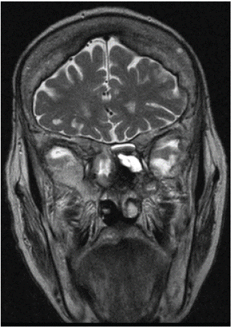
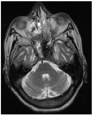
An eighty-one year old female with medical history of diabetes type II, rheumatoid arthritis, and hyperlipidemia, was referred to the emergency department of a tertiary re-ferral center with inflammation and drop of her left upper eyelid since the last twenty days. The clinical examination revealed loss of the papillary light reflex on both sides, left upper eyelid drop, anisocoria and ocular movement deficiency of the left eye. In addition, drop of the left nasolabial fold, hypoesthesia of the V3 branch and hyperalgesia of the V1 branch of the left half of her face. Function of the remaining cranial nerves was clinically normal and there were no sensory or motor deficits in the upper and lower extremities, while all tendon reflexes were normal. No palpable cervical lymphadenopathy was rec-ognized and physical and endoscopic examination of the nasal tract, larynx and pharynx were normal. Magnetic Resonance Imaging (MRI) scanning of the head with gadolinium injection was performed on an emergency basis and showed a solid heterogenous contrast intake, invading the clivus, the left cavernous sinus, without the presence of thrombosis of the cavernous sinus, and the interior carotid artery, the superior orbital fissure, and the left side of the pituitary gland, causing displacement of the pituitary gland to the right side (Figure 1,2).

Figure 1: Coronal t2 weighted mri resonance, solid lesion of sphenoid sinus with homogenous contrast intake.

Figure 2: Axial t2 weighted mri resonance, solid lesion of sphenoid sinus with homogenous contrast in-take.
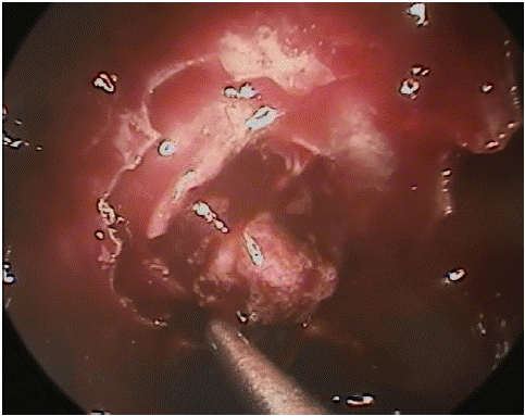
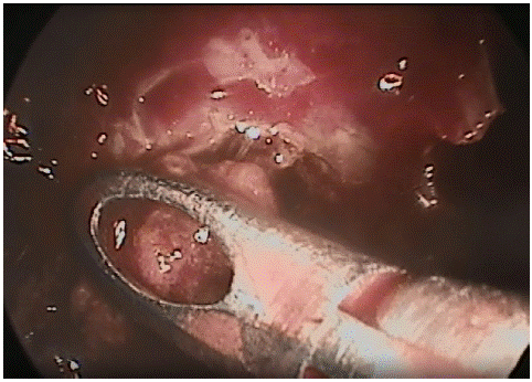
The patient was admitted to the neurological department for further investigation. As no clinical improvement of the patient was noticed, and no clinical diagnosis was reached, on the fifth day of hospitalization, endoscopic transphenoid biopsy was per-formed under general anesthesia (Figure 3, 4). It was performed transnasally with a zero de-gree scope and navigation. The middle turbinate was lateralized gently until the superior turbinate visualized. The natural ostium of the sphenoid sinus was identified medial and posterior to the superior turbinate and approximately 1 cm above the choana. The ostium was extended and eventually most of the anterior wall of the sphenoid was removed with a mushroom punch. The content of the sphenoid sinus was biopsied with a Blakesley for-ceps and anterior nasal packing with neurosurgical patties were placed. The patties were removed during the first postoperative hours. Histopathological examinations including immunohistochemistry and flow cytometry showed CD20+, MUM1+, CD3-, CD5- and di-agnosis of high malignant rate non-Hodgkin DLBCL was confirmed. Additional investi-gation of bone marrow, cerebrospinal fluid and thorax, cervical and abdomen CT showed neither primary state of lymphoma nor metastasis in other anatomical regions. The pa-tient was treated with R-CHOP chemotherapy regimen, consisting of rituximab, cyclophosphamide, doxorubicin, vincristine and prednisone and precautionary for the central nervous system with two courses of intrathecalmethotraxate therapy. Left eye movement was remarkably improved after the first course of R-CHOP chemotherapy. The patient is under chemotherapy treatment and monitoring from the Haematology Department of the University Hospital of Crete.

Figure 3: Enlargement of the sphenoid sinus ostium and excision of the posterior wall of the sphenoid si-nus.

Figure 4: Sphenoid tumor biopsy.
Discussion
Lymphomas are a heterogenous group of malignancies which originate either from nodal or from extranodal sites of the head and neck. Lymphomas are the second most common primary malignancy occurring in the head and neck. They are classified ac-cording to clinical manifestation, cell morphology, immunophenotype, and genetic fea-tures [6]. World Health Organisation (WHO) and European-American Classification of Lymphoid Neoplasms subdivide lymphomas according to cell origin, to B-cell lympho-mas, T-cell lymphomas, Natural-Killer (NK) cell lymphomas, Hodgkin and non-Hodgkin lymphoma [6]. Lymphomas of the sinonasal tract account for 23-31% of the malignant tumors of the nasal and paranasal cavities [1]. Among these, NHLs are uncommon, rep-resenting 3% to 5% of all malignancies [7,8]. Most of the NHLs of the head and neck arise in extranodal sites, such as nasal cavity, paransal sinuses, and lymphoid tissue regions in the Waldeyer’s ring. Frequency of appearance and histological subtype of NHLs may be affected by geographic factors; in Asian populations, tumors of the nasal cavity are more common, while in western populations, paranasal sinuses, and specifically the maxillary, are common sites of appearance [7-9]. Overall, on the other hand, NHLs of paranasal sinus-es are uncommon and account for 0,17%-2% of all NHL cases [2,4]. Primary malignancies, and especially DLBCL of the sphenoid sinus, are extremely rare and only a few cases have been reported in literature.
The sphenoid sinus is located under the middle cranial fossa and is surrounded by significant vascular structures, cranial nerves, optic structures, dura, pituitary gland, and the brainstem. For this reason, sphenoid sinus diseases may appear with significant clin-ical symptoms. In cases of patients with sphenoid lymphoma, nasal obstruction or nasal discharge have been reported in 33,3%, cranial nerve II, III, and IV palsy in 86,7%, pares-thesia of V cranial nerve in 20%, and headache in 40% of cases [2]. On Computed Tomog-raphy (CT) and MRI imaging, lymphomas of the sinonasal tract do not have distinctive characteristics for differential diagnosis. Lymphomas may present with diffuse tumor in-filtration of the nasal walls or as large bulky masses of the sinus walls that may cause re-modeling or erosion of the adjacent bone [10]. Lymphomas may have isointense signal to muscle on T1 and moderate to high signal on T2 [10.] The FDG-PET/CT seems to have significant value for nasal lymphomas, especially for follow up, but not for paranasal sinus lymphomas [10,11]. The contrast enhancement of sinonasal tract lymphomas may not easily be discriminated from other malignancies of nasal cavity and sinuses [10,12].
In conclusion, lymphomas of the sphenoid sinus are extremely rare and there is significant difficulty in diagnosis, often leading to misdiagnosis during early stages of the disease. The main reasons for misdiagnosis are the lack of nasal symptoms, no characteristic clinical appearance, and no distinctive and diagnostic imaging characteristics. Taking into consideration all of these, a high level of clinical suspicion is paramount and ear-ly consideration for biopsy and histopathological examination are indispensable for a definite diagnosis.
References
- Wang Q, Zhou SH. B-cell malignant non-Hodgkin’s lymphoma of the sphenoid sinus cavity: a case report and brief review of the literature. Ann Otolaryngol Rhinol. 2010; 1: 1010.
- Yoshihara S, Kondo K, Ochi A. Diffuse large B-cell lymphoma in the sphenoid sinus mimicking fibrous dysplasia in CT and MRI. BMJ Case Rep. 2014; 2014: bcr2014205272.
- Hatta C, Ogasawara H, Okita J, Kubota A, Ishida M, Sakagami M. NonHodgkin’s malignant lymphoma of the sinonasal tract—treatment outcome for 53 patients according to REAL classification. Auris Nasus Larynx. 2001;28:55-60 Author 2. Book Title B. Publisher: publisher location. 3rd ed. Country. 2008; 154-96.
- Abbondanzo SL, Wenig BM. Non-Hodgkin’s lymphoma of the sinonasal tract, Aclinicopathologic and immunophenotypic study of 120 cases. Cancer. 1995; 75: 281-91.
- Shankland KR, Armitage JO, Hancock BW. Non-Hodgkin lymphoma. Lancet. 2012; 380: 848-57.
- Weber AL. Rahemtullah A. Hodgkin and non-Hodgkin lymphoma of the head and neck: clinical, pathologic, and imaging evaluation. Neuroimag Clin N Am. 2003; 13: 371-92.
- Azarpira A, Ashraf MJ. Primary lymphoma of nasal cavity and paranasal sinuses. Lab Med. Fall 2012; 43: 2994-299.
- Quraishi MS, Bessell EM, Clark D, Jones NS, Bradley PJ. Non-Hodgkin’s lymphoma of the sinonasal tract. Laryngoscope. 2000; 110: 1489-92.
- Fasunla AJ, Lasisi AO. Sinonasal malignancies: a 10-year review in a tertiary health institution. J Natl Med Assoc. 2007; 99: 1407-10.
- Eggesbo HB. Imaging of sinonasaltumours. Cancer Imaging. 2012; 7: 136-52.
- Karantanis D, Subramaniam RM, Peller PJ, Lowe VJ, Durski JM, Collins DA, et al. The value of [(18)F]fluorodeoxyglucose positron emission tomography/computed tomography in extranodal natural killer/T-cell lymphoma. Clin Lymphoma Myeloma. 2008; 8: 94-9.
- Lee FK, King AD. Dynamic contrast enhacement magnetic resonance imaging (DCE-MRI) for differential diagnosis in head and neck cancers. Eur J Radiol. 2011; 81: 784-8.