
Research Article
Austin J Forensic Sci Criminol. 2015; 2(4): 1036.
Application of Entomology in Some Medicolegal Issues
El-Mehy I¹, Sief A², Soliman E¹, Hassan NA¹* and Alrouf TA¹
¹Department of Forensic Medicine, Tanta University, Egypt
²Department of Entomology, Tanta University, Egypt
*Corresponding author: Neven Ahmed Hassan, Department of Forensic Medicine and Clinical Toxicology, Tanta University, Egypt
Received: June 29, 2015; Accepted: August 06, 2015; Published: August 09, 2015
Abstract
As the application of the forensic entomology has been strongly criticized for years, the present study was carried out on fifteen cases representing all putrefied cases in different seasons and habitats with larval infestation referred to the Forensic Medicine Directorate in Middle Delta. The aim of the present study was to identify and record the different species of insects that are frequently associated with human corpses to establish data base for the potential use of insects as postmortem interval indicators.
The maximum number of cases was in the age group 10-20 years, and the least number was encountered in the age group less than 10 years and over 50 years.
A relatively larger number of victims (73%) were males while female corpses were (27%) in this study. Nine of the reported cases (60%) were exposed, while, the other six cases were equally distributed between aquatic locations and burial (20% for each).
The maggots recovered from the fifteen decomposing human remains belong to family Calliphoridae and Sarcophagidae (Diptera).
The blow fly Chrysomya albiceps was the most common species invading nine cases (60%), followed by Sarcophagidae family (four cases with a percentage of 26.7%). The least number was Psychoda and Lucilia sericata which infested only one case for each (6.7%).
This study revealed that, Chrysomya albiceps was found to be a highly indicative species to corpses found in urban (6 cases) and rural (3 cases) areas representing 67% and 33% on sequence during the warmer seasons. While both Sarchophagidae and Dermistidae were equal in both urban and rural areas. Lucilia sericata and Psychoda species were only present in rural areas in this study.
In the present study Chrysomya albiceps was attracted to corpses in spring (5 cases), summer (2 cases) and winter (2 cases). Sarcophaga species larvae were infesting dead bodies in both spring (3 cases) and fall (1 case) representing 75% and 25% respectively. Luicilia sericata larvae were collected from 1 case in winter. Psychoda species and Dermestidae larvae were collected from cases in spring only.
In the present study, 3 cases were found to be of interest to forensic entomology. The first case concerned a corpse of young female killed by smothering, the second was exhumated for autopsy and the third one was partially submersed and killed by strangulation and found near a water canal coast. The implications for estimation of postmortem interval by entomological methods were discussed.
The results of this study demonstrated the usefulness of testing larvae associated with decomposed remains for toxicological analyses in cases number 12 & 14 as the cause of death was carbamate and organophosphorus toxicity respectively.
This study concluded that, in death cases where insects are associated with the remains, it can be used for detection of postmortem interval and the cause of death in some cases. The forensic entomologist should cooperate with the forensic pathologist; from the visual observations of the cadaver in the scene, through the collection of insects and temperature data up to the final report with the interpretation of entomological and other biological evidence.
Keywords: Entomology; Chrysoma albiceps; Sarchophagidae; Dermistidae; Lucilia sericata
Introduction
Dead bodies undergo a variety of changes, which eventually return the tissue components to the food chain. These changes are brought about by chemical (mostly enzymatic) processes, bacterial and fungal attack and predation by many varieties of animal life, from tigers to bluebottle [1].
The sequence of putrefactive changes can be divided into four stages or phases (discoloration, bloating, liquefaction and advanced decay or skeletonization). These phases are markers for diagnostic purposes indicating time elapsed since death [2].
Insects are extremely abundant and ubiquitous organisms. Since arthropods are the largest and most important biological group on earth, they can be found in a wide variety of locations including crime scenes [3]. They are attracted to natural orifices in particular, the facial orifices to feed and lay eggs but, when wounds present, they are usually more attractive. So, insects can be used to determine presence, position, and patterns of wounds [4].
Forensic entomology is based on the analysis of the insects and other invertebrates, which sequentially colonized a corpse as decomposition progresses [5,6].
Entomo-toxicology is a relatively new branch of forensic entomology. The potential use of insects for detecting drugs and other toxins in decomposing tissues has been widely demonstrated in death investigations [7].
Entomologists with forensic experience can deduce minimum or probable times of death from the stage of development of blowfly maggots and other time-related insect colonizers. Hundreds of arthropod species are attracted by corpses, primarily flies (Diptera), beetles (Coleoptera), and their larvae, and also mites, isopods, opiliones and nematodes [8]. These insects feed, live, or breed in and on the corpse, depending on their biological preferences and the state of decomposition [3].
Basic research and advanced application of forensic entomology in the US, Russia , Canada , France and Japan as well as casework in other countries like England and India has opened the way to routine casework. Researchers worldwide use entomology in criminal investigations including murder and other high profile cases [3]. Forensic entomology has been used and accepted in courts around the world and gaining international recognition [9].
Facts are not always available from corpses when they are recovered, so the aim of this study is to 1-suggest a protocol for proper collection of specimens for the entomological study, 2- identify and record the larvae recovered from some cadavers in some governorates in Egypt (Elmahala Alkobra, Tanta, Kafr Alzayat, Zefta and Elsanta cities (Gharbia governorate), Kafr Elsheikh city (Kafr Elsheikh governorate) and Shebin Elkom city (Monofia governorate), 3-justify the role of the entomological study of these larvae in the estimation pf postmortem interval as well as a tool to answer some medicolegal questions.
Subjects and Methods
This study was carried out on 15 human putrefied corpses which were found in Middle Delta of Egypt during the period from the first of February 2004 up to the end of January 2006 (24 months). Putrefied corpses in this study referred to the Forensic Medicine directorate in Middle Delta, Ministry of Justice. These corpses were collected from Elmahala Alkobra, Tanta, Kafr Alzayat, Zefta and Elsanta cities (Gharbia governorate), Kafr Elsheikh city (Kafr Elsheikh governorate) and Shebin Elkom city (Monofia governorate).
Sociodemographic data were collected for each corpse as regard age, sex and residence.
Suggested protocol for the proper collection of specimens needed for the entomological study:
1-All data about the condition in which the corpse was found should be as regards environment, the ambient temperature, and nature of the locus in terms of vegetations, trees and undergrowth if outdoors.
2-Maggots if present should be placed live in a tube and if there is a delay in transit to the entomological labs, a fragment of meat should be included for food.
3-Adult flies and eggs should be collected without preservatives.
4-Some pupae, empty pupa cases and eggs should be sent for fixation.
5-Different species of insects on the body should be placed in separate tubes especially if alive.
6-All samples should be labeled or numbered according to the location on the corpse.
7- If outdoors, samples of the soil is collected.
According to Dadour et al. [10] larvae were collected from the bodies either in scene of death or in the mortuary as follows:
The larvae were collected from different parts of the body, natural orifices (such as mouth and nose), or wounds and soil under the corpses or near them. The larvae from each corpse were placed directly into 75% ethyl alcohol. The largest larvae were actively searched for and collected. Representative sample of 50-60 larvae was collected at the autopsy from the maggot mass in each case by a Soft-touch forceps which are useful for the collection of these larvae without damage and preserved immediately in plastic specimen containers. The samples were transported to the Department of Zoology, Entomological Lab, Faculty of science, Tanta University. The collected samples were labeled for further identification.
Morphological examination in the lab
The larvae collected in this study were placed in an aqueous solution of 10% Potassium Hydroxide (KOH) at room temperature for several days. The specimens were then washed in 75% ethyl alcohol for 10 to 15 minutes. While the maggots were in the alcohol, each one was pierced with a sharp pin (on one side) and then flattens it with a small spatula. This was to remove the remains of the body contents and to facilitate the replacement of liquids within the body cavity, in this and succeeding steps. The specimens were transferred to absolute alcohol (100%) for 10 to 15 minutes to dehydrate the specimen. Thereafter the specimens were transferred to xylene and left there overnight. The cleared specimens were placed into a mixture of xylene and canada balsam. Each maggot was mounted separately in the middle of a standard microscope slide. A large drop of canada balsam was dropped in the middle of the slide. Then the maggot was placed carefully on the Canada balsam, and care was given to place each larva on its lateral side. This position is ideal for examining the mouth hooks and anterior spiracles. A cover slip was placed over the specimen. The slides were placed on a slide-dryer. The slides were left to dry in an oven at 40°C for about two weeks. Reports describing the condition of the body when found and detailed autopsy procedures and results also were reviewed. Identifying the right species and instar was done relatively easy based on mouthparts and morphology of the posterior spiracles .Identification was made with a stereomicroscope [11].
Toxicological analysis
Raw samples of larvae were subjected to toxicological examinations by the chemical labs of forensic medicine administration using gas chromatography mass spectrum. The extraction process for samples consists of a series of operating steps according to Clarke [12].
According to Niessen [13] the mass spectrum is used to identify chemicals based on their structure.
Larvae collected from the corpses subjected to identification as regards, species, instar or stage of development, the habitat where the remains were found, expected age, season of discovery and results of toxicological examinations.
Post mortem interval: Estimation was based on comparison of data from autopsy reports (rate of decay), local environmental conditions (temperature, humidity, rainfall) and developmental times for the immature stages of each species of local arthropod and succession patterns using the schedules.
Statistical analysis
The collected data was organized, tabulated and statistically analyzed using SPSS software statistical computer package version 12. For age, the range, mean and standard deviation were calculated. For other variables, the number and percent distribution was calculated. Fisher exact test was used as a test of significance. Significance was adopted at P<0.05.
Results
The present study was carried out on fifteen cases representing all putrefied cases with larval infestation referred to the Forensic Medicine Departments in Middle Delta (Gharbia, Menoufia and Kafr Elsheikh), Ministry of Justice, during the period from the first of February 2004 to the end of January 2006 (24 months).
The corpses were found exposed, buried or in aquatic scene.
The sociodemographic data of the studied cases were observed in Table 1. The age of these cases range from (7-54) with a mean and SD (29.07+-14.22).
![]()
    -Age
    Number of cases
      (n=15)
    Percentage %


    <10
      10-20
      20-30
      30-40
      40-50
      > 50
    1
      4
      3
      3
      3
      1
    6.7 %
      26.6 %
      20 %
      20 %
      20 %
      6.7 %


    -Sex


    - Male
      -Female
    11
      4
    73%
      27%


    -Residence


    Urban
      Rural
    8
      7
    53 %
      47 %
Table 1:  The sociodemographic characters of 15 forensic entomology cases in
Middle Delta, Egypt, from 2004 to 2006.
Seventy three percent of the cases were males and twenty seven percent were females.
As regards the residence (53%) of the cases were found in urban areas, (47%) were found in rural residents.
The maggots recovered from the fifteen decomposing human remains belonged to family Calliphoridae and Sarcophagidae (Diptera) (Table 2). The maggots of Calliphoridae family were found infesting 10 forensic entomology cases (No. 1,2,7,8,10,11,12,13,14 and 15). Meanwhile the larvae of family Sarchophagidae were found in 4 corpses (No. 4,5,6 and 9). The larvae of Dermestidae (Coleoptera) were recorded infesting two of the examined cases (No. 3 and 12). Their numbers were more in highly decayed case (number 3).
![]()
    Entomofauna
      Case No.
    Order
    Family
    Insect
      species


    Case 1
    Diptrea
    Calliphoridae
    Chrysomya albiceps


    Case 2
    Diptrea
    Calliphoridae
    Chrysomya albiceps


    Case 3
    Diptrea  Coleoptera
    Dermestidae
    Psychoda


    Case 4
    Diptrea
    Sacophagidae
    Sarcophga species


    Case 5
    Diptrea
    Sacophagidae
    Sarcophga specie


    Case 6
    Diptrea
    Sacophagidae
    Sarcophga species


    Case 7
    Diptrea
    Calliphoridae
    Chrysomya albiceps


    Case 8
    Diptrea
    Calliphoridae
    Chrysomya albiceps


    Case 9
    Diptrea
    Sacophagidae
    Sarcophga species


    Case 10
    Diptrea
    Calliphoridae
    Chrysomya albiceps


    Case 11
    Diptrea
    Calliphoridae
    Chrysomya albiceps


    Case 12
    Diptrea Coleoptera
    Calliphoridae Dermestidae
    Chrysomya albiceps Dermestid


    Case 13
    Diptrea
    Calliphoridae
    Chrysomya albiceps


    Case 14
    Diptrea
    Calliphoridae
    Chrysomya albiceps


    Case 15
    Diptrea
    Calliphoridae
    Luicilia sericata
Table 2: Entomofauna recovered from human remains in Middle Delta, Egypt, during the period from 2004-2006.
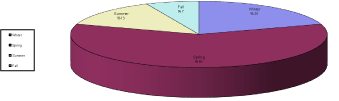
Regarding to the distribution of arthropod larvae species recovered from the examined 15 forensic entomology cases in Middle Delta, Egypt, pie chart 1 demonstrate that Chrysomya albiceps larvae (Calliphoridae) (photomicrograph 1&2) outnumbered the other dipterous species as it was found infesting nine cases (60%), followed by Sarcophaga species (photomicrographs 3&4)(Sarcophagidae) (4 cases = 26.7 %). Dermestidae (photomicrograph 5) infested two cases (13.3%), while Psychoda species (photomicrograph 6) and Lucilia sericata (photomicrographs 7&8) (Calliphoridae) infested only one case for each (6.7%).
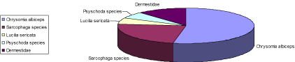
    Pie chart 1:  Distribution of arthropod larvae species recovered from the 15
forenic entomology cases in middle Delta, Egypt.
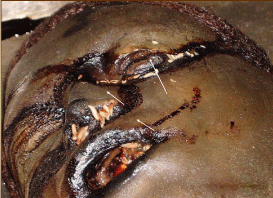
    Photomicrograph 1:  A male corpse aged 18 years in a state of bloated
decay with masses of maggots were found mainly in the natural orifices.
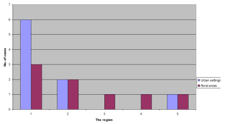
In this study, Chrysomya albiceps was the most common dipterous species that breed in corpses in both urban (6 cases) and rural (3 cases) areas representing 67% and 33% respectively. While both Sarcophagidae and Dermestidae were equal in both urban and rural areas. Lucilia sericata and Psychoda species were only present in rural areas in this study (Table 3 and Histogram 1).
![]()
    Habitat
    Urban(n=8)
    Rural(n=7)
    Total(n=15)
    P


    Arthropod species
    No.
    %
    No.
    %
    No.
    %


    Chrysomya albiceps
    6
    67%
    3
    33
    9
    60.0
    0.315


    Lucilia sericata
    0.0
    0.0
    1
    100
    1
    6.7
    0.467


    Sarcophaga specis
    2
    50%
    2
    50
    4
    26.7
    1.000


    Psychoda species
    0.0
    0.0
    1
    100
    1
    6.7
    0.467


    Dermestidae
    1
    50%
    1
    50%
    2
    13.3
    10.00


    P value significant at p < 0.05
Table 3:  Percentage of the different species of insects collected from 15 forensic
entomology cases in Middle Delta in different habitats.
    Histogram 1:  Number of corpses representing the habitat of species of insects I
in different areas in Middle delta, Egypt. 1-Chrysomia albiceps. 2-Sarcophaga
species. 3-Lucilia sericata. 4-psychoda species. 5-Dermestidae.
As regards to season of examination of the studied 15 forensic entomology cases, 9 cases (60%) were found in spring, 3 cases (20%) in winter, 2 cases (13.3%) in summer and 1 case (6.7%) in fall (Table 4 and Pie chart 2).
![]()
    Spring
    Summer
    Fall
    Winter
    Total


    No.
    %
    No.
    %
    No.
    %
    No.
    %
    No.
    %


    9
    60%
    2
    13.3%
    1
    6.7%
    3
    20%
    15
    100%
Table 4:  Percentage of seasonal distribution of 15 forensic entomology cases in
Middle Delta, Egypt from 2004-2006.
    Pie chart 2:  Percentage of seasonal distribution of the 15 entomology cases
in middle Delta, Egypt from 2004-2006.
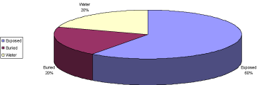
In the present study, the percentage of the exposed cases were (9 cases=60%), while, the other six cases were equally distributed between aquatic locations and burial (20% for each) (Table 5 and Pie chart 3).
![]()
    Body location
    Exposed
    Buried
    water


    Cases
    No.
    %
    No.
    %
    No.
    %


    No. of cases
    9
    60%
    3
    20%
    3
    20%
Table 5:  Percentage of distribution of different cases in accordance to Location
of the body.
    Pie chart 3:  Percentage of distribution of different cases in accordance to
location of the body.
The distribution of different species of arthropods in different cases in accordance to seasons is shown in Table 6 and Histogram 2. In the present study Chrysomya albiceps was attracted to corpses in spring (5 cases), summer (2 cases) and winter (2 cases). Sarcophaga species larvae were infesting dead bodies in both spring (3 cases) and fall (1 case) representing 75% and 25% respectively.
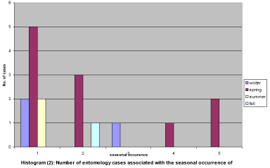
    Histogram 2:  Number of entomology cases associated with the seasonal
occurrence of insect species.
1-Chrysomia albiceps. 2-Sarcophaga species. 3-Lucilia sericata. 4-psychoda
species. 5-Dermestidae.
![]()
    Season
    Winter
    Fall
    Summer
    Spring
    Total
    P


    Larval species
    No.
    %
    No.
    %
    No.
    %
    No.
    %
    No.
    %


    Chrysomya  albiceps
    2
    22.2
      0
    0.0
    2
    22.2
    5
    55.6
    9
    100
    1.000


    Sarcophaga species
    0.0
    0.0
    1
    25
    0
    0.0
    3
    75
    4
    100
    1.000


    Luicilia sericata
    1
    100
    0
    0.0
    0.0
    0.0
    0
    0.0
    1
    100
    0.235


    Psychoda species
    0.0
    0.0
    0
    0.0
    0.0
    0.0
    1
    0.0
    1
    100
    1.000


    Dermestid larvae
    0
    0.0
    0
    0.0
    0
    0.0
    2
    100
    2
    100
    1.000
Table 6:  Percentage of seasonal distribution of the different species of insects
recovered from forensic entomology cases in Middle Delta, Egypt.
Luicilia sericata larvae were collected from 1 case in winter. Psychoda species and Dermestidae larvae were collected from cases in spring only.
In this study, most Chrysomya albiceps collected from corpses were in the 3rd instar (9 cases), followed by the prepupa (post feeding 3rd instar) infesting 4 cases (44.4%). Second instars were found in 2 cases only (22.2%). All larvae of Luicilia sericata and Dermestidae (100%) were in the 3rd instar (Table 7).
![]()
    Species of    the insects
    Stages of    maggots


    1st  instar
    2nd instar
    3rd instar
    Post-feeding    3rd instar
    Total
      No.
      of cases    containing
      different    larval stages


    No.
    %
    No.
    %
    No.
    %
    No.
    %


    Chrysomya albiceps
    0.0
    0.0
    2
    22.2
    9
    100
    4
    44.4
    9


    Sarcophaga species
    0.0
    0.0
    0.0
    0.0
    4
    100
    1
    25
    4


    Lucilia    sericata
    0.0
    0.0
    0.0
    0.0
    1
    100
    0.0
    0.0
    1


    Psychoda    species
    0.0
    0.0
    1
    100
    1
    100
    0.0
    0.0
    1


    Dermestid    larvae
    0.0
    0.0
    0.0
    0.0
    2
    100
    0.0
    0.0
    2
Table 7: Percentage of distribution of larvae instars stages in examined cases in accordance to different arthropod species.
The application of entomological study was used in some cases as a representative for this study
In the studied case number one, a 7-years-old female found, in winter (November) on a heap of mud at the edge of a city in Gharbia governorate. The climatological data revealed that, the mean of maximum temperature of 15°C and the mean of minimum temperature was 9°C. The 3rd instar larvae of Chrysomya albiceps was found on the corpse of this female. The estimated time since death was 15-16 days. This time was also compatible with that estimated during the judicial proceedings which pointed to a time of death 17 days before the body was found.
The only case in this study which was invested with Lucilia sericata. It was a young female, about 12 years old found in January (winter), in an rural area in Gharbia governorate , near a water canal coast with a rob around her neck.
During the autopsy, larvae only found in the buccopharyngeal and other natural orifices of the body (photomicrograph 9) to protect them from bad weather conditions. The age of Lucilia sericata larvae could be estimated as at least 80h from oviposition, to which a minimum of 24h before the adults arrived in the corpse should be added. Thus, the postmortem interval must be at least 4 days before the discovery of the body. In this study the judicial enquiry pointed to a time of 5 days since death, which coincided with the estimation of postmortem interval by entomological study of Lucilia Sericata.
The case number 3 was a male accidentally crushed under the wheels of a moving heavy truck in winter (February) in an agricultural area. Later in beginning of spring, after shallow burial in an old tomb with many cracks in its walls. The body was exhumed for medicolegal investigations. The body showed insect activity by both adult and immature insects (masses of larvae) all over the body. Subsequent identification confirmed the presence of Coleoptera larvae (Dermestidae), and third instar larvae of Psychoda (Diptera). Dermestidae larvae were very numerous and characteristics of the last stages of decomposition when the remains were coming to be dry. They were found in great abundance among the muscular mass and bones. The Postmortem interval estimated from entomological evidence was calculated by mainly taking into account the development of Dermestidae larvae which, takes about 22 days at 28–30°C and 40–50 days at lower temperatures. The estimated time of death was 5 weeks, with temperatures much lower than 28°C. This conclusion is congruent with the data obtained from the presence of Dermestidae larvae in the corpse. The judicial enquiry pointed to a time of death 34 days before the body was found.
The application of entomo-toxicology was used in two cases of carbamate and organophosphorus toxicity as a cause of death (case no. 12& 14 respectively).
Discussion
It is important to note that although the application of the forensic entomology has been strongly criticized for years, it is now gaining acceptance in many countries and offers a great potential of contribution to the legal profession in legal proceedings in many countries [14].
The objective of this study was to identify the arthropod species visiting the carrions, describing it and trying to correlate between decomposition processes, entomological findings and information obtained from judicial proceeding. The summarized results refer to the most important taxa for estimating the postmortem interval.
The maximum number of cases in the present study was in the age group 10-20 years (26.6%). This may be due to that teenagers are more vulnerable to violence and injuries as they are more active, trouble makers with more tendency for aggression [15]. On the other hand, the least number of cases was encountered in the age group less than ten years and more than 50 years. This may be due to the fact that this age group is less mobile out of doors and of less aggression, so, less predisposed to violence.
This study showed that, males outnumbered females (73%) and (27%) respectively with a sex ratio about 3:1. This might be due to the fact that males are more liable to injuries as they are more mobile, more violent, and more liable to be engaged in quarrels and assaults. These findings coincide with the work of El-Moslimani [16].
Arthropods observed in this study follow the same general pattern found in both tropical and temperate areas. Rapid invasion of carrions was carried by adult Diptera (especially Calliphoridae and Sarchophagidae). Four carrion-breeding dipterans (Chrysomya albiceps, Sarcophaga species, Psychoda and Lucilia Sericata) were found in this study which coincide with El- Shenawey et al. [17] who noted that Calliphoridae and Sarcophagidae larvae were the most dominant and abundant insects infesting all the carrions in all seasons as in urban and rural habitats.
Benecke [18] reported that the identification of the arthropod species found at the site of, or on a corpse, is essential but often very difficult if performed by morphological means. One reason being that the order of insects consists of more species than any other form of life which means that there are few experts for a whole insect family or insect order. Another reason for difficulty in insect determination is the minuscule morphological differences between species. In addition, the characteristics of immature larvae are sometimes extremely hard to determine. Although size of larvae can be used to determine which larval stage of development a specific species of fly is in, there is a wide range of sizes that each larval stage can have. This depends on the amount of food available to the larvae have and the ambient and maggot mass temperatures in which they develop, this is why spiracular formation is often used to determine the stage of development of a larvae [19].
On badly decomposed corpses or skeletonized human remains infested with insects, where no classical postmortem changes in soft tissues (hypostasis, cadaveric rigidity, body cooling) are available, only the entomological evidence can provide invaluable aid to the forensic pathologist for predicting the postmortem interval [20].
Interestingly, Chrysomya albiceps larvae were the most dominant and common larvae infesting 60% of the corpses and were present in all seasons except fall as well as rural and urban habitat. Therefore, Chrysomya albiceps is the most important fly species in postmortem interval estimates during death investigation in Middle Delta of Egypt.
Chrysomya albiceps is well known as a warmer species. In Europe, this species extends from southern to northen regions during hot summer. In southeastern Spain the presence of this species is extremely rare in winter [21].
Tantawi et al. [22] have noted the complete absence of Chrysomya albiceps larvae in rabbit carcasses exposed during winter in Alexandria. However, in this study the larvae of this species were found infesting two corpses at the end of the winter season when the temperature was above its normal level during this period. The previous findings were in accordance with Arnaldos et al. [11].
The duration and viability of the larval stage of Chrysomya albiceps at 18, 22, 27 and 32°C were 21.30, 10.61, 5.0 and 4.0 days with corresponding percentage of growth 76.5, 88.5, 98.5 and 99.5%, respectively [20].
The minimal duration of development from oviposition to adult was inversely related to temperature, ranging from 8.3 +/- 0.5 days at 35°C to 19.2 +/- 0.92 days at 20°C. Although eggs hatched after 1.9 +/- 0.16 days at 15°C, larvae did not complete development and frequently died during the first instar stage [21].
In the present study, out of the nine cases invested with Chrysomya albiceps, six (67%) were confined to urban and three (33%) were confined to rural areas, and the four cases invested with Sarcophaga (26%) are equally distributed between urban and rural areas (50%) for each. No habitat specificity can be obtained from this study, because, in Middle Delta of Egypt there is no distinctive discrimination between urban and rural areas. The previous findings were supported by Carvalho and Linhares [23] who found that, Chrysomya albiceps and Sarcophaga is of no value as an indicator of a particular habitat type.
The presence of only Chrysomya albiceps in most of the studied cases is due to predation of Chrysomya Albiceps on other larvae as a result of the aggressive feeding behavior of 2nd and 3rd stage larvae of Chrysomya albiceps clearing all earlier arrivers [24].
The third instar larvae of Chrysomya albiceps was found on the corpse of a female 7 years old in winter. The estimated time since death was 15-16 days, bearing in mind the season in which death occurred according to the study of Arnaldos et al. [11] about the samples reared in the laboratory, at a temperature over 15°C, 3rd instar larvae of Chrysomya albiceps appear on day 3 and remain until day 13. This time was also compatible with that estimated during the judicial proceedings which pointed to a time of death 17 days before the body was found.
In this study, Chrysomya albiceps invested two cases which were buried under the ground. The mechanism by which Phorid flies could reach buried corpses could be explained as follows: adult phorid flies of genera Conicera and Metopina reach the soil depth of 50cm in four days. Also adults of the parasitic wasps of the families Braconidae and proctotrupidae occur in soil depth of at least 50cm. Conversely, bigger fly species, the larvae of which dominate in carrion covered by a thin layer of soil (e.g., Muscina species and the Helomyzid Morpholeria Kertezi Czerny), lay their eggs on the ground surface, and the young larvae migrate into the carrion through the soil [25,26].
In this study, all cases of Sarcophagidae larvae were found in spring and fall. These observations are in agreement with those of Tantawi et al. [21], who mentioned that in Giza province, Egypt, the breeding activity of Sarcophaga reaches its maximum in fall and spring.
Moreover, in summer, high temperatures suppress the breeding activity of Sarcophagidae and in winter cloudy days affect access to human remains by delaying colonization [2].
In the present study Sarcophagidae were infested 4 cases which were on the surface of the ground. On the other hand Campobasso et al. [2] founded in their study that Sarcophagidae were able to colonize rabbit carcasses buried at a depth of 10-20 cm already in March and April.
Moreover growth curves for the larvae and puparia of Sarcophaga haemorrhoidalis were studied. A significant growth of Sarcophagidae was observed at 25°C, but larvae were demonstrated under lower temperatures [27].
Surprisingly, Sarcophagidae may be the only carrion fly larvae found on a body that is physically isolated. This may reflect the fact that, in contrast to other forensically important flies, Sarcophagids deposit live larvae rather than eggs, and these may be dropped in the vicinity of the corpse when adult female cannot physically reach the body [2].
The only case in this study which was invested with Lucilia sericata. It was a young female, about 12 years old found in January (winter). In this study the judicial enquiry pointed to a time of 5 days since death, which coincided with the estimation of postmortem interval by entomological study of Lucilia Sericata. This observation was in agreement with Arnaldos et al. [11], who recorded the presence of Lucilia sericata larvae in the body of a male found in an area in south of Spain in February (winter) with similar climatic conditions. According to Kentner and Streit [28] taking into consideration the prevailing temperatures of the area and the data concerning the development of the Lucilia sericata larvae, their age could be estimated as at least 80h from oviposition, to which a minimum of 24h before the adults arrived in the corpse should be added. Thus, the postmortem interval must be at least 4 days before the discovery of the body.
Lucilia sericata is one of the earliest arriving fly species on remains with oviposition occurring typically only a few hours after death. Adult of Lucilia sericata are frequent in open and sunny habitat [29]. On the other hand Singh and Bharti [30] reported that Lucilia sericata laid eggs on human corpses discovered indoors.
In this study, Dermestidae larvae were collected from two cases (numbers 3 and 12), which were examined in spring, and their numbers were more in the highly decayed case. These findings partially in agreement with those of Kentner and Streit [28], who stated that the Dermestidae were collected during the earliest stages of decomposition in spring and summer, and their numbers increased as the remains began to dry.
In the studied case number 3, the body was exhumed for medicolegal investigations. The body showed insect activity by both adult and immature insects (masses of larvae) all over the body. Subsequent identification confirmed the presence of Coleoptera larvae (Dermestidae), and 3rd instar larvae of Psychoda (Diptera). Dermestidae larvae were very numerous and characteristics of the last stages of decomposition when the remains were coming to be dry. They were found in great abundance among the muscular mass and bones. The Postmortem interval estimated from entomological evidence was calculated by mainly taking into account the development of Dermestidae larvae which, according to Arnaldos et al. [31], takes about 22 days at 28–30°C and 40–50 days at lower temperatures. The estimated time of death was 5 weeks, with temperatures much lower than 28°C. This conclusion is congruent with the data obtained from the presence of Dermestidae larvae in the corpse. The judicial enquiry pointed to a time of death 34 days before the body was found.
The cause of death in two cases was carbamate and organophosphorus previous studies focused on the potential use of insects associated with decomposed remains as alternate specimens for toxicological analyses. In the case of malathione poisoning reported by Guatilake and Goff [32], the development stages of both Chrysoma megacephala and Chrysoma rufifacies were indicative of a minimum postmortem interval of 5 days, whereas the victim had been seen alive 8 days prior to the discovery of the body.
To the best of our knowledge, some papers reviewed that prescription and illegal drugs and toxins can be detected in arthropods. Diptera larvae, in particular those that actively feeding on human bodies provide a potentially valuable source of information in forensic investigations especially in the absence of tissues and fluids normally taken for toxicological analyses [7].
Because the results of such studies are greatly influenced by the available number of corpses investigated, so the present study recommends that in the future many studies should be conducted on a large number of corpses to explore more the ecology and biology of carrion flies in different seasons and habitats at different areas of Egypt.
The scene of death can yield information useful to reconstruct events and circumstances, link a suspect to the victim or scene, establish the credibility of the statements made to investigators by witnesses. Therefore, the forensic entomologist should perform his work at the scene of death even after removal of the body to collect the specimens most relevant to the case study and document the ecological and environmental conditions, thus more accurate estimation of the postmortem interval could be made (Table 8).
![]()
    Subj.
    Age by Ys.
    sex
    Resid.
    Time of death
    St. of put.
    Time of sample collect.
    Ext.wo.
    Amb. Temp.
    Gov.
    Cause of death
    Larvae distrib.
    Tox
    site


    Case no.


    1
    7
    ♀
    (Urban)
    1/2/ 04
    2nd
    18/2/ 04
    nil
    15-9
    Gh.
    Smoth.
    allover
    nil
    Ground surface


    2
    25
    ♂
    (urban)
    28/1/ 04
    2nd
    24/2/ 04
    nil
    14-8
    Gh.
    Myocard infarct.
    allover
    nil
    Partial submer.


    3
    42
    ♂
    (rural)
    20/2/ 04
    2nd
    24/3/ 04
    yes
    22-17
    Gh.
    RTA
    allover
    nil
    buried


    4
    18
    ♂
    (urban)
    2/5/ 04
    2nd
    12/5/ 04
    nil
    31-18
    Ksh.
    Strang.
    Natural
      orifices
    nil
    Ground surface


    5
    22
    ♂
    (rural)
    13/5/ 04
    2nd
    20/5/ 04
    nil
    30-20
    Mnf.
    diabetes
    allover
    nil
    Ground surface


    6
    31
    ♂
    (urban)
    18/5/ 04
    2nd
    1/6/ 04
    yes
    34 -19
    Gh.
    Stab
    allover
    nil
    Ground
      surface


    7
    54
    ♀
    (urban)
    20/6/ 04
    2nd
    28/6/ 04
    nil
    35-22
    Gh.
    Myocard
      infarct.
    allover
    nil
    Ground
      surface


    8
    24
    ♂
    (rural)

    3/7/ 04
    2nd
    22/7/ 04
    nil
    34-23
    Gh.
    Strang.
    allover
    nil
    Ground
      surface


    9
    17
    ♀
    (rural)
    5/10/ 04
    2nd
    12/10/04
    nil
    28-18
    Gh.
    Heart failure.
    Natural
      orifices
    nil
    Ground
      surface


    10
    50
    ♂
    (urban)
    14/3/ 05
    2nd
    24/3/ 05
    nil
    24-14
    Gh.
    Ruptured aortic aneurys.
    allover
    nil
    buried


    11
    37
    ♂
    (rural)
    23/3/ 05
    2nd
    3/4/ 05
    nil
    25-13
    Ksh.
    Strang.
    allover
    nil
    Ground
      surface


    12
    18
    ♂
    (urban)
    27/4/ 05
    2nd
    7/5/ 05
    nil
    26-14
    Gh.
    Carb. poison
    allover
    carb
    Ground
      surface


    13
    34
    ♂
    (rural)
    30/4/ 05
    2nd
    10/5/ 05
    yes
    30-18
    Ksh.
    Head trauma
    allover
    nil
    buried


    14
    45
    ♂
    (urban)
    25/5/ 05
    2nd
    5/6/ 05
    nil
    32-20
    Gh.
    organo ph.
    allover
    organo ph.
    Submer.


    5
    12
    ♀
    (rural)
    26/1/06
    2nd
    31/1/ 06
    nil
    12-7
    Gh.
    strangulation
    Natural
      orifices
    nil
    Submer.
Table 8: Master table showing the case study form.
References
- Knight B. Changes after death. Arnold, editor. In: Simpson’s Forensic Medicine. 11th edn. London. Sydney. Auckland. 1997; 27-39.
- Campobasso CP, Di Vella G, Introna F. Factors affecting decomposition and Diptera colonization. Forensic Sci Int. 2001; 120: 18-27.
- Benecke M. A brief history of forensic entomology. Forensic Sci Int. 2001; 120: 2-14.
- Rodrigez WC, Bassw M. Examination of badly decomposed skeletonized remains-over looked evidence. Thirty-ninth Annual Meeting. American Academy of Forensic Sciences. San Diego, CA. 1987; 112-132.
- Truman LC, Bnnett GW, Butts WL. Scientific Guide to Pest Control Operation. Harvest Publishing Co. Cleveland, Ohio. 8th edn. 1976; 230-256.
- Smith KGV. A Manual of Forensic Entomology. The Trustees British Museum. Natural History and Cornell University. Press. London. 1986; 205-220.
- Introna F, Campobasso CP, Goff ML. Entomotoxicology. Forensic Sci Int. 2001; 120: 42-47.
- Tullis K, Goff ML. Arthropod succession in exposed carrion in a tropical rainforest on O'ahu Island, Hawai'i. J Med Entomol. 1987; 24: 332-339.
- Zhu GH, Xu XH, Yu XJ, Zhang Y, Wang JF. Puparial case hydrocarbons of Chrysomya megacephala as an indicator of the postmortem interval. Forensic Sci Int. 2007; 169: 1-5.
- Dadour IR, Cook DF, Fissioli JN, Bailey WJ. Forensic entomology: application, education and research in Western Australia. Forensic Sci Int. 2001; 120: 48-52.
- Arnaldos MI, García MD, Romera E, Presa JJ, Luna A. Estimation of postmortem interval in real cases based on experimentally obtained entomological evidence. Forensic Sci Int. 2005; 149: 57-65.
- Clarke EGC. Isolation and Identification of Drugs in Pharmaceuticals, Body Fluids and Post-Mortem Materials. Pharmaceutical Press. 5th edn. 1996; 230-234.
- Niessen WM. Current Pracice of Gas Chromatography-Mass Spectrometry. CRC Press Pub. 1st edn. 2001; 423-424.
- Wolff M, Uribe A, Ortiz A, Duque P. A preliminary study of forensic entomology in Medellín, Colombia. Forensic Sci Int. 2001; 120: 53-59.
- Roberts I, Li L, Barker M. Trends in intentional injury deaths in children and teenagers (1980-1995). J Public Health Med. 1998; 20: 463-466.
- El Moslimani RF. A medicolegal study of incised and stab wounds. Thesis Submitted to the Faculty of Medicine Alexandria University in partial fulfillment of the requirements of the degree of Master of Forensic Medicine and Toxicology. 1979; 85-86.
- El Shenawey IE, Abd Alsalam FH, Tantawi TI, Madkor AS, Mahana NM. Insects associated with human corpses in Alexandria, Egypt and their potential use as forensic indicators. Thesis Submitted to the Faculty of Medicine Alexandria University in partial fulfillment of the requirements of the degree of Master of Forensic Medicine and Toxicology. 2004; 100 - 102.
- Benecke M. Random amplified polymorphic DNA (RAPD) typing of necrophageous insects (Diptera, Coleoptera) in criminal forensic studies: validation and use in practice. Forensic Sci Int. 1998; 98: 157-168.
- Joy JE, Herrell ML, Rogers PC. Larval fly activity on sunlit versus shaded raccoon carrion in southwestern West Virginia with special reference to the black blowfly (Diptera: Calliphoridae). J Med Entomol. 2002; 39: 392-397.
- Adams ZJ, Hall MJ. Methods used for the killing and preservation of blowfly larvae, and their effect on post-mortem larval length. Forensic Sci Int. 2003; 138: 50-61.
- Grassberger M, Friedrich E, Reiter C. The blowfly Chrysomya albiceps (Wiedemann) (Diptera: Calliphoridae) as a new forensic indicator in Central Europe. Int J Legal Med. 2003; 117: 75-81.
- Tantawi TI, eL-Kady EM, Greenberg B, el-Ghaffar HA. Arthropod succession on exposed rabbit carrion in Alexandria, Egypt. J Med Entomol. 1996; 33: 566-580.
- Lopes de Carvalho LM, Linhares AX. Seasonality of insect succession and pig carcass decomposition in a natural forest area in southeastern Brazil. J Forensic Sci. 2001; 46: 604-608.
- Faria LDB, Godoy WAC. Prey choice by facultative predator larvae of Chrysomya albiceps, Chrysomya megacephala, Chrysomya putoria and Cochliomyia macellaria (Diptera: Calliphoridae). J. Applied Entomol. 2001; 119: 263-266.
- Nuorteva P. Sarcosaprophagous insect as forensic indicators. Tedechi CG, Eckert WG, editors. In: Forensic Medicine, A Study of Trauma and Environmental Hazards. Saunders Company. Philadelphia, London, Toronto. 1977; 1072-1095.
- Catts EP, Goff ML. Forensic entomology in criminal investigations. Annu Rev Entomol. 1992; 37: 253-272.
- Byrd JH, Butler JF. Effects of temperature on Sarcophaga haemorrhoidalis (Diptera: Sarcophagidae) development. J Med Entomol. 1998; 35: 694-698.
- Kentner E, Streit B. Temporal distribution and habitat preference of congeneric insect species found a rat carrion. J. Pedobiología. 1990: 34: 347–359.
- Tarone AM, Foran DR. Components of developmental plasticity in a Michigan population of Lucilia sericata (Diptera: Calliphoridae). J Med Entomol. 2006; 43: 1023-1033.
- Singh D, Bharti M. Further observations on the nocturnal oviposition behaviour of blow flies (Diptera: Calliphoridae). Forensic Sci Int. 2001; 120: 124-126.
- Arnaldos MI, Romera E, Presa JJ, Luna A, García MD. Studies on seasonal arthropod succession on carrion in the southeastern Iberian Peninsula. Int J Legal Med. 2004; 118: 197-205.
- Gunatilake K, Goff ML. Detection of organophosphate poisoning in a putrefying body by analyzing arthropod larvae. J Forensic Sci. 1989; 34: 714-716.