
Clinical Image
Austin J Gastroenterol. 2014;1(3): 1012.
Condyloma Acuminatum Diagnosed by Dyed and Magnified Endoscopy
Ashikari K, Chiba H*, Suto T, Seki S, Takahashi A, Ida T, Morohashi T and Goto T
Department of Gastroenterology, Omori Red Cross Hospital, Japan
*Corresponding author: :Chiba H, Department of Gastroenterology, Omori Red Cross Hospital. 4-30-1, Chuo, Ota-Ku, Tokyo, 143-8527, Japan
Received: July 28, 2014; Accepted: July 30, 2014; Published: Aug 01, 2014
Keywords
Condyloma Acuminatum; Rectal tumor; Magnified endoscopy
Condyloma acuminatum is caused by human papilloma virus, and occurs more often in homosexual males or immune suppressed patients. The shape is sometimes similar to flat elevated adenomatous lesions, including lateral spreading tumors. There are little reports about the magnified or dyed endoscopic findings of Condyloma acuminatum. An 80-year-old male was admitted because of hematochezia and emergency endoscopy was performed. We found the whitish and accumulated granular tumor surrounded the anal canal. The tumor was stained densely with an iodine stain, which meant the tumor was not squamous cell carcinoma (Figure 1). In magnified endoscopy with narrow band imaging, enlarged and elongated loop capillary pattern without irregularity was found (Figure 2). According to these findings, we diagnosed with Condyloma acuminatum and performed giant biopsies by snare for collecting a tissue definitely. Pathological findings showed the Condyloma acuminatum with koilocytosis.
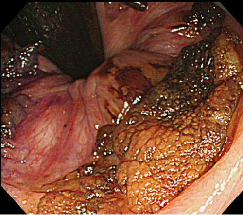
The flat elevated tumor was stained densely and uniformly.
    Figure 1:  Endoscopic finding (after an iodine stain)
 The flat elevated tumor was stained densely and uniformly.
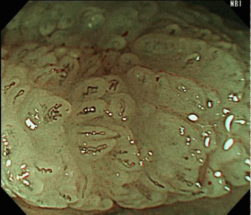
Enlarged and elongated loop capillary pattern without irregularity was found.
    Figure 2:  Endoscopic finding (magnified endoscopy with narrow band imaging)
 Enlarged and elongated loop capillary pattern without irregularity was found.