Clinical Image
Gastrointest Cancer Res Ther. 2016; 1(2): 1008.
Rare Gastric Submucosal Mass: Metastatic Melanoma
Lu X, Wu J, Liu L and Cao D*
Department of Radiology, The First Hospital of Jilin University, China
*Corresponding author: Dr Dianbo Cao, Department of Radiology, The First Hospital of Jilin University, 71 Xin Min Zhu Street, Chang Chun, China
Received: September 02, 2016; Accepted: September 07, 2016; Published: September 09, 2016
Keywords
Gastric metastatic tumors; Melanoma; Treatment
Clinical Image
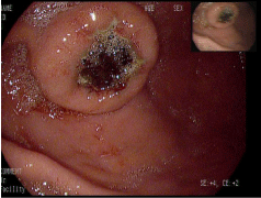
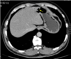
A 50-year-old man complained of fatigue and intermittent black stool over the past 5 days and came to our hospital to search for the source of gastrointestinal bleeding. He denied any intake of nonsteroidal anti-inflammatory medications and had a surgical history of cutaneous malignant melanoma of left heel 1 year previously. Systemic physical examination including ophthalmologic and dermatologic investigation was unremarkable. The hematological and biochemical profiles were normal limits except for mild decrease of blood red cell with the value to 3.93×10¹². A colonoscopy was negative, and endoscopy revealed a 2cm mass in diameter with central ulcer on the anterior wall of the distal gastric body (Figure 1). Due to the presence of brown blood clot on the surface of ulcer, biopsy for the lesion was cancelled. This outcome was felt to be the cause of his black stool since abdominal computerized tomography showed no other gastrointestinal abnormalities except for gastric occupying lesion (Figure 2). The patient underwent partial gastrectomy and histopathologic evaluation of the resection specimen confirmed malignant melanoma with tumor-free resection margin. Lymph nodes were also negative for metastases. Associated with past history of cutaneous malignant melanoma, the diagnosis of gastric metastatic melanoma was finally made. No additional metastasis or recurrence has been detected in the patient 12 months after discharge.
Metastasis to the stomach is uncommon, with a reported incidence of 0.2–0.7 % based on clinical and autopsy findings. Gastric metastatic tumor may come from an array of primary malignancies such as breast cancers followed by lung cancer, esophageal cancer, renal cell carcinoma and malignant melanoma. Hematogenous route may be responsible for its occurrence in our patient. The clinical manifestations of metastasis in the stomach are variable. Endoscopic and radiological evaluations are key to discriminate between primary gastric cancer and metastatic tumors to the stomach. As known, the presence of gastric metastasis is associated with advanced disease and the survival period is extremely short. When there is a risk of bleeding, tumor perforation and/or a solitary metastasis, surgical resection may be strongly recommended as the first choice, as described in our case.

