Abstract
Introduction: Congenital Common Bile Duct (CBD) webs are extremely rare abnormalities of the extrahepatic ducts with approximately 10 cases reported in the literature. The age at presentation and the clinical symptomatology of these anomalies depend on the grade of the biliary obstruction. These webs usually exhibit early in life as obstructive jaundice, dilation of the proximal biliary tree or even spontaneous perforation of the extrahepatic duct. Some of these congenital webs are partially developed and remain asymptomatic until adulthood.
Case Report: 28 year female patient presented with cholestatic pattern jaundice for 2 months. On evaluation found to have dilated CBD with IHBRD on USG. On further imaging studies, CT revealed horizontal web like projection from distal CBD suggestive of web with similar findings on MRCP. ERCP showed horizontal filling defect on cholangiogram with dilated CBD. Endoscopic Ultrasound examination revealed horizontal hyperechoic structure at distal CBD with proximally dilated CBD and IHBRD. Dilatation was performed using Soehendra Biliary Dilation Catheter with significant improvement in her symptomatology.
Conclusion: Our case remains the first of its kind in which EUS characterisation of CBD web is described. Though rare congenital anomalies remain an important cause of young patients presenting with obstructive jaundice. Treatment for such cases remain Endoscopic dilatation or surgical by-pass in whom endoscopic treatment fails.
Keywords: Common Bile Duct Web; Obstructive Jaundice; Soehendra Biliary Dilation Catheter; Endoscopic Ultrasound
Abbreviations
Hb: Hemoglobin; TLC: Total Leucocyte Count; AST: Aspartate Transaminase; ALT: Alanine Transaminase; ALP: Alkaline Phosphatase; USG: Ultrasonography; IHBRD: Intra-Hepatic Biliary Radicle; CBD: Common Bile Duct; GB: Gall Bladder; MRCP: Magnetic Resonance Cholangio-Pancreatography
Case Report
28 year old female presented with yellowish discoloration of eyes with high coloured urine followed by itching and clay coloured stools, without any history of fever or other constitutional features for 2 months. No significant history of pain abdomen, abdominal distension, signs or symptoms of GI bleeding, anorexia or weight loss was there. History of complimentary and alternative medication use for around 1 month after onset of jaundice without any improvement. On investigation, Hb 11.3g/dl, TLC 16,900/μl, Platelet count of 321 × 109/μl, Bilirubin (Total/Direct) 18.8/13.8, AST 37, ALT 31, ALP 305, Protein/Albumin of 6.9/3.1. USG abdomen revealed normal sized liver with moderate central and peripheral IHBRD. Dilated CBD of 2cm at porta with smooth tapering seen at distal CBD. GB normally distended. CT scan was done which revealed linear enhancing web like projection measuring 10mm arising from lateral wall of distal CBD approximately 2cm from opening at ampulla with significantly dilated CBD, CHD and moderate central and peripheral IHBRD. MRCP was planned next, which showed marked dilatation of intra and extra hepatic biliary tree, proximal CBD of 2.4 cm with abrupt change in calibre of intra-pancreatic CBD, with linear T2 hypointense lesion, likely web (Figures 1,2).
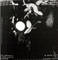
    Figure 1: T2-MRI demonstrating hypointense lesion in intra pancreatic CBD
with resultant proximal CBD dilatation.
    Figure 2: T1 MRI demonstrating hyperintense lesion in distal CBD causing
proximally dilated CBD.
EUS was performed which revealed horizontal hyperechoic structure of 7mm in distal CBD with dilated proximal CBD, measuring 2.5 cm at porta and intra-pancreatic CBD of 5 mm (Figure 3).
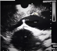
    Figure 3: EUS demonstrating hyperechoic structure “web” in distal CBD with
proximal CBD dilatation.
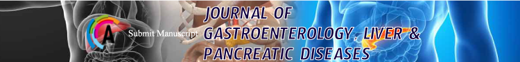
Patient then planned for ERCP for biliary drainage for alleviation of her symptoms. Cholangiogram revealed dilated CBD measuring 2.5 cm with horizontal band like filling defect in distal CBD (Figure 4). Dilatation was performed using Soehendra Biliary Dilation Catheter of 7, 8.5 & 10 Fr. 10 Fr × 10 cm stent was placed post dilatation. Serum CA 19-9 was normal.
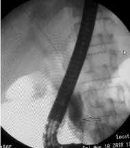
    Figure 4: Retrograde cholangiogram demonstrating horizontal band like
filling defect in distal CBD.
Post ERCP follow up, patient had declining serum bilirubin level with significant improvement in itching and clay stools.
Discussion
Embryologically, the bile ducts in the developing phase become obliterated by epithelial concrescence or proliferation. Later these solid structures become vacuolated, leading to formation of a lumen. Recanalization of the lumen of the biliary tree usually starts at the end of the fifth week of gestation [1]. Congenital CBD webs develop due to incomplete re-canalization of these solid structures.
The described associated abnormalities with these webs include anomalous hepatic duct of the caudate lobe, anomalous junction of the pancreato-biliary ductal system, choledochal cyst or hepatic fibrosis, choledocholithiasis, primary sclerosing cholangitis or idiopathic. Our case represents the first to report EUS characteristics of choledochal web, with no previous reports till date.
Webs may be partial or complete and the degree of obstruction determines whether patient will be symptomatic or not. Treatment is either Endoscopic dilatation or surgical by-pass [2]. Dolar et al. described a congenital web of the CHD in an adult patient, which was not associated with jaundice [3]. Chapoy et al. described a similar case to the one described by Dolar et al. but their patient was only 40 months old.4 The association of CHD ‘diaphragms’ and biliary obstruction has been described by both Fisher et al. and Devanesan et al., who postulated that small perforations in these diaphragms allowed some degree of biliary drainage and thus a delayed presentation of obstructive jaundice [5,6]. Ravi K et al. In their report mentioned about successful endoscopic therapy of choledochal web by Balloon Dilatation [7]. Kim et al described a case of intra-hepatic choledochal web which was treated by balloon dilatation [8]. Gulliver et al. described common bile stones to be associated with the web in a significant number of patients [9]. Our patient did not, however, have bile duct stones, but clearly had a web present in the CBD and had significant improvement in clinical and biochemical parameters post endoscopic treatment.
The physiologic implications of web of the extrahepatic biliary tree are not necessarily the same as for other causes of extrahepatic biliary obstruction. While the web may ultimately cause obstruction, it is likely that forward drainage from the liver will be undisturbed in the vast majority of cases. Initially the patient may be asymptomatic or may present with vague and nonspecific symptoms such as abdominal pain, nausea and vomiting. Early on in the disease process, one may only demonstrate elevations of transaminases and alkaline phosphatase, together with ductal dilatation, but without obstructive jaundice. However, in the setting of inflammation in the region of the porta hepatis, or the passage of a small calculus, the web will almost certainly become obstructed and result in obstructive jaundice with or without the association of cholangitis. These webs are probably congenital and only present later in life due to an associated abnormality.
While the standard imaging studies such as ultrasonogram and computerized tomography may reveal bile duct dilatation, they are unlikely to reveal the presence of a web. Endoscopic Retrograde Cholangiopancreatography may be more successful in adequately delineating the presence of a web. With the increasing usage of Magnetic Resonance Cholangiography, this anatomical abnormality may be more commonly diagnosed in the future. Moreover, role of Endoscopic Ultrasound in choledochal web has not yet been described in world literature. Our case remains first of its kind to describe the same.
References
- Ando H Embryology of the biliary tract. Dig Surg. 2010; 27: 87-89.
- Siegel JH. Endoscopic retrograde cholangiopancreatography: technique, diagnosis and therapy. New York: Raven Press.1992; 38-39.
- Dolar ME, Ates KB, Dalay AR, Caner ME, Sasmaz N, Sahin B. Congenital stricture of the common hepatic duct due to a web: An unusual case without jaundice. Hepatogastroenterology. 1993; 40: 194-195.
- Chapoy PR, Kendall RS, Fonkalsrud E, Ament ME. Congenital stricture of the common hepatic duct: An unusual case without jaundice. Gastroenterology. 1981; 80: 380-383.
- Fisher MM, Chen S, Dekker A. Congenital diaphragm of the common hepatic duct. Gastroenterology. 1968;54: 605-610.
- Devanesan J, deBlasi HP, Sable R, Hoe R, Kinkahabwala M. Congenital hepatic duct obstruction with perforate diaphragms. Arch Surg. 1978; 113: 1452-1455.
- Ravi K, Jose F, Harshad D. Successful endoscopic therapy of an obstructing common bile duct web. GastrointestEndosc. 2001;53: 126-128.
- Kim HI, Lee SO, Jeong YW, Kim SH, Kim IH, Kim SW, et al. A case of intrahepatic choledochal web that was diagnosed by percutaneous transhepatic cholangioscopy and it was treated with balloon dilatation: review of the korean cases. Korean J GastrointestEndosc. 2009;39: 319-323.
- Gulliver DJ, Baker ME, Putnam W, Baillie J, Rice R, Cotton PB. Bile duct diverticula and webs: Nonspecific cholangiographic features of primary sclerosing cholangitis. AJR. 1991; 157: 281-285.