
Case Report
Austin Gynecol Case Rep. 2016; 1(1): 1006.
Septic Pelvis Thrombophlebitis in Postpartum Period: Diagnosis by Clinical and Magnetic Resonance Imaging Findings
Busse Filho KR, Campanharo FF, Araujo Júnior E*, Martins Santana EF and Moron AF
Department of Obstetrics, Paulista School of Medicine - São Paulo Federal University (EPM-UNIFESP), Brazil
*Corresponding author: Edward Araujo Júnior, Department of Obstetrics, Paulista School of Medicine - São Paulo Federal University (EPM-UNIFESP), Rua Belchior de Azevedo, 156 apto. 111 Torre Vitoria, São Paulo-SP, Brazil
Received: September 15, 2016; Accepted: December 12, 2016; Published: December 14, 2016
Abstract
Septic Pelvic Thrombophlebitis (SPT) is a rare condition in the postpartum period which is characterized by persistent fever, despite the antimicrobial therapy, and diffuse abdominal pain and in the legs. SPT is more frequent after caesarean section delivery. The diagnosis can be performed by ultrasound, computed tomography and magnetic resonance imaging, being this last imaging diagnostic method of chosen because it is better to visualizing of soft tissue, edema and inflammatory signs. The treatment is based on heparin and oral anticoagulation drug until six months after delivery. We showed a case of primiparous which developed SPT after caesarean section delivery and presented good prognosis with heparin therapy.
Keywords: Septic pelvic thrombophlebitis; Cesarean section; Postpartum; Heparin; Magnetic resonance imaging
Introduction
Septic Pelvic Thrombophlebitis (SPT) still is a condition under diagnosed and poorly managed. The 1960’s brought a more clinical approach, and with the advent of broad-spectrum antibiotics regimens, development of newer diagnostic tools and a better understanding of the pathophysiology of disease, mortality and incidence significantly decreased [1]. Nowadays the incidence is growing again with more caesarean sections being performed. We present a case to illustrate how SPT may clinically present, be diagnosed and treated.
Case Presentation
A 34-year-old gravida 2 para 0 pregnant woman at 38 6/7 weeks gestation was admitted to our service in labor with clear signs of chorioamnionitis (maternal and fetal tachycardia, fever, foul smelling amniotic fluid and uterine tenderness). During the labor conduction, signs of acute fetal distress were observed (persistent fetal tachycardia) and a caesarean section delivery was performed. A male newborn, birth weight of 3615g and Apgar score of 9/9 was delivered. In the postpartum, woman received a broad-spectrum antibiotic therapy (penicillin, gentamicin and metronidazole) and uterotonic agents, with clinical improvement. On the 4th day postpartum, however, the patient was still febrile, despite the antimicrobial therapy, and with a diffuse abdominal pain that was worse in the lower levels of the abdomen. Pelvic and transvaginal ultrasonography were performed without significant findings, raising suspicion of SPT. Anticoagulation with enoxaparin 1mg/kg twice a day was initiated. She underwent a magnetic resonance angiography, which confirmed the clinical suspicion, finding a filling defect in the middle third of the right internal iliac artery suggestive of thrombus (Figure 1). After 2 days of anticoagulant therapy with enoxaparin, patient became afebrile, and then decided to keep warfarin for anticoagulation maintenance. Later, the patient had no further complications in the puerperium, being discharge home with a target International Normalized Ratio (INR) between 2 and 3. The warfarin therapy was maintained during 6 months after delivery.
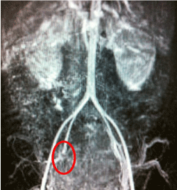
    Figure 1:  Angio-magnetic resonance imaging in the coronal view with T1-
weighted sequences after contrast injection, showing filling defect in the
middle third of the right internal iliac artery suggestive of thrombus (red circle).
Discussion
The pelvic plexus is a common site of postpartum thrombosis. A study found that the overall incidence of SPT was 1:3000 deliveries, being 1:9000 after vaginal delivery and 1:800 after caesarean section [2]. In the presence of a strong clinical suspicion of pulmonary embolism, with Doppler negative lower limb, we must continue the investigation [3]. Differential diagnoses of SPT are the following: endometritis, pelvic abscesses, urinary infection, wound infection, deep vein thrombosis, pulmonary embolism and drug induced fever.
Symptoms suggestive of SPT include prolonged puerperal fever, defined as more than five days of fever regardless of appropriate antimicrobial treatment, commonly associated with constant vague lower abdominal pain. Physical examination often reveals few signs, with the overall condition of the patient preserved [4]. The risk factors identified in this case included the caesarean delivery and the presence of pelvic infection. Back pain and leg weakness after regional anesthesia for caesarean section has been described as alert sign of SPT [5]. Usually, patients become afebrile after a mean of 4.7 ± 2.1 days of heparin therapy [6].
Imaging studies may be used to evaluate the extension of thrombosis and to determine the duration of anticoagulation therapy, ranging from 1 week to 6 months. A study performed in 1996 [6] concluded that imaging methods could not help in the diagnoses of SPT and suggests other criteria. Other study compared Magnetic Resonance Imaging (MRI), Computed Tomography (CT) and ultrasound, both MRI and CT proved to be comparable, but MRI is better at visualizing of soft tissue, edema and inflammatory signs. Ultrasound is not useful at diagnosing SPT, but may have a role at excluding other differential diagnosis [7]. Ultrasound has limited sensibility to identify the echogenic material into the internal iliac artery. On the other hand, CT and MRI demonstrate a filling defect into the internal iliac defect [8]. MR direct thrombus imaging allows the direct visualization of the thrombus in the legs, without intravenous contrast. In the diagnosis of deep vein thrombosis, MR direct thrombus imaging showed sensibility of 96% and specificity of 96% compared to ultrasound and ascendant phlebography [9]. Other advantage of MRI over CT is the absent of ionizing radiation.
References
- Dunnihoo DR, Gallaspy JW, Wise RB, Otterson WN. Postpartum ovarian vein thrombophlebitis: a review. Obstet Gynecol Surv. 1991; 46: 415-427.
- Brown CE, Stettler RW, Twickler D, Cunningham FG. Puerperal septic pelvic thrombophlebitis: incidence and response to heparin therapy. Am J Obstet Gynecol. 1999; 181: 143-148.
- James A; Committee on Practice Bulletins—Obstetrics. Practice bulletin no. 123: thromboembolism in pregnancy. Obstet Gynecol. 2011; 118: 718-729.
- Garcia J, Aboujaoude R, Apuzzio J, Alvarez JR. Septic pelvic thrombophlebitis: diagnosis and management. Infect Dis Obstet Gynecol. 2006; 2006: 15614.
- French RA, Cole C. An “enigmatic” cause of back pain following regional anaesthesia for caesarean section: septic pelvic thrombophlebitis. Anaesth Intensive Care. 1999; 27: 209-212.
- Witlin AG, Mercer BM, Sibai BM. Septic pelvic thrombophlebitis or refractory postpartum fever of undetermined etiology. J Matern Fetal Med. 1996; 5: 355- 358.
- Twickler DM, Setiawan AT, Evans RS, Erdman WA, Stettler RW, Brown CE, et al. Imaging of puerperal septic thrombophlebitis: prospective comparison of MR imaging, CT, and sonography. AJR Am J Roentgenol. 1997; 169: 1039-1043.
- Laifer-Narin SL, Kwak E, Kim H, Hecht EM, Newhouse JH. Multimodality imaging of the postpartum or posttermination uterus: evaluation using ultrasound, computed tomography, and magnetic resonance imaging. Curr Probl Diagn Radiol. 2014; 43: 374-385.
- Kelly J, Hunt BJ, Moody A. Magnetic resonance direct thrombus imaging: a novel technique for imaging venous thromboemboli. Thromb Haemost. 2003; 89: 773-782.