Case Report
Austin Gynecol Case Rep. 2017; 2(2): 1014.
A Case of Uterine Leiomyoma Presenting as Atypical Pseudo-Meigs’ Syndrome
Moukit M¹*, Ait E¹ Fadel F¹, Kouach J¹, Chenana A¹, Guelzim K¹, E¹ Hassani ME¹, Babahabib A¹, Moussaoui RD¹ and Dehayni M²
¹Department of Obstetrics and Gynecology, Military Training Hospital Mohammed V, Morocco
²Pole of Gynecology and Visceral surgery, Military Training Hospital Mohammed V, Morocco
*Corresponding author: Mounir Moukit, Department of Obstetrics and Gynecology, Military Training Hospital Mohammed V, Hay Riyad, 10100, Rabat, Morocco
Received: January 23, 2017; Accepted: March 27, 2017; Published: April 03, 2017
Abstract
Benign uterine leiomyomas presenting as pseudo-Meigs’ syndrome with elevated CA125 are rare, and only few cases have been reported in the literature. We report a case of a 37-year-old lady, presented for progressive abdominal distension. Physical examination was difficult due to a massive pelvic mass reaching to the umbilicus. Transvaginal ultrasound revealed a giant echogenic mass swimming in a peritoneal effusion. The preoperative serum CA125 was 1218 UI/ml. Laparotomy with complete excision of the mass was done and sent for histopathological examination, confirming the diagnosis of a uterine leiomyoma. The definitive diagnosis of atypical pseudo-Meigs’ syndrome was made by confirming the fact that peritoneal effusion disappeared, and CA125 level returned to normal after myomectomy.
Keywords: Uterine leiomyoma; Ascites; CA125; Atypical Pseudo-Meigs’ syndrome
Introduction
Pseudo-Meigs’ Syndrome (P-MS) caused by leiomyoma was first described by Brown, in 1998 [1]. Since then, fewer than 15 cases have been reported in the literature. Here, we present a case of giant pedunculated subserosal leiomyoma presenting as atypical Pseudo- Meigs’ syndrome with elevated CA125 level.
Case Report
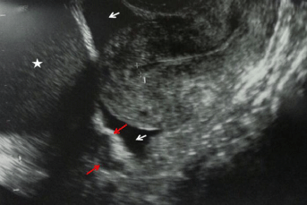
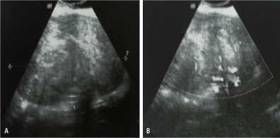
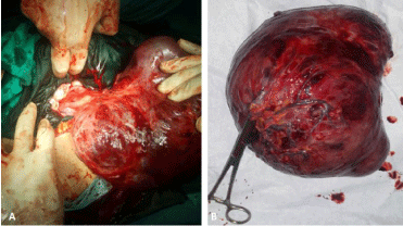
A 37-year-old Moroccan lady presented in our department with the history of progressive abdominal distension. She denied any genito-urinary symptoms. On admission, the patient was afebrile, with normal vital signs. She was on the second day of the menstrual cycle. Physical examination was difficult due to a massive pelvic mass reaching to the umbilicus; mild tenderness was found on deep palpation of the right lower abdominal quadrant. Systemic examination did not show any abnormality. Transvaginal ultrasound revealed a heterogeneous lobulated mass, occupying the entire pelvic cavity, extended to the abdomen with a peduncle attached to the uterus, suggestive of pedunculated subserosal leiomyoma (Figures 1 and 2). Left and right ovaries were normal with middle amount of free fluid in the peritoneal cavity. Laboratory studies showed no abnormalities except for a serum CA125 level of 1218 UI/ml (normal <35 UI/ml). An exploratory laparotomy was planned for diagnostic and therapeutic purposes. A large firm mass measuring 20×21 cm, which originated from the posterior wall of uterus with a long and tenuous fibrotic peduncle was seen (Figure 3A). The uterus, ovaries, bowel and omentum were normal. Complete excision of the mass after aspiration of the ascitic fluid was performed (Figure 3B). Microscopic examination showed uterine leiomyoma with significant hemorrhage, necrosis and areas of myxoid degeneration; mitotic activity and atypical cells were absent. Postoperatively, chest X-ray did not show a pleural effusion. At one month follow-up, she was clinically well, without evidence of ascites re-accumulation and had a normal CA125 level.
Discussion
Pseudo-Meigs’ syndrome is a rare entity characterized by association of a pelvic tumour other than an ovarian fibroma with peritoneal and pleural effusion [2,3]. The majorities of these cases have been ovarian pathologies and rarely associated with uterine leiomyomas [2]. In our case, the patient presented with leiomyoma, peritoneal effusion without hydrothorax, which is extremely rare. This variant in terms of presentation has been called atypical pseudo-Meigs’ syndrome [4]. Various theories have been proposed regarding the mechanisms of peritoneal effusion. Inflammatory reaction of the peritoneum, compression with subsequent congestion of the lymphatics and blood vessels, twisting of the tumour, and hypoalbuminemia are the common proposed theories [3,5,6]. Peroperatively, the subserosal leiomyoma was large, and there was no opportunity for the pedicle to rotate. We suggest that ascites might be due to compression with congestion of the lymphatics and blood vessels. Interstitial fluid secretion from a fibroid surface is also supposed to cause the development of ascites [7]. In our case, the connection between uterine leiomyoma and ascites has been confirmed by the rapid resolution of peritoneal effusion after myomectomy. Clinical, sonographic, computed tomography scan, and Magnetic Resonance Imaging findings of leiomyomas are typical and the diagnosis is usually easy. However, large pedunculated leiomyomas may present with a wide variety of symptoms and unusual appearances, mimicking other gynecologic or non-gynecologic masses. They can only be recognized if they are separate from the ovary and uterus and if their peduncle can be demonstrated on sonography, as was our case. CA125 does not have 100% specificity in the diagnosis of ovarian cancer, and its elevation may also occur following others clinical situations: benign ovarian tumours, pelvic inflammatory disease, endometriosis, uterus fibroma, hepatitis, pancreatitis, peritonitis and peritoneal tuberculosis [8]. In Meigs’ and pseudo-Meigs’ syndromes, mesothelial cells expression of this tumour maker and peritoneal inflammation caused by the large leiomyoma are the primary cause of elevated CA125 level [7,9]. In the present case, decline and normalization of serum CA-125 were detected after surgery confirming the association between raised CA- 125 levels and uterine leiomyomas. Consequently, this case provided an example for the limitation of this tumour marker in predicting pelvic malignancy.

    Figure 1: Transvaginal ultrasound revealing pelvic mass (Asterisk), with a
peduncle attached to the posterior wall of uterus (red arrows) and free fluid in
the peritoneal cavity (White arrows).

    Figure 2: Transvaginal ultrasound objectifying heterogeneous lobulated
mass with dystrophic calcifications, (A) measuring 20x18cm, and (B) flow
signals in Doppler.

    Figure 3: (A) Peroperative view showing a giant pedunculated mass
protruding from the uterus and (B) macroscopic view of the resected
specimen (3750 g).
Conclusion
Although uterine leiomyoma presenting as atypical pseudo- Meigs’ syndrome is extremely rare, it should be considered in differential diagnosis when a patient has a pelvic mass with ascites and an elevated level of CA125, particularly if laparocentesis is negative for malignant cells.
Acknowledgment
We are thankful to the patient who permitted us to present his history in this report.
References
- Brown RSD, Marley JL, Cassoni AM. Pseudo-Meigs’ syndrome due to broad ligament leiomyoma: a mimic of metastatic ovarian carcinoma. Clin Oncol. 1998; 10: 198-201.
- Ollendorff AT, Keh P, Hoff F, Lurain JR, Fishman DA. Leiomyoma causing massive ascites, right pleural effusion and respiratory distress: a case report. J Reprod Med. 1997; 42: 609-612.
- Terada S, Suzuki N, Uchide K, Akasofu K. Uterine leiomyoma associated with ascites and hydrothorax. Gynecol Obstet Invest. 1992; 33: 54-58.
- Ricci G, Inglese S, Candiotto A, Maso G, Piccoli M, Alberico S, et al. Ascites in puerperium: a rare case of atypical pseudo-Meigs’ syndrome complicating the puerperium. Arch Gynecol Obstet. 2009; 280: 1033-1037.
- Amant F, Gabriel C, Timmerman D, Vergote I. Pseudo-Meigs’ syndrome caused by a hydropic degenerating uterine leiomyoma with elevated CA 125. Gynecol Oncol. 2001; 83: 153-157.
- Zannoni GF, Gallotta V, Legge F, Tarquini E, Scambia G, Ferrandina G. Pseudo-Meigs’ syndrome associated with malignant struma ovarii: a case report. Gynecol Oncol. 2004; 94: 226-228.
- Timmerman D, Moerman P, Vergote I. Meigs’ syndrome with elevated serum CA125 levels: two case reports and review of the literature. Gynecol Oncol. 1995; 59: 405-408.
- Ghamande SA, Eleonu B, Hamid AM. High levels of CA125 in a case of a parasitic leiomyoma presenting as an abdominal mass. Gynecol Oncol. 1996; 61: 297-298.
- Lin JY, Angel C, Sickel JZ. Meigs syndrome with elevated serum CA 125. Obstet Gynecol. 1992; 80: 563-566.