
Case Report
Ann Hematol Oncol. 2014;1(3): 1013.
A Rare Case of Concomitant Small Lymphocytic Lymphoma and Kaposi's Sarcoma in the Same Lymph Node Biopsy
Madonna E1*, Avilia S1, Catalano L1, De Rosa G1 and Pane F1
1Department of Clinical Medicine, Federico II University, Italy
*Corresponding author: Madonna E, Division of Hematology, Department of Clinical Medicine, Federico II University, Naples, Italy
Received: October 26, 2014; Accepted: November 28, 2014; Published: December 02, 2014
Abstract
Small Lymphocytic Lymphoma (SLL) and its leukemic variant, Chronic Lymphocytic Leukemia (CLL), are the most common B-cell lymphomas in western countries.
In CLL/SLL, cutaneous or mucous membranes lesions occur in up to 25% of patients: infection or hemorrhage are the most common aetiologies, but they can also be an early manifestation of skin malignancy, as, when compared with normal population, in CLL/SLL skin cancer risk is increased eightfold.
Herein, we describe a case in which Kaposi Sarcoma (KS) and SLL were contemporary diagnosed in the same lymph node biopsy: concomitant diagnosis of KS and CLL/SLL is rare, and even rarer is the observation of the coexistence of both diseases in the same biopsy.
Keywords: Small Lymphocytic Lymphoma; Chronic Lymphocytic Leukemia; Kaposi Sarcoma
Case Presentation
A 61 years-old male was referred to our Haematology Division in May 2012, for diffuse asymptomatic lymphoadenopathy, appeared about one month earlier. He had not been exposed to immunosuppressive therapy nor had congenital or acquired immunodeficiency. Peripheral blood counts were: Hb 14.8 g/dL, WBC 6.250/μL, ANC 2.625/ μL, Platelets 163.000/ μL, Lymphocytes 3000/ μL.
CT scan showed multiple enlarged nodules on both sides of diaphragm, mainly in abdomen (para-aortic, maximum size: 28 mm).
After one week, the patient also showed small purple-blue cutaneous lesions on the left leg, associated with venous stasis, lymphedema, and hyperkeratosis. Color Doppler ultrasonography excluded vascular abnormalities.
Cervical lymph node biopsy showed diffuse architectural effacement due to infiltration of small monomorphous lymphocytes, with round nuclear contours and scant cytoplasm, without significant mitotic activity. Surface Immunophenotyping profile was: CD19+, CD5+/-, CD23+, CD20+/-, CD22+, lambda+. A small area with a multicentric proliferation of spindle-shaped, Human-herpesvirus positive (HHV8+) cells, with erythrocyte extravasations, was also evident in the sub capsular zone. Histological diagnosis was Chronic Lymphocytic Leukemia (CLL)/Small Lymphocytic Lymphoma (SLL) associated with Kaposi Sarcoma (KS) (Figure 1). Histhology of skin nodules was not performed. Bone marrow aspiration and biopsy revealed marked hypercellularity with nodular infiltration by small CD20+ and faintly CD5+ lymphocytes, supporting the diagnosis of SLL.
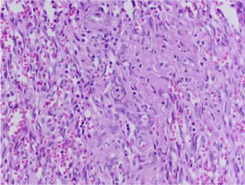
    Figure 1:  Lymph node biopsy. Vascular spaces containing erythrocytes. Note
the mitosis (encircled) (HE, x400).
Lymphocyte counts were normal at differential; anti-hepatitis B virus antibodies (HBsAb; HBcAb IgG) were found in the serum. HBV prophylaxis was performed with lamivudine. Work up for hepatitis C and HIV were negative.
The patient received six courses of anthracycline, vincristine, cyclophosphamide and rituximab every 3 weeks (CHOP-21), which caused disappearance of lymph nodes. However, skin lesions got worse (Figure 2). Significant local edema appeared, and the patient started local intralesional electro chemotherapy, combining the administration of a cytotoxic drug (bleomycin) followed by the application of high intensity electric pulses in the tumor lesions7. Unfortunately, there was no improvement. The patient then received oral etoposide (50 mg every day for three consecutive week) remaining in stable conditions about one year after the diagnosis. At the time of this report (September 2013) skin lesions are all over his legs, causing pain, disfigurement and functional disability: patient refuses any other treatment or hospitalization (Figure 3).
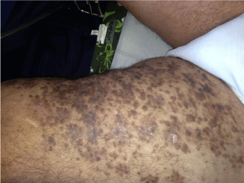
    Figure 2:  Lesion on the left leg, after six course of CHOP-R, and before
starting intralesional electro chemotherapy.
    Figure 3:  Lesion on the left leg, after intralesional electro chemotherapy, got
worse. Patient continued oral etoposide.
Discussion
The association of KS with CLL/SLL has been frequently described, but the observation of the coexistence of both diseases in the same lymph node biopsy, with a mixed histology, is exceedingly rare.
KS is a spindle-cell, low-grade malignant, vascular tumor associated with HHV-8. HHV8, however, is necessary but not sufficient for sarcoma development: other factors, such as immunosuppression, have been shown to play a major role. Four types of KS are known: (1) epidemic or AIDS-related KS, (2) KS in immunocompromised patients, (3) Classic KS (CKS), and (4) endemic or African KS; all four types are more frequently seen in men than in women. HHV-8 has been demonstrated to be the etiological agent of all forms of KS.
CLL and SLL are CD5+ve lymphoproliferative disorders sharing common lymphocyte morphology, immunophenotype, and histological and biological features, the only difference being where the cancer occurs (bloodstream or lymph nodes). SLL is usually associated with lymphoadenopathy, while peripheral or bone marrow lymphocyte number is usually normal. Patients with CLL/SLL do not usually show B-symptoms at diagnosis (fever, night sweats, weight loss, tiredness) [1,2].
There is a general consensus about the increased risk of developing a second cancer in patients with CLL/SLL. Two reviews found that the most frequent second tumors are melanoma and non-melanoma skin cancer, lung and respiratory tract cancer, oral and pharynx, prostate and kidney cancers and lymphoma [3,4].
The mechanisms linking CLL/SLL to KS are unknown. One hypothesis is based on the role of an ineffective T-cell response to a HHV-8 latent infection (imbalanced T-cell subpopulation), as the intrinsic immunosuppression of CLL/SLL can predispose to infection. Another important factor is represented by drug-related immunosuppression, but other risk factors probably exist. However, the possibility that the two diseases derive from different mutations in different progenitor cells cannot be ruled out [5].
CKS is not life threatening, since many therapeutic options are available, but in our patient it was unresponsive to different chemotherapy lines. Prognostic data about KS in CLL/SLL are scanty, but several studies confirm that secondary skin cancers are in general more aggressive [6]. Best therapeutic strategy for such cases is a problem still needing to be addressed: one option could be the contemporary administration of systemic and intralesional chemotherapy, employing options efficacious for both diseases. Timing of administration is another open question, as upfront association of systemic chemotherapy with intralesional; bleomycinbased electrochemotherapy could be considered [7-10].
If the concomitant presence of SLL and KS in the same lymph node biopsy is a negative prognostic factor, is still unknown.
In conclusion, patients presenting with lymphoadenopathy and multiple bluish lesions or small isolated skin or mucous membranes nodules distributed over the lower and upper extremities, the trunk or the neck, besides skin lymphoma should raise the possibility of the coexistence of two malignancies, as in this case, in which sarcoma developed simultaneously to the diagnosis SLL.
Pathogenesis and possible risk factors as well as the therapeutic options still remain open questions.
References
- Oscier D, Fegan C, Hillmen P, Illidge T, Johnson S, Maguire P, et al. Guidelines on the diagnosis and management of chronic lymphocytic leukaemia. Br J Haematol. 2004; 125: 294-317.
- Cottoni F, Masia IM, Cossu S, Montesu MA, Pardini S, Massarelli G. Classical Kaposi's sarcoma and chronic lymphocytic leukaemia in the same skin biopsy. Report of two cases. Br J Dermatol. 1998; 139: 753-754.
- Wiernik PH. Second neoplasms in patients with chronic lymphocytic leukemia. Curr Treat Options Oncol. 2004; 5: 215-223.
- Molica S. Second neoplasms in chronic lymphocytic leukemia: incidence and pathogenesis with emphasis on the role of different therapies. Leuk Lymphoma. 2005; 46: 49-54.
- Hisada M, Biggar RJ, Greene MH, Fraumeni JF Jr, Travis LB. Solid tumors after chronic lymphocytic leukemia. Blood. 2001; 98: 1979-1981.
- Robak E, Robak T. Skin lesions in chronic lymphocytic leukemia. Leuk Lymphoma. 2007; 48: 855-865.
- Di Monta G, Caracò C, Benedetto L, La Padula S, Marone U, Tornesello ML, et al. Electrochemotherapy as "new standard of care" treatment for cutaneous Kaposi's sarcoma. Eur J Surg Oncol. 2014; 40: 61-66.
- Brambilla L, Miedico A, Ferrucci S, Romanelli A, Brambati M, Vinci M, et al. Combination of vinblastine and bleomycin as first line therapy in advanced classic Kaposi's sarcoma. J Eur Acad Dermatol Venereol. 2006; 20: 1090-1094.
- Zidan J, Robenstein W, Abzah A, Taman S. Treatment of Kaposi's sarcoma with vinblastine in patients with disseminated dermal disease. Isr Med Assoc J. 2001; 3: 251-253.
- Nini G, Papa G, Gatti S, Bianchi L, Carrozzo AM, Iraci S, et al. [Kaposi's sarcoma and lymphoproliferative neoplasms]. G Ital Dermatol Venereol. 1988; 123: 425-429.