
Case Report
Ann Hematol Oncol. 2015;2(3): 1028.
Biatrial Double-Hit Lymphoma Presenting with Superior Vena Cava Syndrome
Farè E1, Mussetti A1, Matteucci P1, Cabras AD2, Testi A2, Arendar I3, Morosi C4 and Guidetti A1,5*
1Hematology Department, Fondazione IRCCS Istituto Nazionale dei Tumori, Milano, Italy
2Department of Pathology and Laboratory Medicine, IRCCS Istituto Nazionale dei Tumori, Milano, Italy
3Cardiology Unit, IRCCS Istituto Nazionale dei Tumori, Milano, Italy
4Radiology Division, Fondazione IRCCS Istituto Nazionale dei Tumori, Milano, Italy
5Department of Pathophysiology and Transplantation, University of Milano, Milano, Italy
*Corresponding author: Guidetti A, Hematology Department, Fondazione IRCCS Istituto Nazionale dei Tumori, Via Venezian, 1- 20133– Milano, Italy
Received: December 15, 2014; Accepted: February 17, 2015; Published: February 19,2015
Abstract
Cardiac involvement is very rare in lymphomas and is typically characterized by a poor prognosis, largely due to delay in diagnosis and cardiac complications or venous/arterial thromboembolism.
This unusual presentation and other extranodal localizations are more likely to be found in aggressive lymphomas rather than indolent diseases. In this report we present the case of a patient with heart involvement by a biatrial double mass conditioning superior vena cava syndrome. Histological findings consistent with double hit lymphoma could explain the very aggressive behaviour and the extranodal tropism of this lymphoma. We are treating this patient with an intensified chemotherapy according to EPOCH regimen under carefully clinical and cardiological monitoring.
Lymphoma; Double-Hit; C-Myc; Heart; Atrial Mass
Case Presentation
We report the case of a 67-year-old man affected by a double hit lymphoma presenting with a cardiac bi-atrial involvement at diagnosis.
Patient’s medical history was positive for previous tobacco use and single-vessel coronaric heart disease.
In July 2014 he presented to his cardiologist with a three-month history of jugular compulsion. He performed an electrocardiography examination, an ergometric test and a coronarography without significant findings. In August 2014 he developed shortness of breath and face-neck oedema. Thus, the cardiologist ordered a chest Computed-Tomography (CT) scan that revealed a mediastinal mass in continuity with two hypodensities in the left and right cardiac atria.
The patient underwent a Positron Emission Tomography with Computed Tomography (PET/CT) which showed abnormally increased 18F-FDG uptake in the right hilar mass, involving the mediastinum and the atria (maximum standardized uptake value, SUVmax, 29) and pathological paratracheal and subcarinal lymphadenopathies (SUVmax24) (Figure 1).
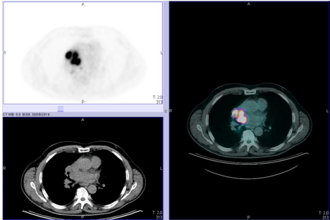
    Figure 1:  PET and PET/CT showing pathological increased uptake of the
intra-atrial masses.
Eventually, Adiagnostic Fine-Needle Aspiration Cytology (FNAC) of the hilar mass under endoscopic ultrasound guidance was performed. Histological findings were consistent with Diffuse Large B-Cell Lymphoma (DLBCL). On immunohistochemistry analysis, the large atypical lymphocytes were positive for CD20, BCL-2, BCL- 6, CD10 stains and Ki-67 was higher than 90%. He subsequently started a steroid therapy. In October 2014 the patient was referred to our center in order to complete staging assessments and to start a cytoreductive treatment.
On admission to our hospital the patient presented with overt signs of superior vena cava syndrome: oedema of face and neck, bilateral jugular distention in standing position and changed voice tone due to vocal chords oedema. No venous collateral circulation was visible on the anterior chest wall.
Vital parameters were normal. Cardiac auscultation revealed no pathological sounds. Both blood counts and standard biochemical tests were normal. Of notice, Lactate Dehydrogenase (LDH) level was normal too (347 UI/L; normal value < 460 UI/L).
Contrast-enhanced total body CT scan confirmed the presence of a polycyclic expansion invading both left atrium (intra-atrial mass of 27 cm) and right atrium (42 mm) where it obstructed the superior cavoatrial junction. Azygos vein ectasia and paravertebral venous collaterals were present. There were no other radiological abnormalities in other anatomical districts (Figure 2).
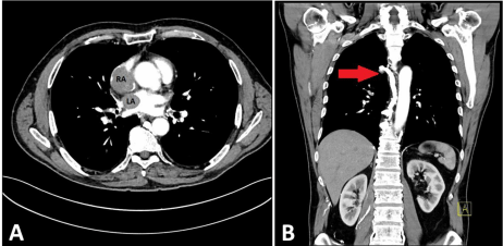
    Figure 2:  Contrast enhanced CT scan. Axial section (A): the lymphoma’s
mass in the Left Atrium (LA) and in the Right Atrium (RA). Coronal section (B):
the dilated and tortuous azygos vein (red arrow) which drain superior vena
cava blood flow into the paravertebral collaterals.
Considering the extremely rare disease localization and in order to better assess cardiac involvement we also performed a transthoracic echocardiography (Figure 3). The examination properly visualized an echogenic solid mass in the left atrium (24 x 15 mm) between the top of the atrium and the interatrial septum, closed to the connection with the superior pulmonary vein. The right atrium was occupied by a polylobed mass (diameter 40 x 31 mm) which obstructed the venous return from superior vena cava. Ventricular ejection fraction, segmentary kinesis and global systolic function were normal
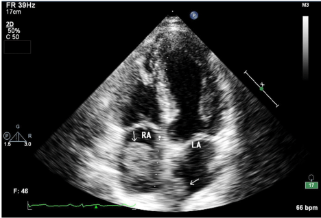
    Figure 3:  Two dimension (2D) echocardiography. It points out the Right and
Left Atria (RA and LA), occupied by the neoplastic nodules (arrow).
Bone marrow biopsy resulted negative for lymphoma infiltration.
The anatomopathological revision confirmed the diagnosis of DLBCL, germinal center like. Revised immunohistochemistry confirmed CD20, Bcl-2 and Bcl-6 positivity and showed over 70% c-Myc expression in the lymphoid cells. Fluorescent in Situ Hybridization (FISH) demonstrated the presence of bcl2 and bcl6 translocation whereas was negative for c-Myc rearrangements (Dual color break-apart probe for c-myc, blc2, bcl6) (Figure 4).
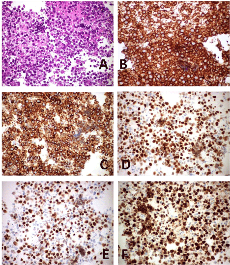
    Figure 4:  Hematoxylin and Eosin staining slide (A). Immunohistochemical
staining demonstrating intense positivity of CD20 (B), BCL2 (C), BCL6 (D),
C-MYC (E) and MIB-1 (F).
Thereby, a diagnosis of double hit DLBCL (by means of immunohistochemical analysis), Ann Arbor stage IVA, revised International Prognostic Index 2, because of age and stage, wasperformed.
In consideration of a very likely risk of cardiac perforation following chemotherapy, we decided to perform a mild debulking cytoreductive therapy with a single dose of doxorubicin 50 mg/sm before proceeding to immunochemotherapy. At starting therapy, we considered the risk of venous/arterial thromboembolism, of cardiac rhythm changes and tumor lysis syndrome and we started adequate therapy with aspirin plus low molecular weight heparin, allopurinol and hydration. The first chemo-treatment has been administered during continuous electrocardiographic monitoring. Chemotherapy was well tolerated without cardiac complications.
Two weeks later, the patient started immunochemotherapy with dose-adjusted EPOCH (Etoposide, Prednisone, Vincristine, Cyclophosphamide, and Doxorubicin) plus Rituximab. He is currently on treatment with initial clinical evidence of response.
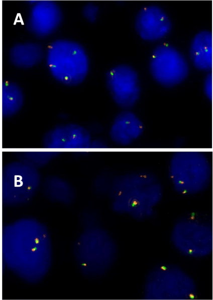
    Figure 5:  Fluorescent In Situ Hybridization (FISH) with dual color probe
proves the presence of bcl2 (A) and bcl6 (B) translocations in lymphoma B
cells.
Discussion
Extranodal involvement in lymphomas has always been associated with a worse prognosis [1]. This feature could be present in almost every lymphoma sub type; however, aggressive diseases have a higher probability to present with such a rare phenotype. In particular, DLBCL, Mantle Cell Lymphoma (MCL) and Peripheral T Cell Lymphoma (PTCL) represent the subgroups with the highest rate of extranodal involvement [2]. The gastrointestinal tract, liver, bone, lungs and central nervous system represent the extra lymphatic regions/organs that are more likely to be sites of extranodal localization of disease [1].
Primary cardiac lymphoma represents only 1,5% of extranodal lymphomas while cardiac localization of systemic lymphoma could be present in up to 15% of patients as derived from post-mortem pathological studies [3]. In this very specific context, the most common cardiac involved sites are represented by right atrium followed by right ventricle, left ventricle, and atrial septum [4]. A biatrial involvement is almost unique and has been reported only by a few case reports in the whole medical literature (Table 1). Cardiac lymphoma localization has a poor prognosis essentially related to cardiovascular complications (pulmonary embolism, cerebrovascular ischemic disease, cardiac failure, cardiac perforation, myocardial infarction) [5].
    Reference
    Year
    Age (years)
    Sex
      (M/F)
    Clinical presentation
    Histology
    Treatment
    Outcome, OS
    Cause of death


    Somers [9]
    1960
    66
    F
    Dyspnea
    NS
    No treatment
    Dead, 1 month
    NS


    Moore [10]
    1992
    80
    M
    Dyspnea, dysphagia
    NS
    No treatment
    Dead, 1 month
    NS


    Chao [11]
    1995
    57
    M
    Dyspnea, atrial    fibrillation
    B cell    lymphoma (NS)
    Chemotherapy    (NS)
    Alive, NS
    /


    Jurkovich [12]
    2000
    75
    M
    Dyspnea, facial    swelling, hemoptysis
    DLBCL
    Chemotherapy (NS)
    Alive, NS
    /


    Mejhert [13]
    2000
    59
    M
    Dyspnea
    NS
    Chemotherapy
    Dead, 10    months
    NS


    Saotome [14]
    2002
    69
    M
    Dyspnea
    NS
    Chemotherapy
    Dead, 14 weeks
    NS


    Zakynthinos [15]
    2004
    48
    M
    Superior cavae    vena syndrome
    DLBCL (bcl2 +    on IHC)
    None
    Dead, 8 days
    Inferior vena    cavae thrombosis, sepsis


    Nascimento [16]
    2007
    40
    F
    Dyspnea,    bradycardia
    Follicular lymphoma
    CHOP
    Alive, 120 months
    /


    Lin [17]
    2010
    42
    M
    Syncopes
    DLBCL
    R-CHOP
    Dead, 2 months
    Sudden cardiac    death


    Cho [5]
    2012
    63
    F
    Dyspnea, facial    swelling
    DLBCL
    R-CHOP
    Alive, NED
    /


    Hajj-Chahine [18]
    2014
    70
    NS
    NS
    NS
    NS
    NS
    NS
Table 1: Cardiac biatrial involvement in lymphoma: literature case reports.
Double-Hit Lymphomas (DHLs) are a subclass of lymphomas belonging to the heterogeneous DLBCL group. Morphologically they consist in large B cell lymphoid cells. Immunophenotypically they are similar to classic DLBCL (usually germinal center type). Clinically, DHLs present with signs of hyper proliferation (B symptoms, compression symptoms) or bulky disease. Moreover they have a Ki-67 proliferation rate higher than 90%. These features make them similar to Burkitt Lymphoma (BL). The most distinguishing and diagnostic feature of DHLs is represented by positivity to c-myc (proliferative stimulus) plus bcl2 rearrangements (antiapoptotic mechanism) by means of FISH. Recently, Green et al. have showed that also an Immunohistochemistry (IHC) positive for C-MYC or BCL2/BCL6 over expression, in the absence of a positive FISH, is a negative prognostic factor [6]. This is probably due to a more common presence of non-classic gene rearrangements that are not detected by standard FISH probes. Also epigenetic phenomena leading to gene over expression could play a role in this setting. Even if the prognosis of these double-hit “expression” lymphomas is not as worse as FISH-positive DHLs, many centers are treating both entities in the same way.
The more we learn to diagnose DHL, the more we find that they are not just in and between DLBCL and BL. They do have their own phenotypical features. In our clinical experience we noticed that DHLs have a more common extranodal presentation compared to DLBCL and BL. Our observation is confirmed by the few series reporting the incidence of extranodal disease in DHLs [7,8]. The incidence of extranodal involvement at diagnosis in this disease group ranges from 30% to 50% that is much higher compared to DLBCL (20%) (1) or BL (3-5%) [2]. This is of great interest when we consider that the presence of c-myc or bcl2/bcl6 rearrangements alone are not sufficient to generate this phenotype. In fact, also follicular lymphoma, usually characterized by bcl2 rearrangement, has a very low rate of soft tissue involvement (5-10%) [2]. Based on these observations, we could hypothesize that the sum of hyperproliferative and antiapoptotic signals conferred by altered C-MYC and BCL2/BCL6 expression are sinergic in making neoplastic cells independent by lymphoid (or bone marrow) microenvironment. This could eventually lead to a tropism to extranodal tissues and to soft tissue proliferation.
Conclusion
In conclusion, our report describes a rare case of biatrial lymphoma with a double mass conditioning superior vena cava syndrome. Besides their rare incidence, lymphoma with cardiac involvement represents a clinical challenge for the physician. In the presence of such a rare localization, an accurate pathological assessment including search for c-myc and bcl2/bcl6 rearrangements or over expression should be performed. Moreover, cardiologic and radiologic assessments represent helpful tools in order to better understand the bounds between the mass, the cardiac tissues and the vascular elements. Due to serious events correlated with the critical localization of the lymphoma mass, treatment of these patients requires carefully management. In this specific setting, a preferable chemotherapy does not exist but some reports describe a more favorable outcome with more aggressive therapies [8]. Even though the risk of cardiovascular complications is high, a DHL diagnosis could lead to an aggressive, and hopefully more effective therapeutic approach.
Acknowledgements
We are grateful to the departments of Radiology and Cardiology for the images presented in this paper.
References
- Takahashi H, Tomita N, Yokoyama M, Tsunoda S, Yano T, Murayama K, et al. Prognostic impact of extranodal involvement in diffuse large B-cell lymphoma in the rituximab era. Cancer. 2012; 118: 4166-4172.
- Krol AD, le Cessie S, Snijder S, Kluin-Nelemans JC, Kluin PM, Noordijk EM. Primary extranodal non-Hodgkin's lymphoma (NHL): the impact of alternative definitions tested in the Comprehensive Cancer Centre West population-based NHL registry. Ann Oncol. 2003; 14: 131-139.
- Mukai K, Shinkai T, Tominaga K, Shimosato Y. The incidence of secondary tumors of the heart and pericardium: a 10-year study. Jpn J Clin Oncol. 1988; 18: 195-201.
- Bussani R, De-Giorgio F, Abbate A, Silvestri F. Cardiac metastases. J Clin Pathol. 2007; 60: 27-34.
- Cho JS, Her SH, Park MW, Kim HD, Baek JY, Youn HJ, et al. A butterfly-shaped primary cardiac lymphoma that showed bi-atrial involvement. Korean Circ J. 2012; 42: 46-49.
- Green TM, Young KH, Visco C, Xu-Monette ZY, Orazi A, Go RS, et al. Immunohistochemical double-hit score is a strong predictor of outcome in patients with diffuse large B-cell lymphoma treated with rituximab plus cyclophosphamide, doxorubicin, vincristine, and prednisone. J. Clin. Oncol. 2012; 30: 3460–3467.
- Petrich AM, Gandhi M, Jovanovic B, Castillo JJ, Rajguru S, Yang DT, et al. Impact of induction regimen and stem cell transplantation on outcomes in double-hit lymphoma: a multicenter retrospective analysis. Blood. 2014; 124: 2354-2361.
- Oki Y, Noorani M, Lin P, Davis RE, Neelapu SS, Ma L, et al. Double hit lymphoma: the MD Anderson Cancer Center clinical experience. Br J Haematol. 2014; 166: 891-901.
- SOMERS K, LOTHE F. Primary lymphosarcoma of the heart: Review of the literature and report of 3 cases. Cancer. 1960; 13: 449-457.
- Moore JA, DeRan BP, Minor R, Arthur J, Fraker TD. Transesophageal echocardiographic evaluation of intracardiac lymphoma. Am Heart J. 1992; 124: 514-516.
- Chao TY, Han SC, Nieh S, Lan GY, Lee SH. Diagnosis of primary cardiac lymphoma. Report of a case with cytologic examination of pericardial fluid and imprints of transvenously biopsied intracardiac tissue. Acta Cytol. 1995; 39: 955–959.
- Jurkovich D, de Marchena E, Bilsker M, Fierro-Renoy C, Temple D, Garcia H. Primary cardiac lymphoma diagnosed by percutaneous intracardiac biopsy with combined fluoroscopic and transesophageal echocardiographic imaging. Catheter Cardiovasc Interv. 2000; 50: 226–233.
- Mejhert M, Muller-Suur R. Primary lymphoma of the heart. Scand Cardiovasc J. 2000; 34: 606-608.
- Saotome M, Yoshitomi Y, Kojima S, Kuramochi M. Primary cardiac lymphoma--a case report. Angiology. 2002; 53: 239-241.
- Zakynthinos E, Tassopoulos G, Haritos C, Kitsanta P, Pyros J, Roussos C, et al. Huge biatrial primary cardiac B-cell lymphoma resulting in bilateral atrioventricular valve obstruction. Leuk Lymphoma. 2004; 45: 2339-2342.
- Nascimento AF, Winters GL, Pinkus GS. Primary cardiac lymphoma: clinical, histologic, immunophenotypic, and genotypic features of 5 cases of a rare disorder. Am J Surg Pathol. 2007; 31: 1344-1350.
- Lin JN. Cardiac lymphoma with first manifestation of recurrent syncope-a case report and literature review. Int Med Case Rep J. 2010; 3: 1-6.
- Hajj-Chahine J, Uzan C, Corbi P. Primary B-cell cardiac lymphoma presenting as a biatrial mass. Asian Cardiovasc Thorac Ann. 2014.