Clinical Image
Ann Hematol Oncol. 2017; 4(4): 1143.
Gelatinous Bone Marrow Transformation in a Patient with Eating Disorder
Rivas-Delgado A¹ and Rozman M²*
¹Department of Hematology, Hospital Clinic, Spain
²Department of Pathology, Hematopathology Unit, Hospital Clinic, Spain
*Corresponding author: Rozman M, Department of Pathology, Hematopathology Unit, Hospital Clínic, Villarroel 170 08036 Barcelona, Spain
Received: February 23, 2017; Accepted: March 08, 2017; Published: March 20, 2017
Clinical Image
A 41-year-old male presented with weakness and weight loss. He denied drug abuse, fever or diarrhoea. The physical examination revealed only cachexia. Blood count showed hemoglobin 116 g/L, leukocytes 1.98x10⁹/L and platelets 230x109/L. Gastrointestinal pathology, infections and autoimmune diseases were ruled out.
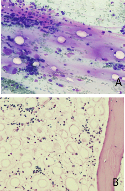
The bone marrow aspirate (Figure 1A) and biopsy (Figure 1B) showed decreased non-dysplastic hematopoiesis without evidence of malignancy, and a deposition of amorphous eosinophilic material consistent with a gelatinous transformation.
After psychiatric evaluation, the patient was diagnosed of an eating disorder not otherwise specified.
Gelatinous bone marrow transformation, with fat cell atrophy and deposition of eosinophilic stromal material (likely hyaluronic acid), leading to impaired haematopoiesis can be observed as a consequence of a generalized illness including anorexia nervosa, neoplasms, malabsorption and infections. The bone marrow study is mandatory for the diagnostic workup of pancytopenia, showing features highly demonstrative of this condition. The pathophysiology of the bone marrow changes is unknown, but the blood and bone marrow alterations resolve after nutritional support.
