Clinical Image
Ann Hematol Oncol. 2017; 4(11): 1180.
Central Nervous System Localisation of Multiple Myeloma
Marjolein W.M. van der Poel¹*, Michel van Gelder¹, Myrurgia A. Abdul-Hamid², Martijn W. Smulders¹ and Aniek de Coninck¹
¹Department of Internal Medicine, Maastricht University Medical Centre, Netherlands
²Department of Pathology, Maastricht University Medical Centre, Netherlands
*Corresponding author: Marjolein W.M. van der Poe, Department of Internal Medicine, Maastricht University Medical Centre, Maastricht, Netherlands
Received: October 17, 2017; Accepted: November 08, 2017; Published: November 23, 2017
Clinical Image
An 80-year-old woman presented to the emergency department with diffuse skeletal pain. She had a history of symptomatic multiple myeloma (MM) for which she had been treated with melphalan, prednisone and bortezomib (MPV). During MPV treatment the monoclonal (M) protein (IgG kappa) had decreased from 25.45 g/L to 9.74 g/L and kappa free light chains had decreased from 1610 mg/L to 571 mg/L. More importantly, the hemoglobin level had increased from 10.5 g/dL to 11.7 g/dL (normal range 11.7-15.6 g/dL) and the creatinine improved from 137 umol/L to 97 umol/L (normal range 50-100 umol/L). After 6 courses of MPV, treatment was discontinued because there was no further decrease in M-protein or free light chains. After discontinuing MPV the M-protein and kappa free light chains steadily increased. Since the MM was asymptomatic at that time there was no indication to start second-line therapy.
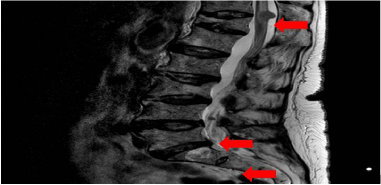
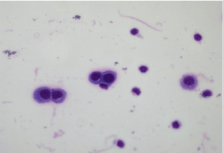
Six months after discontinuing MPV the patient presented to the emergency department with bone pain. Laboratory assessment showed normal hemoglobin and serum calcium levels and a normal kidney function. A PET CT scan was performed, which demonstrated osteolytic bone lesions throughout the skeleton. The M-protein had increased to 18.94 g/L and kappa free light chains to 1790 mg/L, confirming progression of the multiple myeloma. A few days later, the patient complained of weakness in the lower extremities. This was confirmed by neurological examination, as well paresthesias of the right lower extremity. An MRI scan of the vertebral spine showed abnormalities compatible with leptomeningeal metastases (Figure 1). In the differential diagnosis a metastatic carcinoma was considered, however there were no signs of a primary carcinoma on the total body PET-CT scan. A lumbar puncture was performed and cytological analysis of the cerebrospinal fluid (CSF) demonstrated plasma cells that proved to be CD138 positive by immunohistochemistry (Figure 2). Fluorescent activated cells sorting (FACS) of the CSF showed that these plasma cells were all cytoplasmic kappa positive, proving their monoclonality (Figure 3), hence confirming the diagnosis of leptomeningeal metastases from MM. Because of the neurological complications the patient started with radiotherapy. Systemic or intrathecal treatment was not initiated.
Central nervous system (CNS) localisation is a rare but severe complication of MM. It effects fewer than 1% of patients and after diagnosis the median overall survival ranges from 2 to 8 months. The available data on CNS localization in MM are sparse. Treatment options for CNS localization of MM mentioned in the literature are systemic treatment, intrathecal therapy and radiotherapy. There is evidence that some anti-MM agents can cross the blood-brain Figure 1: Leptomeningeal metastases on MRI scan. barrier, such as thalidomide and pomalidomide, but not bortezomib. However, due to a paucity of data treatment is challenging and there is no standard of care.

Figure 1: Leptomeningeal metastases on MRI scan.

Figure 2: Cytology of CSF showing atypical plasmacytoid cells.

    Figure 3:  FACS of CSF proves the presence of monoclonal plasma cells (i.e.,
large (high forward scatter (FSC)) strongly CD38 and uniformly cytoplasmic
kappa light chain positive).
Acknowledgement
Marjolein W.M van der Poel wrote the paper. Michel van Gelder performed the FACS analysis of the CSF, provided the FACS images and revised the paper critically. Myrurgia A. Abdul-Hamid performed the cytological analyses of the CSF and provided the images of the CSF cytology. Martijn W. Smulders and Aniek de Coninck both revised the paper critically. All authors approve of the submitted and final version of the paper.