
Case Report
Austin J Med Oncol. 2015; 2(2): 1019.
Benign Pulmonary Metastasizing Leiomyoma of Uterus Presenting as Malignancy of Unknown Origin, A Diagnostic Dilemma
Latif MF*, Sirelkhatim MS, Osman B and Azam F
North Wales Cancer Treatment centre, Betsi Cadwaladr University Health Board, UK
*Corresponding author: Latif MF, North Wales Cancer Treatment centre, Betsi Cadwaladr University Health Board, Bodelwyddan, LL18 5UJ, UK
Received: December 24, 2014; Accepted: September 01, 2015; Published: September 15, 2015
Abstract
Benign metastasizing leiomyoma (BML) is a rare condition characterized by well circumscribed, multiple nodules of smooth muscle cells predominantly involving lungs and other soft tissue organs. Up to 120 cases have been reported since Steiner first did in 1939. It usually affects women of reproductive age with previous hysterectomy for benign leiomyoma. Time interval between initial surgery and development of BML varies significantly with many occurring years after hysterectomy. Most patients are diagnosed incidentally after chest radiograph or computed tomography (CT) scan for other indications. These patients are usually misdiagnosed as malignancy of unknown origin and a delay in obtaining histological diagnosis can cause significant psychological trauma to the patient. Optimum treatment is controversial but hormone manipulation is generally considered the first approach. We report a case of a 54 year old woman with previous hysterectomy who presented with abdominal pain and vomiting. CT scan reported bowel obstruction and multiple lung nodules.
Keywords: Benign metastasizing leiomyoma; Leiomyoma of uterus; Carcinoma of unknown primary
Case Presentation
A 54 year old postmenopausal woman was admitted with a history of abdominal pain, vomiting and constipation. She had hysterectomy 11 years ago for benign uterine polyps. Other medical problems included anxiety, irritable bowel syndrome, depression, recurrent bilateral breast cysts and benign subcutaneous lipomatosis. She was a non-smoker with no history of alcohol intake and was on regular citalopram for depression. She lived with her husband and 3 children. There was no history of malignancy in the family. Clinical examination revealed tender epigastrium with no guarding or rigidity and bowel sounds were present. Rest of the systemic examination was unremarkable. Abdominal and chest radiographs on admission showed distended bowel loops suggestive of small bowel obstruction and pulmonary nodules, respectively. An urgent CT scan of chest reported multiple pulmonary nodules highly suspicious of metastases. CT scan of abdomen and pelvis showed evidence of small bowel obstruction with multifocal abnormal thickening of small bowel (Image1,2). Due to the absence of chest symptoms, pulmonary function tests were not performed. Other investigations including renal functions tests, liver functions tests, bone profile, ascitic fluid examination from laparotomy and tumour markers including CA125 and CA19.9 were all normal. Absence of fever and normal neutrophil count and C reactive protein levels ruled out the possibility of infection. Auto-immune disease as a cause of pulmonary nodules was excluded by negative rheumatoid factor and auto-antibodies.
The patient was informed about the possibility of incurable metastatic cancer and was referred to the acute oncology team. She underwent laparotomy for bowel obstruction, which revealed distended loops of small bowel with obstruction secondary to a band of adhesion in the right iliac fossa to the site of previous hysterectomy. A 1 cm cyst in the left ovary was also seen. The rest of the abdominal viscerae were unremarkable and no obvious primary or secondary intra-abdominal malignancy was identified. Biopsy of small bowel was not taken due to absence of any suspicious lesion. Acute oncology team arranged a CT guided biopsy of lung lesions. It was not conclusive and non-diagnostic. Her case was discussed in the Lung cancer multidisciplinary team (MDT) meeting and a repeat lung biopsy was recommended. A repeat CT guided lung biopsy was reported as benign with no significant inflammation or granulomata. The patient was informed about the biopsy results and it was reiterated that the possibility of malignancy still existed as there was a high suspicion of malignant metastatic disease radiologically. She and her family were struggling to cope with this uncertain situation and difficulties in obtaining a diagnosis. They were extremely anxious and were not sure about consenting for further diagnostic interventions. After a few lengthy consultations, she agreed for an open lung biopsy which was reported as fascicles of bland, fusiform cells with elongated nuclei with morphological features of smooth muscles. Immunohistochemistry confirmed strong expression of smooth muscle actin. These appearances were consistent with smooth muscle tumours. There were occasional nuclear pleomorphism but no mitosis and no morphological features of malignancy. This histology was compared with the previous hysterectomy specimen. Both of the histological specimens were reviewed and discussed in MDT meeting. They were found to be similar in appearance with smooth muscle tumours and the final histological diagnosis of benign metastasizing leiomyoma was confirmed.
Discussion
BML is a very rare condition, first described in 1939 by Steiner [1], whereby multiple nodules develop in lungs. He reported a 36-year-old female with uterine leiomyomas who presented with cor pulmonale secondary to numerous pulmonary metastases. At autopsy the pulmonary metastases were grossly and histologically benign and were identical to the uterine tumours. He believed that they resulted from pulmonary spread of a histologically benign uterine tumour.
BML can also rarely involve skin, bone, mediastinum, lymph nodes, heart, and retroperitoneum [2]. Majority of patients tend to be asymptomatic, however in a minority symptoms including shortness of breath, cough, and chest pain can be debilitating and potentially fatal [2,3]. There seems to invariably be a history of leiomyoma in the past and the condition is often diagnosed months up to 20 years after hysterectomy, typically affecting women in their late reproductive age [4].
BML is often found incidentally in radiological examinations such as chest radiographs and CT scan. The appearances pose diagnostic difficulties, varying from solitary lesions to multiple lesions mimicking pulmonary metastases from malignant tumours. There can be cavitation of lesions and rarely may be accompanied by pneumothorax [3]. They can be 0.2-8 cm in size, and are usually non-calcified and non-enhancing with intravenous contrast administration [3,4].
Histologically, there are few if any mitotic figures which differentiates it from leiomyosarcomas. Oestrogen and progesterone receptors are present in these lesions [5]. The tumour possesses many morphological, immunohistochemical, and molecular features characteristic of a benign neoplasm despite its potential for metastastic spread. Lesions tend to regress in size after menopause and during pregnancy [6].
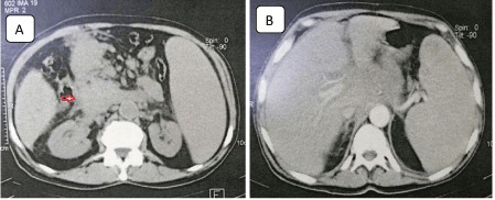
Figure 1: Chest X-ray showing BML lung nodules.
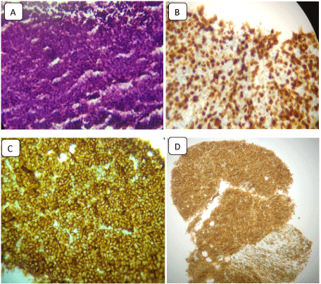
Figure 2: CT scan of chest showing multiple lung nodules.
The nodules are thought to arise from haematogenous metastases of the original leiomyoma at the time of surgical removal [2]. Differential diagnoses include lung secondaries from a malignant tumour, granulomatous conditions such as sarcoidosis, rheumatoid nodules, auto-immune diseases and arterio-venous malformations [3,4]. The patient in this case report had normal inflammatory markers and vasculitic screen, thus excluding infection and vasculitis respectively. CT scan of the chest and abdomen did not show any evidence of a primary tumour or metastatic lesions apart from the suspicious pulmonary nodules. Tumour markers were negative. Laproscopy findings were again normal with normal ascitic fluid analysis. There was no evidence of granulomatous disease from the histology of the lung lesions [7].
Management of BML is individualized as there is no standard treatment owing to its rarity. Treatment options include surgical resection as well as hormonal manipulation. The latter has been successful and includes aromatase inhibitors, progesterone, gonadotropin-releasing hormone analogues and selective oestrogenreceptor modulators [8-10].
These patients are often initially misdiagnosed as malignancy of unknown origin. This creates an immense anxiety and distress in patients and their families as prognosis associated with malignancy of unknown origin is poor.
Conclusion
BML is usually asymptomatic and the lesions are found incidentally on imaging. They pose diagnostic difficulties and the clinician needs to bear in mind the possibility of BML in the differential diagnoses, especially if there was history of hysterectomy. This should be communicated to the patients and families to avoid distress and psychological trauma, as well as unnecessary investigations. Pathologically BML has the same histology of the parent leiomyoma and has a favourable prognosis.
References
- Steiner PE. Metastasizing fibro leiomyoma of the uterus: Report of a case and review of the literature. Am J Pathol. 1939; 15:89–110.7.
- Lim SY, Park JC, Bae JG, Kim JI, Rhee JH. Pulmonary and retroperitoneal benign metastasizing leiomyoma. Clin Exp Reprod Med. 2011; 38:174-177.
- Fasih N1, Prasad Shanbhogue AK, Macdonald DB, Fraser-Hill MA, Papadatos D, Kielar AZ, et al. Leiomyomas beyond the uterus: unusual locations, rare manifestations. RadioGraphics. 2008; 28: 1931-48.
- Caminati A, Cavazza A, Mirenda MR, Harari S. A 69-year-oldfemale with multiple, bilateral pulmonary nodules. Eur Respir Rev.2011; 20: 56-59.
- Jautzke G, Muller-Ruchholtz E, Thalmann U. Immunohistological detection of estrogen and progesterone receptors in multiple and well differentiated leiomyomatous lung tumors in women with uterine leiomyomas. Pathol Res Pract. 1996; 192: 215–223.
- Horstmann JP, Pietra GG, Harman JA, Cole NG, Grinspan S. Spontaneous regression of pulmonary leiomyomas during pregnancy. Cancer. 1977; 39: 314–321.
- Hague WM, Abdulwahid NA, Jacobs HS, Craft I. Use of LHRH analogue to obtain reversible castration in a patient with benign metastasizing leiomyoma. Br J ObstetGynaecol. 1986; 93: 455-60.
- Cohen JD, Robins HI. Response of “benign” metastasizing leiomyoma to progestin withdrawal. Case report. Eur J Gynaecol Oncol. 1993; 14: 44-5.
- Wentling GK, Sevin BU, Geiger XJ, Bridges MD. Benign metastasizing leiomyoma responsive to megestrol: case report and review of the literature. Int J Gynecol Cancer. 2005; 15: 1213-7.
- Taveira-DaSilva AM, Alford CE, Levens ED, Kotz HL, Moss J. Favorable response to antigonadal therapy for a benign metastasizing leiomyoma. Obstet Gynecol. 2012; 119: 438-442.