Special Article - Gynecologic Oncology Case Reports
Austin J Med Oncol.2016; 3(1): 1026.
Immature Teratoma with Extra-Ovarian Deposits of Mature Teratoma–Case Report and Review of the Literature
Haran G¹*, Pomerantz M¹, Ganor-Paz Y¹, Kravtsov V², Kidron D¹, Fishman A¹ and Bruchim I¹
¹Department of Obstetrics & Gynecology, Meir Medical Center, Israel
²Department of Pathology, Meir Medical Center, Israel
*Corresponding author: Haran G, Division of Gynecologic Oncology, Meir Medical Center, Israel
Received: May 02, 2016; Accepted: May 30, 2016; Published: May 31, 2016
Mature teratoma is the most common type of germ cell tumor of the ovary, consisting of 10-20% of ovarian neoplasms. Mature teratoma can occur at any age, but is more common in the child-bearing years. Transformation to a malignant, immature teratoma is reported in 0.17%-2% of cases. Deposits of mature teratoma in the omentum are even rarer, with only 32 cases described in literature. Only 4 cases of co-existing omental and ovarian teratoma have been described. The current case report presents a 42-year-old female who was admitted to the Emergency Department at Meir Medical Center, Kfar Saba, Israel with abdominal pain. Trans-vaginal ultrasound revealed an enlarged, complex, left ovarian mass, 120x105x132 mm with low resistance index. On exploratory laparotomy, a large ovarian mass was identified, with enlarged lymph nodes in the retroperitoneum. Unilateral salpingo-ophorectomy was performed. Pathology was consistent with an immature teratoma in the left ovary, co-existent with implants of mature teratoma in the omentum and Douglas` pouch. We hereby present a unique case of immature teratoma with mature extra-ovarian deposits.
Keywords: Mature teratoma; Immature teratoma; Omental deposits
Mature teratoma is the most common histologic type among the ovarian germ cell tumors. About 10-20% of ovarian neoplasms are of this origin [1]. Mature teratoma can appear at any age, but is more common in the reproductive age group. It can contain all three germ cell layers- endoderm, ectoderm and mesoderm. Only 10% to 17% are bilateral and most cystic tumors are unilateral [1].The incidence of malignant transformation of mature teratomas is 0.17% to 2% of reported cases [1]. Deposits of mature teratoma in the omentum are even rarer, with only 32 cases previously reported in literature and only 4 cases of co-existing omental and ovarian teratoma have been described [2]. The first omental mature teratoma was described by Lebert in 1734 [3], but to the best of our knowledge, no case of ovarian immature teratoma, with distant mature deposits has been reported.
A 42-year-old female presented to the emergency room at Meir Medical Center, Kfar Saba, Israel with a history of abdominal pain for the last month, escalating in the days before admission. Abdominal tenderness was noted on admission and gynecological exam revealed a non-tender mobile mass in the Douglas pouch. Trans-vaginal ultrasound revealed an enlarged, complex, left ovarian mass, 120x105x132 mm with low Resistance Index (RI) of 0.41 and classical sonographic signs of left adnexal torsion. Following a detailed explanation regarding the working diagnosis of ovarian tumor, the patient signed an informed consent for cystectomy/ salpingoophorectomy and possible hysterectomy and staging procedures. Onexploratory laparotomy, a largeovarian mass was identified with enlarged retroperitoneal lymph nodes. The tumor ruptured during mobilization of the left adnexa and unilateral salpingo-ophorectomy was performed. Frozen section was consistent with an ovarian,immature teratoma. Therefore, total abdominal hysterectomy, pelvic and para-aortic lymphnode dissection and infracoloic omentectomy were performed. Alphafetoprotein taken before surgery was elevated to 219.5 ng/ml, while other ovarian tumor markers were normal.
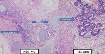
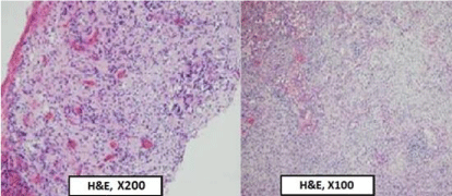
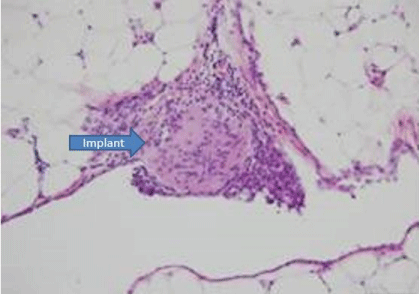
Final histopathologic examination found a large ovarian cyst with a smooth surface consisting of hair and solid components. Microscopically, the findings were consistent with immature cystic teratoma composed of numerous wide areas of embryonic-appearing neuroectodermal elements, grade 2-3 (Figure 1). The left parametrium and Douglas space contained foci of mature glial tissue (Figure 2).The right parametrium had no significant changes. No metastasis was seen in 14 lymph nodes. There was a fibroid uterus and the fallopian tubes had no significant changes. The omentum contained a few mature glial implants (Figure 3). The cytology of the ascitic fluid contained numerous, reactive, mesothelial cells, lymphocytes and macrophages.

    Figure 1:  Left ovary: immature cystic teratoma, areas of mature and immature
embryonic-appearing neuroectodermal elements. H&E, X40, X100.

    Figure 2:  Douglas space: mature glial tissue (implant). H&E, X200 and left
parametrium: mature glial tissue (implant). H&E, X100.

Figure 3: Omentum mature glial tissue (implant). H&E, X100.
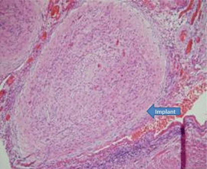
Following a diagnosis of ovarian, high grade, immature teratoma, adjuvant chemotherapy with Bleomycin, Etoposide and Cisplatin (BEP) was administered. During the third chemotherapy cycle, the patient was admitted to the emergency room with right abdominal pain and high fever. CT scan revealed signs of appendicitis with no evidence of other intraperitoneal findings. The patient underwent laparotomy and diffuse spread of small peritoneal implants was seen in the pelvic side wall and on the bowel mesentery. An appendectomy and biopsies of the lesions were performed. Pathology revealed acute fibrinopurulent appendicitis and mature glial implants in the mesentery of the appendix and pelvis (Figure 4). The implants were consistent with mature teratoma.

    Figure 4:  Mesoappendix: mature glial tissue (implant), associated with acute
appendicitis and periappendicitis. H&E, X100.
The patient completed 4 cycles of BEP. Alphafetoprotein at the end of treatment was within the normal range (Table 1 describes the relationship between the aFP levels and image scans and the stage of treatment.) She continues surveillance at the Gynecologic Oncology Clinic every three months.
    Stage    of treatment
    On    diagnosis
    On    first BEP course
    Before    appendectomy


    a    fetoprotein level (ng/ml)
    219.5
    3.2
    1.6


    CT    Scan

    Signs of    appendicitis with no evidence of other intraperitoneal findings


    Pelvic    US
    Left    ovarian mass, 120x105x132 mm with low resistance index (RI) of 0.41 and    classical sonographic signs of left adnexal torsion
Table 1: Levels of a fetoprotein and image scans on the different stages of treatment.
Malignant transformation of mature teratoma is a rare event. Squamous cell carcinomas are the most commonly associated cancer [1]. Rarely, other histologic types arise in a mature cystic teratoma, including adenocarcinoma, basal cell carcinoma, adenosquamous carcinoma, thyroid carcinoma, sebaceous carcinoma, malignant melanoma, sarcoma, carcinoid tumor, and neuroectodermal tumor(0.2-1.4%) [1]. The presenting symptoms of malignant immature teratoma are the same asthose of the mature type and include abdominal pain and distention. Sixteen percent of patients will experience adnexal torsion, 1-2% ruptures and 1%infection. Mature cystic teratomas are usually detected 15–20 years before they undergo secondary, malignant transformation [4]. Cytogenetic abnormalities might precede histological changes and it has been suggested that prolonged exposure to various carcinogens in the pelvic cavity might cause malignant changes to immature teratoma [5]. During early fetal development, germ cells from the yolk sac migrate along the hindgut (route of the mesentery) toward the genital ridge (primitive gonad). These totipotential cells may give rise to a variety of tissues originating from the three primitive embryonic layers. Migration along the hindgut explains how teratomas can develop in multiple locations [6]. Omental teratoma is the most common extra-gonadal teratoma and is reported among women and men. It is typically found in women of reproductive age, but may also appear in young girls and in older women [6]. A review of the literature indicates that only 32 cases of teratoma of the greater omentum have been reported to date and among these, this is the twelfth case that contained teratoma in both the omentum and ovary [2]. To the best of our knowledge, this is the only report of a case where the omental teratoma was mature, while the ovarian teratoma was immature. Table 2 reviews 11 other cases with co-existent teratoma in the ovary and omentum, most of which were located in the left ovary, although no such observation of side preference was reported in the literature [1-3,7-8,10-15]. When treatment was mentioned it was surgical in all cases, and none mentioned treatment with chemotherapy. In all but one case, the ovarian teratoma and the omental deposits were mature type teratoma. The only two exceptions were Smith et al. [7] who described a case of transformation of ovarian teratoma to poorly differentiated mucinous adenocarcinoma and Mandal & Badhe who described malignant transformation of mature teratoma to well- differentiated squamous cell carcinoma [1].
    Study
    Age
    Presentation
    Size    of mass (cm)
    Pathology
    Surgery
    Chemo-    therapy


    Ovary
    Omentum


    Lazarus    and Rosenthal (1931) [3]
    48
    NS
    NS
    NS
    Teratoma    L ovary; R ovary not mentioned
    NS
    NS


    Judd and    Fulcher (1933) [9]
    52
    NS
    NS
    NS
    Teratoma    L ovary; R ovary not mentioned
    NS
    NS


    Printz    et al (1973) [10]
    23
    NS
    NS
    NS
    Teratoma    L ovary; R ovary small
    NS
    NS


    Bell and    Demopoulos (1980) [11]
    53
    NS
    19x14x10
    4.5
    Teratoma    L ovary; R ovary small
    TAH+BSO
    NS


    Kearney    (1983) [12]
    70
    Postmortem    exam
    3
    7x5x3.5
    Teratoma    L ovary; R ovary not identified
    Postmortem
    None


    Compton,    et al. (1985) [13]
    39
    NS
    10x10
    8x7
    Teratoma    L ovary; R ovary small
    L    oophorectomy+ excision of RUQ cystic mass
    NS


    Rails    (1987) [14]
    45
    NS
    8x9
    5x5
    Teratoma    L ovary; R ovary normal
    TAH+BSO
    NS


    Smith et    al. (1990) [7]
    68
    Weight    loss
    15x13
    6x5
    Omental-    mature teratoma, L ovary teratoma poorly differentiated mucinous    adenocarcinoma
    TAH+BSO,    cytoreduction; omentectomy, appendectomy, and common iliac and para-aortic    LND
    None


    Khoo et    al. (2008) [15]
    29
    Abdominal    pain
    NS
    NS
    Mature    teratoma
    Laparoscopic
      ovarian    cystectomy and cytoreduction
    NS


    Mandal    & Badhe (2012) [1]
    56
    Chronic    abdominal distention
    13x10x9
    NS
    Mature    teratoma with deposits of Sq cell carcinoma
    NS
    NS


    Hegde    (2014) [2]
    26
    Abdominal    pain
    3x3
    7x7
    Omental    benign mature cystic teratoma. Ovarian dermoid cyst
    Laparotomy    and ovarian cystectomy, with omental cytoreduction
    None


    Current    case
    42
    Abdominal    pain
    12x10.5x13.2
    NS
    L ovary    immature cystic teratoma omental mature teratoma
    TAH+USO,    omentectomy, LND
    BEP
Table 2: Review of reports of patients with ovarian teratoma with omental lesions.
We present a unique case of immature teratoma with extraovarian mature deposits. It could be argued that this is a case of intragonadal and extra-gonadal mature teratoma that had undergone malignant transformation at the ovarian site. However, this theory does not explain the extensive, extra-ovarian spread and growth of the mature teratoma.
Three main theories have been proposed in the literature to explain the process of extra-ovarian mature teratomas [8]: 1) Originating from displaced germ cells, 2) Developing in a supernumerary ovary of the omentum and 3) Auto-amputation of an ovarian dermoid cyst with secondary implantation into the greater omentum. Unfortunately, there is no evidence to support any of these theories. Of note, the third theory does not explain our case, where the ovarian origin was immature and the deposits were mature. We can only speculate that our case could be explained by the first theory, and that displaced germ cells from the ovary were seeded in the abdomen, resulting in widely spread mature teratoma deposits.
The patient required chemotherapy because immature teratomas are potentially malignant. We used the BEP (bleomycin, cisplatin, etoposide) protocol and the patient completed four cycles. We continue surveillance every three months.
To conclude, we report a unique and interesting case of mature teratoma deposits with an ovarian immature teratoma. Obviously, time will tell if those deposits will become malignant.
- Mandal S, Badhe BA. Malignant transformation in a mature teratoma with metastatic deposits in the omentum: a case report. Case Rep Pathol. 2012; 2012: 568062.
- Hegde P. Extragonadal omental teratoma: a case report. J Obstet Gynaecol Res. 2014; 40: 618-621.
- Lazarus JA, Rosenthal AA. Synchronous dermoid cyst of great omentum and ovary. Ann Surg. 1931; 93: 1269.
- Hackethal A, Brueggmann D, Bohlmann MK, Franke FE, Tinneberg HR, Münstedt K. Squamous-cell carcinoma in mature cystic teratoma of the ovary: systematic review and analysis of published data. Lancet Oncol. 2008; 9: 1173-1180.
- Rim SY, Kim SM, Choi HS. Malignant transformation of ovarian mature cystic teratoma. Int J Gynecol Cancer. 2006; 16: 140-144.
- Ali AA, Sall I, El Kaoui H, Bouchentouf SM, El Hjjouji A, El Fahssi M, et al. Teratoma of the greater omentum. Can J Surg. 2009; 52: E54-55.
- Smith R, Deppe G, Selvaggi S, Lall C. Benign teratoma of the omentum and ovary coexistent with an ovarian neoplasm. Gynecol Oncol. 1990; 39: 204- 207.
- Kubosawa H, Iwasaki H, Kuzuta N, Suzuki H, Iura H. Adenocarcinoma with peritoneal dissemination secondary to multiple mature teratomas of the omentum. Gynecol Oncol. 2006; 101: 534-536.
- Judd. S Fulcher OH. Dermoid cyst of the abdomen. Surg Clin North Am. 1933; 13: 835-842.
- Printz JL, Choate JW, Townes PL, Harper RC. The embryology of supernumerary ovaries. Obstet Gynecol. 1973; 41: 246-252.
- Bell DA, Demopoulos RI. Benign cystic teratoma in the omentum: a mechanism of its development. Diagn Gynecol Obstet. 1980; 2: 205-208.
- Kearney MS. Synchronous benign teratomas of the greater omentum and ovary. Case report. Br J Obstet Gynaecol. 1983; 90: 676-679.
- Compton AA, Tandan A, Fleming WP. Coexistent benign teratomas of the omentum and ovary. A case report. J Reprod Med. 1985; 30: 209-210.
- Rails PW, Hartman B, White W, Radin DR, Halls J. Computed tomography of benign cystic teratoma of the omentum. J Comput Assist Tomogr. 1987; 11: 548-549.
- Khoo CK, Chua SYI, Siow YMA, Chern SBM. Extragonadal dermoid cysts: a review and case report. Singapore J Obstet Gynecol. 2008; 39: 17-20.