
Research Article
Austin J Microbiol. 2015;1(1): 1001.
Evaluation of Suspected Leishmania Samples in University Hospital Laboratory Between 2001 and 2013
Keramettin Yanik1*, Nevzat Unal2, Adil Karadag3,Gumral Alakbarova4, Kemal Bilgin5 and Murat Hokelek6
1Department of Medical Microbiology, Ondokuz Mayis University, Turkey
2Microbiology Laboratory, Samsun Maternity and Child Health Hospital, Turkey
3Health Services Trade High School, Ondokuz Mayis University, Turkey
4Department of Medical Microbiology, Istanbul University Cerrahpasha Medical Faculty, Turkey
*Corresponding author: Keramettin Yanik, Ondokuz Mayis University, Faculty of Medicine, Department of Medical Microbiology, 19 Mayis University, 55139,Samsun, Turkey
Received: October 10, 2014; Accepted: March 10, 2015; Published: March 11, 2015
Abstract
Objective: Leishmaniasis, Leishmania spp., is a zoonotic parasitosis transmitted by Phlebotomus species. It has many vertebrate vectors and is common in Central and South American regions, North Africa, the Middle East, and Central and South Asia. It is also common in the Southeastern Anatolian region of Turkey, including the Eastern Mediterranean. In this study, serum samples of patients with suspected visceral leishmaniasis sent to our laboratory were evaluated retrospectively to determine the prevalence of anti-Leishmania IgG.
Materials and Methods: This study included serum samples taken from patients admitted to various departments between 2001 and 2013 at university hospital. One-hundred eighteen serum samples were included. Leishmania- IgG were determined with an ELISA (NovaTec, Germany) according to the manufacturer’s instructions.
Results: Of the 118 suspected cases, 70 (59%) were male and 48 (41%) were female. Fifty-three (45%) of these patients were younger than 18 years. Most samples were submitted by paediatric departments, followed by internal medicine clinics. Four (80%) of the cases, who tested positive were children and one (20%) was an adult. Microscopic examination of Giemsa-stained bone marrow smears of the Leishmania IgG ELISA-positive patients revealed amastigotes of L. donovani.
Conclusion: Leishmaniasis may present with a variety of clinical symptoms. Hepatosplenomegaly should be considered in Leishmania cases, especially in those aged 18 years and younger who present with fever. The use of the Leishmania-IgG ELISA can be advantageous for rapid diagnosis.
Introduction
Leishmaniasis, belonging to the genus Leishmania, is a parasitic disease. Humans may show various systemic symptoms and syndromes. Leishmaniasis has several different clinical manifestations: ulcerative skin lesions, destructive mucosal inflammation, and disseminated visceral infection (kala azar). Visceral Leishmaniasis (VL), which is caused by Leishmania donovani, L. infantum, and L. chagasi, is frequently seen in children and can lead to death. Phlebotomus species are a vector for the parasite and play a role in transmission to humans. The most common symptoms of patients who present to clinics are fever, loss of appetite, weight loss, and splenomegaly. A physical examination is important to determine the size of the spleen, which is increased in patients with leishmaniasis. Leishmaniasis may be accompanied by lymphadenopathy and, less frequently, hepatomegaly. Leishmaniasis is common in Central and South American regions, North Africa, the Middle East, and Central and South Asia [1]. It is also common in the Southeastern Anatolian region of Turkey, including the Eastern Mediterranean. L. infantum is responsible for VL in all regions of Turkey [2]. This infection can be confused with many diseases, resulting in incorrect diagnosis and treatment of patients [3]. The differential diagnosis in such cases should consider to this diseases. In diagnosing the disease, it is important to be aware of the prevalence of the different species of Leishmania specific to particular regions in the country [4]. The aim of the present study was to evaluate serum samples of patients with suspected VL that were sent to our hospital to determine the prevalence of anti-Leishmania IgG in Samsun (Turkey, Northhern). This is the first study to describe the prevalence of this zoonosis in Samsun (Turkey, Northhern). Regarding the prevalence information about the frequency of this zoonosis putting out the first time to publication.
Materials and Methods
This study included serum samples taken from patients admitted to various departments between 2001 and 2013 at university hospital. One-hundred eighteen serum samples were tested with a Novalis A Leishmania IgG ELISA tests (NovaTec, Germany). After receiving the serum samples in the laboratory, they were tested using these tests, according to the manufacturer’s instructions. Bone marrow samples were obtained from cases that tested positive. Thin smears were prepared and fixed in methanol for 3 min thin smears were prepared and fixed in methanol for 3 min. The preparations were immersed in a Giemsa stain for 30 min, followed by immersion in phosphate buffer and washing. They were then air-dried. Microscopic examinations were performed at 100× objective.
Results
One-hundred eighteen suspected serum samples were examined during the study period. Seventy (59%) were male, and 48 (41%) were female. Fifty-three (45%) of these patients were younger than 18 years. The average age was one and six years. Of the samples, 4.2% were Leishmania-IgG positive. 80% patients who tested positive were male, and %20 were female (Table 1). Figures 1,2,3 shows the results of the microscopic examination of the Giemsa stained smear of the Leishmania IgG ELISA-positive patients, showing L. donovani amastigotes in the bone marrow. The majority of samples were submitted by the Department of Child Health and Disease, followed by internal medicine clinics. Of the patients who tested positive, four (80%) were children, and one (20%) was an adult (Table 2).
![]()
    Age n (%)

    Gender 118

    Positive 5 (4.2)

    Negative 113 (95.8)


    Underage 18

    Male 32(27.2)

    3(9,4)

    29


    53 (45)

    Female 21 (17.8)

    1(4,8)

    20


    18 years and over

    Male 38(32.2)

    0

    38


    65 (55)

    Female 27(22.8)

    1(3,7)

    26


    n: number of    patients, (%): percent
Table 1: Distribution by age and gender suspected patients with Leishmaniasis.
![]()
    Clinics n(%)

    Pediatrics
      53 (45)


    Internal Medicine
      54 (45.7)


    Dermatology4(3.4)
    Infection Diseases
      4(3.4)


    Chest Diseases
      3(2.5)


    Positive: 5(4.2)

    4(3.4)

    1(0.8)

    -
    -

    -


    Negative: 113(95.8)

    49(41.6)

    53(44.9)

    4(3.4)
    4(3.4)

    3(2.5)


    n: number of    patients, (%): percent
Table 2: Distribution of samples according to clinic.
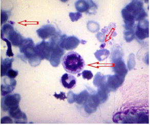
    Figure 1:  Giemsa stained bone marrow smear preparations in L. donovani
amastigotes.
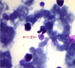
    Figure 2:  Leishmania donovani amastigote form to leukocyte cytoplasm and
outside in Giemsa stained bone marrow smear.
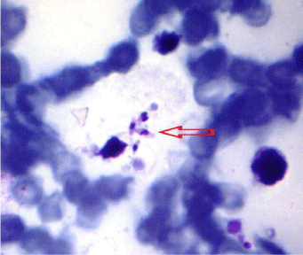
    Figure 3:  Leishmania donovani amastigote form in Giemsa stained bone
marrow smear.
Discussion
VL is a zoonotic disease, which is endemic in more than 80 countries worldwide. Untreated rates up to 100% have been reported, resulting in death [3]. According to one study, Leishmaniasis is associated with about 70,000 deaths per year and about 2.4 million disability-adjusted life years. 90% of visceral leishmaniasis occurs in India, Bangladesh, Nepal, Sudan, and Brazil [4]. Patients in poor countries are most often affected [1]. According to another study, approximately 1.5–2 million children and adults per year develop symptomatic disease, and the incidence of infection is substantial when subclinical infections are included. In Turkey, according to data from the Ministry of Health, 511 cases were reported between 1989 and1996 and 222 between 1997 and 2003 [2,3]. In 2007, Kuk et al. report to an eight-year old female patient who had hepatosplenomegaly was referred to Firat Medical Centre by the Bingol State Hospital. Leishmania amastigotes were emphasized to seen in a smear prepared from bone morrow and stained with Giemsa. Leishmania IgG ELISA and Formol Gel test were positive [3]. They observed Leishmania amastigotes in a Giemsa-stained bone marrow smear.
In study covering 2000–2003, Tanir et al. detected 19 cases of pediatric VL [5]. All the patients presented with fever and weakness. Nine also had pneumonia, and two had a urinary tract infection. Dursun et al. studied 101 children with VL who were admitted to Akdeniz University Hospital during a 20-year period [6]. The median age of the patients was 3 years (range: 5.5 months–13 years). The most common symptoms at presentation were fever, pallor, and abdominal distension. Splenomegaly was found in all the patients, and hepatomegaly was present in 98%. In laboratory tests, anemia (96%), leukopenia (74%), and thrombocytopenia (56%) were the most common findings. Thirty-three (33%) of the patients were pancytopenia on admission. A bone marrow smear was positive for Leishmania in 91% of the patients. Three of the patients died because of secondary infections and hemorrhage, and relapse occurred in two patients. In Spain, from 1 July 2009 to 31 December 2012, 542 cases of leishmaniasis were reported in Madrid to the Epidemiological Surveillance Network, of which 446 (82.3%) met the outbreak case definition: 160(35.9%) cases had VL, and 286(64.1%) cases had cutaneous leishmaniasis. Of the patients, 117(73.1%) were male, and 43(26.9%) were female. The age of the patients with VL ranged from 2 months to 95 years. The mean age was 40 years. Some patients were foreigners. Of these, 44 had visceral forms. Thirty-two patients (20% of all VL cases) were born in sub-Saharan Africa (mostly in Equatorial Guinea and Nigeria). Most cases were laboratory confirmed. L. infantum was identified as the causative agent [7].
Leishmaniasis is endemic in the south of France where the causative agent is L. infantum. The average number of autochthonous cases reported per year is 22.6. Most cases (84.5%) are VL. Of 317 autochthonous human leishmaniasis cases, 268 (84.5%) were VL, 39 (12.3%) were cutaneous leishmaniasis, and 10 (3.1%) were mucocutaneous leishmaniasis. The ratio of males to females was 1:8, and the disease affected mostly adults (222 cases; 70%). Among those, 50 (19.3%) patients were more than 60 years, and 73 (23%) were less than five years. The mean age of the patients was 35.5 years, and the median age was 39 years (range from one to 89 years) [8]. In the present study, the analysis of the data from 2007 to 2012 showed that 62% (90/145) of the VL cases were male and that the disease was more common among those aged 20-60 years. In 19.3% (28/145) of cases, the patient was older than 60 years (8). In our study, five (4.2%) of the 118 patients with suspected leishmaniasis who underwent Leishmania serum IgG ELISA screening were positive for VL. One positive case was an adult (>18 years), and four were pediatric patients. As noted in this study and in many other studies, more children than adults tested positive. In our study, most of the samples came from the department of pediatrics. The second most common source was internal medicine clinics.
When we compared the results from studies in Europe with ours, there are similarities and differences. We detected leishmaniasis in pediatric patients, as well as in Spain patients was male (73.1%) with age 40 years [7]. In the incidence of the disease in Spain compared to Turkey, the rate of VL in Spain was 35.9% [7], whereas it was only 4.2% in our study. A comparison should be drawn between the findings in France and the findings in Turkey.The highest rate of VL was reported in France (84.5%) [8]. The causative pathogen of leishmaniasis in our study was L. donovani, whereas it was frequently L. infantum in Europe [7].
In conclusion, leishmaniasis is endemic in many countries. VL is common in rural areas of Turkey. However, according to the results of the present study, it is not common Samsun (Turkey, Northhern). The possibility of hepatosplenomegaly should be considered in patients with leishmaniasis, especially in those younger than 18 years. Routine bone marrow aspiration and lymph node analysis can be used to diagnose VL. A positive diagnosis is based on the presence of amastigotes and promastigotes in direct microscopic examinations Novy-MacNeal-Nicolle (NNN) medium. The use of serological tests, such as Leishmania-IgG ELISA, can aid rapid diagnosis of the disease.
References
- Desjeux P. Leishmaniasis: current situation and new perspectives. Comp Immunol Microbiol Infect Dis. 2004; 27: 305-318.
- Ozensoy S, Ozbel Y, Turgay N, Alkan MZ, Gul K, Gilman-Sachs A, et al. Serodiagnosis and epidemiology of visceral leishmaniasis in Turkey. Am J Trop Med Hyg. 1998; 59: 363-369.
- Kuk S, Poyrazoglu G, Arslan FN, Erensoy A, Akarsu SA. Pediatric case of visceral leishmanisis. Firat Medical Journal. 2007; 12: 237-238.
- Murray HW, Berman JD, Davies CR, Saravia NG. Advances in leishmaniasis. Lancet. 2005; 366: 1561-1577.
- Tanir G, Taylan Ozkan A, Daglar E . Pediatric visceral Leishmaniasis in Turkey. Pediatr Int. 2006; 48: 66-69.
- Dursun O, Erisir S, Yesilipek A. Visceral childhood leishmaniasis in southern Turkey: experience of twenty years. Turk J Pediatr. 2009; 51: 1-5.
- Arce A, Estirado A, Ordobas M, Sevilla S, García N, Moratilla L, et al. Re-emergence of leishmaniasis in Spain: community outbreak in Madrid, Spain, 2009 to 2012. Euro Surveill. 2013; 18: 20546.
- Lachaud L, Dedet JP, Marty P, Faraut F, Buffet P, Gangneux JP, et al. Surveillance of leishmaniases in France, 1999 to 2012. Euro Surveill. 2013; 18: 20534.