Clinical Image
J Mol Biol & Mol Imaging. 2014;1(1): 2.
18F-FDGPET/CT in A Case of Blastic Plasmacytoid Dendritic Cell Neoplasm with Initial Dissemination
Young Seok Cho1, Seok Jin Kim2, Young-Hyeh Ko3, Byung-Tae Kim1 and Kyung-Han Lee1*
1Department of Nuclear Medicine, Sungkyunkwan University School of Medicine, Korea
2Department of Medicine, Sungkyunkwan University School of Medicine, Korea
3Department of Pathology, Sungkyunkwan University School of Medicine, Korea
*Corresponding author: Kyung-Han Lee, Department of Nuclear Medicine, Samsung Medical Center, Sungkyunkwan University School of Medicine, 50, Irwon-dong, Gangnam-gu, Seoul 135-710, Korea
Received: July 22, 2014; Accepted: July 23, 2014; Published: July 24, 2014
Abstract
An 18-year-old male presented with hyperemic skin nodules on the face, arm, trunk and thigh. 18F-FDG PET/CT showed increased uptake in the cutaneousnodules, but images also revealed hyper metabolic lesions in the nasopharynx, nasal cavity and lymph nodes, and diffusely increased bone marrow activity. Biopsies from the skin lesion, nasal cavity, and bone marrow revealed blastic plasmacytoid dendritic cell neoplasm. This case highlights the utility of FDG PET/CT in identifying metastatic lesions in a case initially presenting with a disseminated phase of this exceedingly rare hematopoietic malignancy.
Keywords: Blastic plasmacytoid dendritic cell neoplasm; 18F-FDG; PET; CT
Acknowledgement
This work was supported by the Samsung grant (#GL1-B2-191-1).
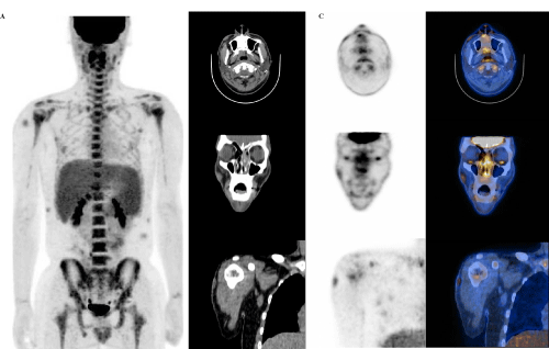
Figure 1: An 18-year-old male presented with a several-month history of skin lesions that began as facial brown colored macules and progressed to epidermal cystic masses of trunk. The patient also complained of nasal obstruction and rhino rhea. Multiple hyperemic nodules were visible and palpable on the face, arm, trunk and thigh. Laboratory data disclosed mild norm chromic normocytic anemia (hemoglobin, 4.4 g/dl) with thrombocytopenia (39 x 103/μl).18F-FDG PET/CT was performed to provide further information. Three-dimensional maximum intensity projection PET images displayed multiple hypermetabolic skin lesions and diffusely increased activities in the bone marrow and spleen (A). On transaxial and coronal images (B, CT; C, PET; D, fusion) the nodular skin lesions in the face and arm had maximal standardized uptake values (mSUVs) of 2.6 and 2.4, respectively. Also noted were hypermetabolic lesions in the nasopharynx (mSUV, 8.6) and nasal cavity (mSUV, 4.3).
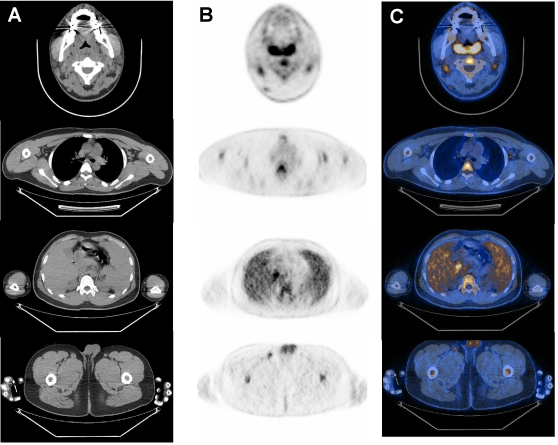
Figure 2: Multiple lymph nodes were shown to have elevated FDG activity including those in cervical (top row), auxiliary (second row), abdominal (third row) and inguinal regions (bottom row). A-CT; B-PET; C-fusion images.
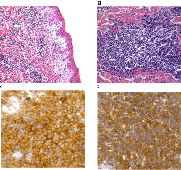
Figure 3: He staining of tissue biopsied from the skin lesion of the arm showed dermal lymphocytic infiltrates between collagen bundles extending into the subcutis (A, x100). Medium-sized infiltrating cells with fine dispersed chromatin are seen on greater magnification (B, x400). These cells stained positive for CD4 (C, x 400), CD123 (D, x 400), and CD56 (not shown). Biopsies of the bone marrow and nasal cavity lesion, as well as the skin lesion, led to the diagnosis of blastic plasmacytoid dendritic cell neoplasm. This is an exceeding rare, highly aggressive hematopoietic malignancy categorized under acute myeloid leukemia by the World Health Organization [1-4]. The malignancy is characterized by an indolent onset manifested as asymptomatic cutaneous lesion. Tumor dissemination usually occurs later or at recurrence and includesinvolvement of the bone marrow and lymphatic system [5-8].
References
- Petrella T, Bagot M, Willemze R, Beylot-Barry M, Vergier B, Delaunay M, et al. Blastic NK-cell lymphomas (agranular CD4+CD56+ hematodermic neoplasms): a review. Am J Clin Pathol. 2005; 123: 662-675.
- Cota C, Vale E, Viana I, Requena L, Ferrara G, Anemona L, et al. Cutaneous manifestations of blastic plasmacytoid dendritic cell neoplasm-morphologic and phenotypic variability in a series of 33 patients. Am J Surg Pathol. 2010; 34: 75-87.
- Kharfan-Dabaja MA, Lazarus HM, Nishihori T, Mahfouz RA, Hamadani M. Diagnostic and therapeutic advances in blastic plasmacytoid dendritic cell neoplasm: a focus on hematopoietic cell transplantation. Biol Blood Marrow Transplant. 2013; 19: 1006-1012.
- Tsunoda K, Satoh T, Akasaka K, Ishikawa Y, Ishida Y, Masuda T, et al. Blastic plasmacytoid dendritic cell neoplasm : report of two cases. J Clin Exp Hematop. 2012; 52: 23-29.
- Lim D, Goodman H, Rademaker M, Lamont D, Yung A. Blastic plasmacytoid dendritic cell neoplasm. Australas J Dermatol. 2013; 54: e43-45.
- Angelot-Delettre F, Garnache-Ottou F. Blastic plasmacytoid dendritic cell neoplasm. Blood. 2012; 120: 2784.
- Chen J, Zhou J, Qin D, Xu S, Yan X. Blastic plasmacytoid dendritic cell neoplasm. J Clin Oncol. 2011; 29: e27-29.
- Munoz J, Rana J, Inamdar K, Nathanson D, Janakiraman N. Blastic plasmacytoid dendritic cell neoplasm. Am J Hematol. 2012; 87: 710.


