Clinical Images
Austin J Musculoskelet Disord. 2015;2(1): 1016.
Microgeodic Disease that Can Mimic Rheumatoid Arthritis
Syuichi Koarada1*, Mitsunori Komine2 and Yoshifumi Tada1
1Department of Rheumatology, Saga University, Japan
2Tsuruta Rehabilitation Clinic, Japan
*Corresponding author: Koarada S, Department of Rheumatology, Faculty of Medicine, Saga University, 5-1-1 Nabeshima, Saga 849-8501, Japan
Received: December 15, 2014; Accepted: March 17, 2015; Published: March 19, 2015
Clinical Images
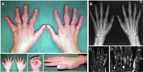
The patient, a 17-year-old man, presented with a year history of swelling (Figure 1A) and mild pain of fingers. The symptoms have not been worse for a year. Bilateral involvement of Proximal Interphalangeal (PIP) joints may be mistaken for the inflammation due to rheumatoid arthritis. However, range of motion in the joints of fingers was not restricted. Although Posterior Anterior (PA) view of the hand revealed swelling of PIP joints, no joint space narrowing and erosions in the digits were found (Figure 1B). Laboratory analyses showed normal. His erythrocyte sedimentation rate was 6 mm/ hour and C-reactive protein level was 0.02 mg/dl. Serum Anti–cyclic Citrullinated peptide Antibodies (anti-CCP Abs) and Rheumatoid Factor (RF) were negative. Soft tissue swelling without synovitis of fingers was shown on gadolinium-enhanced T1-weighted Magnetic Resonance Imaging (MRI) (Figure 1C). Multifocal lesions of bone marrow edema affecting most of the phalanges were revealed on T2-weighted Short-Tau Inversion Recovery (STIR) sequences in MRI (Figure 1D). However, there were no erosions and synovitis. Ultrasonography (US) confirmed the absence of synovitis. These features suggested the diagnosis of microgeodic disease. Microgeodic disease is a rare disease of the bone and soft tissues. The pathogenesis of microgeodic disease appears to be a transient disturbance of the peripheral circulation caused by cold exposure. In most cases, the symptoms and the radiographic changes return to normal within several months without any treatment. He was also free of symptoms after several months.

    Figure 1:  (A) Photographs of the hands. (B) Plain radiograph; Posterior
Anterior (PA) view of the hands. (C) Gadolinium-enhanced T1-weighted
Magnetic Resonance Imaging (MRI) (white arrows). (D) T2-weighted Short-
Tau Inversion Recovery (STIR) sequences in MRI (open arrows).