
Special Article - Nephrology & Dialysis
Austin J Nephrol Hypertens. 2014;1(4): 1016.
An Unusual Cause of Renal Amyloidosis Secondary to Teratoma
Nadir Zemraoui1*, Omar Maoujoud2
1Department of Nephrology & Dialysis, Ibn Sina Military Hospital, Marrakech – Morocco
2Department of Nephrology & Dialysis, 1st Medical & surgical Military Hospital, Agadir – Morocco
*Corresponding author: Nadir Zemraoui, Department of Nephrology & Dialysis, Ibn Sina Military Hospital, Marrakech – Morocco
Received: September 24, 2014; Accepted: September 26, 2014; Published: September 30, 2014
Introduction
Nephrotic syndrome can be primary, being a disease specific to the kidneys, or it can be secondary, being a renal manifestation of a systemic general illness, we report rare cause of nephrotic syndrome, related to fortuitous discovery of an unusual image on x-ray.
Case Report
A 28-year old female presented to the hospital with a six-weeks history of slowly progressive bilateral lower extremity edema and weight gain with abdominal pain, nausea, vomiting,. His past medical history was only significant for hypertension. On physical examination the patient had a blood pressure of 170/100 mmHg, a 4+ bilateral lower extremity edema up to the thighs, the rest of the exam was otherwise unremarkable. Laboratory evaluation revealed a nephritic syndrome: albumin 16 g/L, total protein 40 g/L, proteinuria 14g/24 h. Urea nitrogen 5.6 mmol/L, creatinine 80 μmol/L, On ultrasound the right kidney measured 12.5 cm, the left kidney 12.6 cm. A renal biopsy was performed and histological examination showed homogenous massive amyloid deposits in glomeruli and vessel walls. This material produced apple-green birefringence under polarized light, by Congo red staining. The amyloid stained strongly with anti- AA but not with anti-κ or anti-λ antiserum. Plain X-ray abdomen showed a large 5cm mass containing calcification and compressing the intestinal gas to the left side (Figure 1). Abdominal computed tomography with intravenous contrast revealed an intraperitoneal mass with heterogeneous enhancement with fluid, soft tissue mixed with gross calcifications component and displacing aside the bowel (Figure 2). Surgical exploration revealed encapsulated mass with solid component surrounded by skin and composed of adipose tissue, skeletal muscle and cartilage undergoing endochondral ossification. Evolution has been marked by a gradual regression of nephrotic syndrome after surgery. At 12-month follow-up, the patient presented in good general condition free of proteinuria.
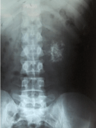
Figure 1: Plain X-ray abdomen showed a large 5cm mass containing calcification and compressing the intestinal gas to the left side.
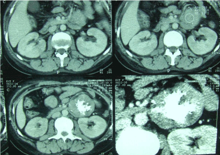
Figure 2: Abdominal computed tomography with intravenous contrast revealed an intraperitoneal mass with heterogeneous enhancement with fluid, soft tissue mixed with gross calcifications component and displacing aside the bowel.
Conclusion
In the presented case amyloidosis affected kidneys and manifested as nephrotic syndrome, Research of chronic inflammatory or disease was negative, the regression of the nephrotic syndrome after removal of the teratoma, suggested a link between the two diseases.