
1Tissue Typing Laboratory, University Hospital Halle/Saale, Germany
2Department of Transfusion Medicine, University Hospital Göttingen, Germany
*Corresponding author: Schlaf G, Tissue Typing Laboratory, University Hospital, Halle/Saale, Magdeburger Strasse 16, D-06112 Halle (Saale), Germany
Received: October 28, 2014; Accepted: November 26, 2014; Published: November 30, 2014
Citation: Schlaf G, Pollok-Kopp B and Altermann WW. Solid Phase-Based Cross-Matching in Order to Avoid Kidney Allografting against Donor-Specific Anti-HlA Antibodies: Long Term Experience with a Procedure Allowing Highly Reliable Diagnoses. Austin J Nephrol Hypertens. 2014;1(6): 1028.
Citation: Bucci J and Hansen KE. Should we treat Secondary Hyperparathyroidism in Patients with Pre-Dialysis Chronic Kidney Disease?. Austin J Nephrol Hypertens. 2015; 2(4): 1046. ISSN : 2381-8964
Transplant recipients with sensitizing events such as previous transplants, blood transfusions or pregnancies often develop antibodies which are directed against Human Leukocyte Antigen (HLA)-molecules of the donor tissue. These preformed Donor-Specific Anti-HLA Antibodies (DSA) represent a high risk of allograft failure due to antibody-mediated hyper-acute or acute rejection. In order to select recipients without these detrimental DSA, the Complement- Dependent Cytotoxicity Assay (CDC) was developed more than forty years ago and established as standard crossmatch technique. However, as a functional i.e. vitality assay it detects only those antibodies which exert their detrimental function through their complement-activating features and fails to identify DSA which lack complement-fixing activity although these may as well be detrimental for the allograft survival. Furthermore, the outcome strongly depends on the availability of isolated vital lymphocytes sometimes not available of a given donor. In addition, pharmacological treatment as well as underlying diseases may lead to artificially positive results of the CDC demonstrating this assay’s general insufficiency under certain circumstances. As a consequence alternative solid phase-based crossmatch assays which function independently of the cell quality had been generated in order to detect DSA and were implemented in our laboratory about nine years ago. We here review the results of these assays and the conclusions to be drawn for special groups of patients on the kidney waiting list of our tissue typing laboratory. The data clearly demonstrate the superiority of the alternative Enzyme-Linked Immunosorbent Assay (ELISA)- based techniques when compared with the CDC-crossmatch not leading to valid results under various circumstances.
Keywords: Allograft; Crossmatch; Donor-specific antibodies; Human leukocyte antigen; Kidney; Rejection
CDC: Complement-Dependent Cytotoxicity Assay; CM: Crossmatch; DTE: Dithioerythritol; DTT: Dithiothreitol; DSA: Donor-Specific Antibodies; ELISA: Enzyme-Linked Immunosorbent Assay; FACS: Fluorescence-Activated Cell Sorter; HLA: Human Leukocyte Antigens; MHC: Major Histocompatibility Complex; moAb: Monoclonal Antibody; PBL: Peripheral Blood Lymphocytes; PRA: Panel Reactive Antibodies; SLE: Systemic Lupus Erythematosus
More than forty years ago Patel and Terasaki described for the first time, that antibodies, which are directed against antigens of donor tissues are clearly associated with hyperacute rejections in recipients of renal allografts [1]. Subsequent studies provided evidence that antibodies which are directed against highly polymorphic human Major Histocompatibility Complex (MHC) antigens, the so-called Human Leukocyte Antigens (HLA) represent the dominating reason for hyperacute and acute rejections of allografts [2,3]. These DSA are thus regarded as a contra-indication for grafting according to the guidelines of most countries and supranational societies (e.g. Eurotransplant) which supervise the allocation of organs. In order to avoid grafting against these highly harmful DSA the so-called crossmatch procedure was developed in the late sixties of the last century which appeared to be an effective predictor of short-term survival of renal allografts. As standard technique for the detection of DSA the Complement-Dependent Lymphocytotoxicity Crossmatch (CDC-CM) was established and hitherto represents the standard technique of nearly all tissue typing laboratories. As a functional assay it is exclusively based on the detection of alloantibodies which exert their detrimental function through their complement-fixing and activating features via the classical pathway of complement activation finally leading to the lyses of donors’ allografted cells and tissues. The CDC-CM, however, fails to detect those DSA which lack complement-activating features although these may as well be harmful for the allograft. Furthermore, this assay is characterized by its low sensitivity which led to the introduction of its modification by additionally using secondary anti-human immunoglobulin antibodies (AHG-enhanced CDC-CM), thus leading to an increase in the activation of complement [4,5]. Alternatively, Flow Cytometry- Based Cross-Matching (FACS-CM) was implemented, the results of which have to be carefully interpreted due to another methodical drawback [6,7]. All over the years the outcome has artificially been influenced by the “irrelevant” binding of the recipient’s antibodies to Fc-receptors highly expressed on B-cells, thus leading to many false positive results especially of B-cell cross-matching [8,9]. This severe artefact of “unspecific” binding of antibodies through their Fc-parts may first be reliably overcome by the procedure recently proposed by using heat-denatured rabbit serum to block Fcγ receptors thus highly reducing the background caused by non-specific IgG-binding [10]. Anyway a striking drawback which holds true for CDC-based as well as FACS-based cross-matching is that both assays do not lead to valid results if only cells of poor quality (vitality) are available. Consequently novel CM-methods which are characterized by complete independence of the cell quality were generated in the past. Two of these assays used in our laboratory had been developed using the design of enzyme-linked immunosorbent assays (ELISA) which were first the Antibody Monitoring System (AMS) HLA class I/II ELISA (GTI Diagnostics, Waukesha, USA) and afterwards the AbCross HLA class I/II ELISA (BioRad/Biotest, Dreieich, Germany). Instead of vital cells both assays use detergent-extracted HLA molecules derived from a given donor’s cells or tissues which are immobilized to pre-coated monoclonal capture antibodies. These are directed against monomorphic epitopes of HLA class I or II molecules, respectively. Due to the first commercial availability of the AMS-ELISA already in the year 2005 as precursor design of the afterwards diagnostically implemented micro variant (from 2006), this assay was first established in our tissue typing laboratory [8]. We used this assay for various diagnostic approaches lacking adequate cellular donor material, thus not resulting in valid outcomes of the CDC-CM [9,11,12]. Furthermore, we modified this ELISA-based procedure to be usable for acellular donor tissue leading to the first crossmatch procedure exclusively using the outer corneal rims as the only material usually available as retain samples from given donors [13]. However, after the discontinuation of the AMS-ELISA by the manufacturer in the year 2013 solely for commercial reasons, we had to replace it by the alternative AbCross crossmatch ELISA after strongly modifying the original workflow given in the manufacturer’s instructions. We here review methodical features, drawbacks and diverging diagnostic outcomes of the conventional CDC-CM as compared with ELISA-based crossmatch results of special groups of patients on our kidney waiting list observed by us for the last nine years. Furthermore, we critically discuss the unfavourable situation for a considerable number these patients who are deprived of a kidney allograft due to CDC-based cross-matching dictated as the current mandatory procedure but highly susceptible to various disruptive factors and artefacts.
All of the patients under investigation in these comparative analyses were examined between February 2006 and June 2014 in the Tissue Typing Laboratory of the University Hospital Halle (GHATT) for the detection and specification of anti-HLA antibodies in general (so-called antibody screening/monitoring). According to the guidelines of the European Federation for Immunogenetics (EFI) all patients of the kidney waiting list have to be screened quarterly to avoid donations of HLA phenotypes against which antibodies are identifiable in the prospective recipient. The identification of a given recipient’s antibody specificity leads to the exclusion of this corresponding donor’s HLA-phenotype. This approach named virtual cross-matching results in the a priori exclusion of “forbidden HLA-antigens” but has, for reasons discussed below, not the capacity to replace the practical de facto crossmatch by directly investigating the recipient’s serum for antibodies against donor’s cell-derived material in order to reliably exclude DSA.
At least quarterly the recipient’s sera were generally screened for anti-HLA class I antibodies using the QuikScreen ELISA (BioRad) and for anti-HLA class II antibodies using the B-Screen ELISA (BioRad). Serum samples positive in this first screening procedure were consecutively investigated for antibody identification/ specification using the DynaChip HLA antibody analysis technique (Invitrogen/Dynal, Bromborough, UK) until 2011. This miniaturized chip-based technique was the only completely automated system available for the detection and specification of anti-HLA antibodies. In its second generation design 106 positions of each microchip were covered with HLA class I and 48 positions with HLA class II molecules of different single donors, respectively. Thus, apart from a number of single HLA class II DQ-antigens immobilized on some positions this assay provided a resolution at the single donor/single ID level but not at the single antigen level. In spite of this reduced level of resolution the combination of the single donors’ HLA class I or II antigens, respectively, led to the reliable identification of the patient’s antibody specificities in most cases (about 80%) especially if the immunization level/PRA level (see below) was not too high. After the discontinuation of this system by the manufacturer in 2011 for commercial reasons, however, the Luminex technique was implemented in our laboratory. Due to lacking alternative methodical procedures this technique currently represents the dominating tool for anti-HLA antibody specification. Technical aspects, features and drawbacks of this technique have in detail been reported and discussed elsewhere [12,14,15]. In order to perform virtual cross-matching (see above) first the DynaChip and afterwards the Luminex-based techniques were used. The general degree of anti-HLA sensitization which is termed “panel reactive antibodies” (% PRA) was defined using both techniques at the adequate single donor (single ID) level of resolution. This value has originally been defined by CDC-based cell tray analyses (see below) defined as CDC-based reactivity against either a cell panel of Peripheral Blood Lymphocytes (PBL) or a cell panel derived from Chronic Lymphatic Leukemia (CLL) patients. To correctly define the % PRA any panel of HLA phenotypes has to comprise all of them occurring in a given recipient’s population and, furthermore, has to represent those phenotypes’ frequencies. The statistical PRA-value which is quarterly determined for all patients on the kidney waiting list indicates the likelihood of a positive de facto crossmatch. It cannot be equated with DSA. However, a high PRA-percentage easily offers the identification of those patients who have to be monitored and crossmatched very carefully due to a high HLA-specific pre-immunization status.
Prior to grafting all patients are investigated for DSA by the standard CDC-CM procedure elsewhere described in a detailed manner [8,9,12]. In spite of its drawbacks which will be the major point of discussion this procedure has been accepted for years and due to its suitability for the routine task of any laboratory represents the standard procedure to circumvent grafting in the presence of highly deleterious DSA. In accordance with the EFI-guidelines this test is performed in our laboratory not only using the whole fraction of PBL but also using separated T-cells and B-cells. The cells are fractionated through the use of tetrameric antibody technique crosslinking unwanted cells to red blood cells and eliminating them via density gradient centrifugation (System RosetteSep, Stemcell Technologies via CellSystem Biotechnology GmbH, St. Katharinen, Germany). Cells which had been recognized by DSA and have consequently been attacked by the complement-derived membrane attack complex are stained red through the inflow of the DNA-intercalating dye ethidium bromide whereas vital cells which have not been recognized are stained green by the uptake of acridine orange. The intensity of the complement reaction is categorized through the percentaged indication of dead (red coloured) cells using a score system of the National Institute of Health (Washington, USA) as shown: score 1: <10% (negative), score 2: 10-20% (doubtfully positive), score 4: 20- 50% (weakly positive), score 6: 50-80% (positive), and score 8: 80- 100% (strongly positive). To get reliable results also of faint antibody-mediated reactions the background caused by dead cells should not exceed 10%. It is noteworthy that antibodies detectable in this vitality assay must belong to the so-called cytotoxic i.e. complement-activating isotypes IgM, IgG1 and IgG3. Other isotypes are not detectable through the use of the CDC-CM.
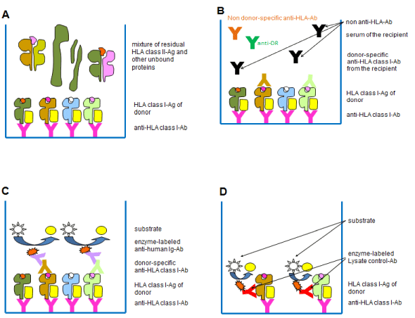
As procedures of ELISA-based cross-matching, first the Antibody Monitoring System (AMS) class I/II ELISA (GTI, Waukesha, USA) was implemented in our laboratory until its discontinuation in 2013 when it had to be replaced by the alternative AbCross HLA class I/ II ELISA (Biotest/BioRad, Dreieich, Germany). In order to lead to faster results and to improve the general applicability for routine task the very laborious workflow of the AbCross-ELISA was completely modified to lead to a workflow which was very similar to that of the former AMS-ELISA. The principle of work is demonstrated in the flow scheme (Figure 1). Detergent lysate of a given donor’s leukocytes/tissue including the HLA class I and class II molecules as target molecules has to be filled into the wells of ELISA strips (GTI) or Terasaki plates (BioRad) where pre-coated monoclonal capture antibodies which are directed against monomorphic epitopes immobilize the extracted HLA class I or class II molecules, respectively (Figure 1A). After this first incubation step the recipients’ sera are pipetted onto the immobilized donors’ HLA molecules. Possible Donor-Specific Anti-HLA Antibodies (DSA) serve as detection antibodies in this sandwich assay (Figure 1B). In the third incubation step the samples are incubated with enzyme-conjugated secondary anti-human IgG (alternatively anti-human IgG/M/A) antibodies which induce the final substrate reaction (Figure 1C). The so-called lysate controls (positive controls) are of high relevance since they provide evidence that a sufficient amount of the donors’ HLA molecules has been immobilized to reach a significant signal (Figure 1D). To be classified as positive the value of a given recipient’s serum has to exceed two-fold the value of the negative control serum. ELISA-based cross-matching was implemented in our tissue typing laboratory about nine years ago. It has hitherto been used as a reliable diagnostic tool for cross-matching in order to reinvestigate nearly all samples characterized by doubtful or invalid outcomes of the conventional CDC-CM.
Humanized monoclonal antibodies have increasingly been used for the pre-conditioning of AB0 blood group-incompatible recipients of living kidney donations (anti-CD20/Rituximab) or for the therapy of acute rejection episodes (anti-CD25/Basiliximab). Rituximab which had originally been implemented to administer a therapy against B-cell non-Hodgkin’s lymphoma [16] was afterwards used to counteract humoral rejections in general but especially to eliminate naturally occurring anti-AB0 antibodies highly deleterious in blood group-incompatible donations [17-19]. Three of the four kidney transplant centres which are in contract with our tissue typing laboratory currently perform AB0 blood group incompatible kidney donations using the therapeutical anti-CD20 antibody Rituximab where as one center stopped this procedure in 2011 due to severe side effects of the Rituximab application. A total of 27 donations under Rituximab application have been performed until now (Table 1). According to the German transplantation law and the guidelines of the different transplant centers at least two de facto i.e. practical crossmatches have to be performed prior to the living kidney donation to avoid grafting against DSA. Right from the beginning of AB0 blood group-incompatible living kidney grafting in the year 2006 it was evident for us that CDC-based cross-matching is highly influenced by the therapeutical moAb Rituximab. Table 1 provides an overview over all 27 cases transplanted under the application of Rituximab clearly demonstrating the problem to perform a reliable CDC-based crossmatch. All of the cases shown were characterized by CDC-based crossmatches of B-cells strongly or maximally attacked (i.e. by scores between 6 and 8). The scores were observable although the great majority of the patients (24 out of 27 i.e. 89%) did not exhibit any anti-HLA antibodies as shown by a PRA value of 0%. Only three of the patients were characterized by anti-HLA antibodies as is visible by relatively low PRA-levels between 4% and 18%. However, it is noteworthy that these three patients did not exhibit DSA as was clearly demonstrated by virtual cross-matching in best accordance with ELISA-based cross-matching. Thus, the data provide clear evidence that the positive CDC-based crossmatch outcomes were not the result of donor-specific anti-HLA antibodies but were due to the therapeutic Rituximab as part of the recipients’ sera. The results of table 1 become clear as this therapeutic moAb belongs to the IgG1 isotype which is capable of inducing the activation of the complement system responsible for positive reactions in the functional CDC-based assays. The clearly positive outcomes, thus, represented the recognition of the target cells by the therapeutic Rituximab leading to lyses of isolated B-cells with scores between 70% and 100% (score 6-8) or the lysed fraction of B-cells out of total PBL with scores between 2 and 2/4 depending on individual percentages of B-cells (5-15% of PBL). Also the fraction of isolated T-cells appeared slightly positive (scores 1/2 to 2) due to an apparent drawback of the RosetteSep cell separation system by Stemcell Technologies. This puzzling factor of remarkably residual B-cells in the fraction of isolated T-cells was presented and discussed previously and has apparently not been stopped by the manufacturer until now [20]. Concludingly both ELISA-based crossmatch systems, the AMS and the AbCross, have resulted in plausible and reliable results in accordance with virtual antibody analyses and represent an adequate procedure to circumvent artefacts caused by the use of Rituximab.
The other therapeutic moAb Basiliximab (Simulect) which is directed against the alpha-chain of the interleukin 2 receptor (CD25) [21-23] artificially influenced the CDC-CM-based outcomes of all three cell populations under investigation more or less evenly. It was more complex to investigate an artificial Basiliximab-mediated effect (Basiliximab posttransplant group in the lower part of Table 1) on the outcome of the CDC-CM since three of the patients under investigation were indeed characterized by anti-HLA antibodies (positive PRA values) and they all were retrospectively investigated for anti-HLA antibodies as a consequence of bioptically and clinically proven rejection episodes. Including the results of virtual cross-matching, however, shows that the ELISA-based crossmatch results were plausible in contrast to the data of the CDC-CM. Although the PRA-level of patient 2 was very high (86%) no donor-specific antibodies were demonstrable both by ELISA-based and by virtual cross-matching. The conclusion drawn was that indeed no donor-specific anti-HLA antibodies existed and HLA molecules were most probably not the rejection target. In the same context patient 1 of this group was important as she/he did not show any anti-HLA antibodies in general (PRA = 0%) which was in best accordance with the negative ELISA-based crossmatch outcomes of PBL, isolated T-and B-cells strongly suggesting that these data are artificially positive. The CDC-based crossmatch results i.e. the overall positivity of the patients 3 and 4 were again not plausible in best accordance with the results of virtual and ELISA-based cross-matching. Both procedures clearly exhibited only anti-HLA class II DSA. It must be concluded that the positive outcomes of CDC-based cross-matching comprising clear signals of PBL and isolated T-cells were rather the consequence of the complement activation induced by the therapeutic Basiliximab antibody which belongs to the IgG1 isotype. Although the number of four cases is very limited these cases plausibly show that ELISA-based cross-matching represents a valid procedure to circumvent artificial CDC-based crossmatch outcomes which for all these four patients was applied to avoid the loss of a kidney allograft after clinically apparent rejection episodes. As a matter of course fresh donor cells have to be available for any CDC-based cross-matching. Thus, the comparative investigations including both crossmatch procedures were limited to living kidney donations for which vital PBL of a given donor were again available for consecutive investigations after kidney allografting. Specifically due to this situation only four cases were suitable for their comparative analyses in this group of patients.
As type III immune complex disease Systemic Lupus Erythematosus (SLE) leads to clinically relevant nephritis in about 50% of patients during the first years of this disease. In about one fifth of these patients the renal damage proceeds to terminal renal failure thus requiring long term dialysis or kidney allografting [24-26]. As was recently published by us two prospective female recipients suffering from SLE and destined for a living and a cadaver kidney donation, respectively, exhibited positive CDC-based crossmatch outcomes although for both patients no immunizing historical events were known [27]. Furthermore, solid phase-based antibody screening or differentiation analyses in contrast to cell tray-based (i.e. CDC-dependent) ones (introduced in section 2.2.) never led to positive results. Additionally, the cadaver kidney offer was characterized by complete compatibility at the level of low (two digit) resolution typing not only for the antigens A-B-DR (resulting mismatch scheme 0-0-0) but also for the HLA class I Cw and the HLA class II DQ-antigens. Immediate reruns of the CDC-CM using the AMS-crossmatch ELISA resulted in clearly negative outcomes. Thus, both transplantations were performed based on the knowledge that the underlying SLE-disease of a given recipient may artificially influence the CDC-based crossmatch outcome and that ELISA-based cross-matching is not or hardly susceptible to the putative disruptive factors involved as discussed below. Both transplantations were successfully performed in 2010 with hitherto existing follow up times of 48 and 51 months, respectively, and characterized by no immunological complications [27].
After gaining experience with solid phase-based cross-matching between 2005 and 2008, the approach of introducing a cadaver kidney donation by neglecting CDC-based and solely considering ELISA-based crossmatch outcomes was on the whole no more followed than two times in the years 2009 and 2010 for judicial reasons. The update of the guidelines of the German Federal Medical Association as amended in December 2010 has clearly defined the CDC-based crossmatch assay as the only procedure allowed for cadaver kidney donations. Prior to this amendment (i.e. until December 2010) the guidelines only claimed to “exclude the existence of cytotoxic anti- HLA DSA” thus allowing the alternative use of ELISA-based cross-matching which detects cytotoxic as well as non-cytotoxic antibodies. However, in spite of its immunological significance any approach as described by us for the female cadaver kidney recipient [27] to enable the grafting of an adequate kidney in spite of an artificially positive CDC-based crossmatch outcome was immediately stopped by us about four years ago to fulfil our duties in accordance with those updated guidelines. In contrast to cadaver kidney donations ELISA-based cross-matching has increasingly been turned from a scientific methodological to an absolute routine approach for living kidney donations for which hitherto no methodical guidelines defined by the German Federal Medical Association exist. It is noteworthy in this context that a living kidney donor is only available for a minority of the recipients suffering from SLE (Table 2). Furthermore, the way of living kidney allografting is often performed in spite of very poor HLA phenotype-matching in contrast to cadaver kidney donations for which well-matched HLA phenotypes lead to quite a high number of allocation points. This holds especially true for countries such as Germany where so-called crossover living donations in order to result in better HLA-matches and to circumvent adverse effects by detectable DSA are not allowed by law. As is clearly visible in table 2, SLE patients who have not a priori been scheduled for a living kidney donation but who have to wait for a cadaver donation in the “regular way” are indeed not in a promising situation due to the obligatory but in the context of the SLE-disease insufficient diagnostic CDC-CM procedure required by law. In this context the diagnostic crossmatch outcomes of 15 patients since January 2011 with unequivocally diagnosed underlying SLE diseases are listed in Table 2. It is clearly visible that positive CDC-CM-based results are in strong contrast to the corresponding ELISA-based crossmatch outcomes and antibody specifications leading to no other conclusion that the CDC-based results are definitively false positive due to the underlying SLE disease. A pathological mechanism leading to these artefacts has recently been proposed [28]. Ten out of fifteen patients have undergone four or more CDC-crossmatches as they were several times listed in the allocation register. They all exhibited positive outcomes to some extent characterized by high scores (i.e. ≥ 6). Since the current guidelines do not allow alternative approaches to circumvent artificially positive CDC-CM outcomes these patients as a matter of fact accumulate on the kidney waiting list without exhibiting a de facto contra-indication for receiving a kidney allograft. Due to the knowledge of the poor chance to receive an organ by regular CDC-CM, three of the patients (#-patients of Table 2) have been transplanted in the meantime by providing a living kidney donor whereas one patient (+-patient of Table 2) is projected for a living donation. As mentioned above the restrictions to mandatorily use CDC-based cross-matching currently hold true only for cadaver allocations (via Eurotransplant) However, although the number of living donations increases due to the striking lack of cadaver kidney offers, for most of the prospective recipients no living donor is available. As is also visible in table 2, a certain area of discretion was used for two patients (§-patients) since the underlying disease as possible source of irritation was known and the CDC-based signal was quite faint i.e. at the border to a positive signal (score 2/4 for B-cells) leading to these patients’ kidney allografting already after the second crossmatch, respectively. There were two patients (∞-patients) who were transplanted after the acute attack of the underlying SLE had passed away and historical sera identified to lead to a false positive crossmatch had been sorted out to use only those sera devoid of “SLE-factors”. To our knowledge, however, this situation represents rather the exceptional case. Most of the sera from SLE patients quarterly collected falsify CDC-based crossmatch results for more than one year highly limiting the “strategy” to wait for a fading of the disruptive SLE-factors.
The impossibility to ignore CDC-based results becomes apparent if pre-immunized patients who are additionally suffering from SLE are concerned. To ignore CDC-based signals is unacceptable in those cases since DSA may additionally exist in combination with false positive SLE-mediated signals thus leading to highest uncertainty regarding this assay’s interpretation. To alternatively draw conclusions on the existence for DSA based only on the virtual crossmatch outcome is not justifiable for the following reason. It is impracticable to definitely exclude all antibody specificities by means of virtual CM which may lead to life- or graft-threatening (hyper-) acute rejections. Thus, the general claim to perform a de-facto crossmatch prior to grafting is, in immunological respects, highly correct. If, however, this de facto crossmatch depends on the CDC-procedure highly susceptible to disruptive i.e. SLE-dependent factors a strongly impaired chance to receive a kidney indeed exists as is clearly shown in table 2. For seven of the fifteen patients with assured SLE diagnosis a kidney has not yet been allocated partly with high numbers of six, eight or nine false-positive crossmatches which for each patient result in an additional time span of several years on the waiting list. Thus, the approaches shown in table 2 (#, $, ∞) must always be regarded as inferior for patients as an adequate diagnostic tool for this group of patients indeed exists to reliably allow regular allocations.
The aspect of medical treatment leading to the falsification CDC-based cross-matching has hitherto rather sparsely been described. A negative influence has recently been published in the context of patients suffering from various forms of leukemia [11]. Patients destined for a transfer of hematopoietic stem cells have regularly not to fulfil the result of a negative crossmatch. This diagnostic approach is not required as the recipient and her/his chosen donor have to be identical at the level of high resolution (four digit) HLA-typing. However, donations are projected where the transfer is performed between two persons not completely HLA-identical or identical for only one HLA-haplotype (haploidentical donation) as is the case between parents and their children. These configurations demand the exclusion of DSA. In this context, false positive CDC-based crossmatch results have been reported to result from the application of the cytostatic agent β-mercaptopurine used to administer a therapy against leukemia whereas alternative ELISA-based cross-matching led to a clear exclusion of DSA again in best accordance with virtual crossmatch analyses.
Due to a higher number of patients concerned the interference of the therapeutic humanized chimeric moAbs Rituximab and Basiliximab mainly with the CDC-CM but also with the flow cytometry-based crossmatch is of higher relevance and was first described about nine years ago [29]. As precursor system of the Micro-AMS ELISA, the Transplant Monitoring System (TMS) (GTI, Waukesha, USA) was the only crossmatch-procedure which was not artificially influenced by those therapeutic moAbs. Due to the old name “TMS-ELISA”, however, we were not aware of the investigations by Book and co-workers [29] for years and indeed independently implemented ELISA-based cross-matching in our laboratory in order to exclude DSA for AB0 blood group-incompatible living kidney donations using the AMS-ELISA already in the year 2006. Right from the beginning of AB0 bloodgroup-incompatible living donations we were aware that CDC-based cross-matching of recipients pre-treated with Rituximab would never result in the detection of DSA but only demonstrate the B-cell depleting activity of this moAb.
With our investigations concerning patients suffering from SLE we point onto the rarely described but in our mind highly important aspect that these patients have a highly reduced chance to get an adequate kidney allograft if the allocation is performed by the CDC-based standard crossmatch procedure as dictated for the laboratories under the authority of Eurotransplant and the European Federation for Immunogenetics. This auto-immune type III (immune complex) disease in conformity with other diseases of this classification represents a highly disruptive factor which in many cases leads to a positive manipulation of CDC-based assays. This holds true for cross-matching (section 2.3.) as well as for cell tray-based antibody monitoring since the technical principle is the same [27,28]. Thus, both CDC-based assays without any additional solid phase-dependent procedure for cross-matching as well as for antibody detection/ specification as a matter of fact deprive SLE-patients of an allograft although no de facto contra-indication exists.
The comparative diagnostic crossmatch outcomes presented here for two groups of patients on the kidney waiting list strongly suggest the use of alternative ELISA-based cross-matching to overcome the general problem of artificially positive CDC-CM results. For this reason the results presented here are not at all in accordance with former attempts by some Eurotransplant authorities to declare CDC-based procedures as “the leading method” and as “gold standard” proposed a few years ago [30]. Quite in contrast to this puzzling proposal the cases reviewed here show general insufficiencies and clear limits of the old fashioned CDC-based assays to lead to valid results under certain recipients’ medical treatment and in case of underlying immune complex diseases. Unfortunately the update of guidelines of the German Federal Medical Association mentioned above as amended in December 2010 has defined the CDC-based crossmatch assay as the only procedure allowed for cadaver kidney donations, and we have immediately stopped any alternative approach to perform ELISA-based cross-matching in the context of cadaver kidney allocations as successfully performed prior to the amendment [27]. Both the publications of the Eurotransplant authorities Doxiadis and co-workers [30] and the chronologically corresponding amendment of the German Federal Medical Association must be regarded as puzzling and anachronistic as the susceptibility of CDC-based assays to disruptive factors has generally been known for years and has increasingly been discussed during the last eight to ten years. Already more than 30 years ago Ozturk and Terasaki reported that autoantibodies and immune complexes such as rheumatoid factors may lead to false-positive results of CDC-crossmatches [31]. They identified “cytotoxic antibodies” which had been detected in patients suffering from various autoimmune diseases such as SLE even without any previous alloimmunization as artefacts. About twenty years later Sumitran-Holgersson described false-positive reactions of CDC-CM due to underlying auto-antibodies as a frequent event [32]. In order to avoid these artefacts the reducing agents dithiothreitol/ dithioerythritol (DTT/DTE) were early introduced to reduce the confounding influence of autoantibodies of the IgM-isotype, and in many cases the interpretability of CDC-CM was apparently improved [33-35]. To this day these two reducing agents have routinely been used to destroy antibodies of the IgM-isotype with the aim of depleting antibodies. However, for about 15 years it has also been well known that autoantibodies which artificially influence CDC-based assay outcomes during autoimmune-mediated diseases such as SLE do not necessarily belong to the IgM-isotype but may also represent lymphocytotoxic IgG (sub-) isotypes (IgG1 and IgG3) which are not destroyed by the concentrations of DTT/DTE used to eliminate IgM-antibodies [32]. Furthermore, HLA-specific alloantibodies of the IgM-isotype have been reported demonstrating the need to detect and not to destroy them with DTT/DTE [36,37].
In conclusion our data strengthen the urgent requirement to implement ELISA-based cross-matching as methodical substitution for CDC-based cross-matching which is essential for special groups of patients observed by us for about nine years. Due to the high susceptibility of the CDC-based procedure to result in artificially positive outcomes for the reasons described and critically discussed here we postulate to generally legitimize the procedure of ELISA-based cross-matching by the certifying societies, the national transplantation laws and the corresponding guidelines. Although representing the standard technique the functional CDC-procedure is far away from a “gold standard” as it fails to comply with the current immunological knowledge and the resulting adequate diagnostic requirements.
Comparison of the outcome of CDC-based cross-matching with ELISA-based cross-matching (AMS- or AbCross-ELISA, respectively) as shown for twentyseven patients treated with anti-CD20 moAb Rituximab and four patients treated with anti-CD25 moAb Basiliximab (Simulect). The outcomes of CDC-based and ELISA-based cross-matching are compared by showing the respective NIH-scores and the corresponding ELISA-based results. Additionally the maximal level of panel reactive antibodies (PRA max.) of each patient is indicated exhibiting the highest historical individual level of immunization against HLA-antigens of the quarterly antibody screening runs.
Patient’s No. |
CDC-CM (NIH-Score) |
ELISA-CM |
Antibody Detection/Specification |
|||
PBL |
T-cell |
B-cell |
Class I |
Class II |
(PRA max) |
|
Rituximab (anti-CD20) group: |
||||||
pre-transplant: |
[AB0-inkompatibel living kidney donations] |
|||||
1 |
2 |
1/2 |
6/8 |
neg. |
neg. |
PRA=0% |
2 |
2 |
1 |
6 |
neg. |
neg. |
PRA=0% |
3 |
2/4 |
1 |
6 |
neg. |
neg. |
PRA=0% |
4 |
2 |
1/2 |
6/8 |
neg. |
neg. |
PRA=0% |
5 |
2/4 |
2 |
8 |
neg. |
neg. |
PRA=0% |
6 |
2 |
1/2 |
8 |
neg. |
neg. |
PRA=0% |
7 |
2/4 |
1/2 |
8 |
neg. |
neg. |
PRA=0% |
8 |
2 |
1/2 |
6/8 |
neg. |
neg. |
PRA=0% |
9 |
2 |
1/2 |
8 |
neg. |
neg. |
PRA=0% |
10 |
2/4 |
2 |
8 |
neg. |
neg. |
PRA = 18% # |
11 |
2 |
1/2 |
6/8 |
neg. |
neg. |
PRA=0% |
12 |
2 |
½ |
6 |
neg. |
neg. |
PRA=0% |
13 |
2/4 |
2 |
8 |
neg. |
neg. |
PRA=0% |
14 |
2 |
½ |
6/8 |
neg. |
neg. |
PRA=0% |
15 |
2 |
½ |
8 |
neg. |
neg. |
PRA = 4% # |
16 |
2 |
1 |
8 |
neg. |
neg. |
PRA = 0% |
17 |
2 |
½ |
6/8 |
neg. |
neg. |
PRA = 0% |
18 |
2 |
1 |
8 |
neg. |
neg. |
PRA = 0% |
19 |
2/4 |
2 |
6/8 |
neg. |
neg. |
PRA = 12% # |
20 |
2/4 |
2 |
6/8 |
neg. |
neg. |
PRA = 0% |
21 |
2 |
½ |
8 |
neg. |
neg. |
PRA = 0% |
22 |
2/4 |
2 |
8 |
neg. |
neg. |
PRA = 0% |
23 |
2/4 |
½ |
6/8 |
neg. |
neg. |
PRA = 0% |
24 |
2 |
1 |
6/8 |
neg. |
neg. |
PRA = 0% |
25 |
2/4 |
2 |
8 |
neg. |
neg. |
PRA = 0% |
26 |
2 |
½ |
8 |
neg. |
neg. |
PRA = 0% |
27 |
2 |
1/2 |
6/8 |
neg. |
neg. |
PRA = 0% |
Basiliximab (anti-CD25) group: |
||||||
post-transplant: |
[(hyper)-acute rejections after living kidney donations] |
|||||
1 |
2/4 |
2/4 |
4 |
neg. |
neg. |
PRA = 0% |
2 |
2/4 |
2/4 |
4/6 |
neg. |
neg. |
PRA = 86% # |
3 |
4 |
4 |
6 |
neg. |
Pos. |
PRA = 12% & |
4 |
6 |
4 |
6/8 |
neg. |
Pos. |
PRA = 20% & |
#: no donor-specific antibodies identifiable by virtual cross-matching despite general pre-immunization (positive PRA-value); &: anti-HLA class II antibodies identifiable by virtual cross-matching; virtual cross-matching always by Luminex- or DynaChip antibody specifications.
SLE patients exhibiting various numbers of positive CDC-based crossmatch outcomes (n) during emergency duties with the score-values of the last one shown. Additional ELISA-based crossmatch results performed in parallel and corresponding solid phase-based anti-HLA antibody specification analyses (without CDC-based cell tray analyses) are presented.
Patient’s ID/ |
CDC-CM (Score) |
ELISA-CM |
Antibody Detection/ |
|||||
Pos. |
CDC-CM (n) |
PBL |
T-cell |
B-cell |
Class I |
Class II |
Specific. |
(PRA max.) |
1 |
(8) |
4 |
2/4 |
6/8 |
Neg. |
Neg. |
Neg. |
(PRA=0%) |
2 |
(6) |
4 |
2/4 |
6 |
Neg. |
Neg. |
Neg. |
(PRA=0%) |
3 |
(4) # |
4 |
2 |
6 |
Neg. |
Neg. |
Pos. |
(PRA=22%)§ |
4 |
(9) |
2 |
1 |
6 |
Neg. |
Neg. |
Neg. |
(PRA=0%) |
5 |
(4) |
2 |
½ |
6 |
Neg. |
Neg. |
Neg. |
(PRA=0%) |
6 |
(3) # |
2/4 |
½ |
6 |
Neg. |
Neg. |
Neg. |
(PRA=0%) |
7 |
(4) |
2 |
½ |
6 |
Neg. |
Neg. |
Pos. |
(PRA=34%)§ |
8 |
(2) $ |
2 |
1 |
2/4 |
Neg. |
Neg. |
Pos. |
(PRA=55%) |
9 |
(3) ∞ |
2 |
½ |
4 |
Neg. |
Neg. |
Neg. |
(PRA=0%) |
10 |
(6) |
2/4 |
2 |
4/6 |
Neg. |
Neg. |
Neg. |
(PRA=0%) |
11 |
(2) # |
½ |
1 |
6/8 |
Neg. |
Neg. |
Neg. |
(PRA=0%) |
12 |
(2) $ |
½ |
1 |
2/4 |
Neg. |
Neg. |
Neg. |
(PRA=0%) |
13 |
(4) + |
4/6 |
4 |
8 |
Neg. |
Neg. |
Neg. |
(PRA=0%) |
14 |
(8) |
4 |
2 |
6/8 |
Neg. |
Neg. |
Neg. |
(PRA=0%) |
15 |
(4) ∞ |
2 |
2 |
4 |
Neg. |
Neg. |
Neg. |
(PRA=0%) |
#: Patients were finally allografted by living kidney donations.
§: No donor-specific antibodies were identifiable using the Luminex– or DynaChip specification assays in spite of general pre-immunization (PRA).
$: Faintly positive reaction of B-cells was ignored due to a full house kidney offer (HLA A-B-DR–mismatch: 0-0-0) and the known underlying disease. Retrospectively performed ELISA-based cross-matching was definitely negative.
+: Living kidney donation projected.
∞: Patients were transplanted after the acute attack of the underlying SLE had passed and historical sera identified as false positive had been sorted out.
Flow diagram of the crossmatch-ELISA for the detection of donor-specific anti-HLA class I antibodies. (A) Binding of the donor’s solubilized HLA class I molecules by monoclonal capture antibodies recognizing a monomorphic epitope on HLA class I molecules. (B) Binding of the donor-specific anti-HLA antibodies out of the recipient’s serum to the HLA molecules of the donor. (C) Binding of enzyme-conjugated secondary anti-human IgG (alternatively anti-human IgG/M/A) antibodies to the immobilized recipient’s donor-specific anti-HLA class I antibodies and subsequent color reaction. (D) Lysate control using an enzyme-conjugated moAb directed against a second monomorphic epitope (AMS-ELISA) or the β2-microglobulin (AbCross-ELISA) for detection in order to confirm the immobilization of a sufficient amount of HLA molecules by the capture antibody to generate a signal. The ELISA-variant for the detection of donor-specific anti-HLA class II antibodies is correspondingly designed.
Austin Publishing Group is an emerging open access publisher specialising in Science, Technology and Medicine is dedicated to serve the biomedical community through its initiatives. Austin Publishing Group is an academic publisher with 100+ peer reviewed open access journals in various subjects such as biomedical, Pharma, Life Sciences, Environmental, Engineering and Management. Austin Publishing Group publishes Open Access eBooks providing free access to vast scientific literature.