
Case Report
Austin J Nephrol Hypertens. 2015;2(2): 1035.
Nephrotic Syndrome as the Initial Presentation of Polycythemia Vera
Shehab T1* and Ismail W2
1Department of Nephrology, Cairo Kidney Center, Egypt
2Department of Pathology, Medical School of BaniSuef University, Egypt
*Corresponding author: Shehab T, Department of Nephrology &Cairo Kidney Center, POB 91 Bab el Louk 11513, Cairo, Egypt
Received: December 01, 2014; Accepted: February 09, 2015; Published: February 11, 2015
Abstract
Polycythemia Vera as an entity of Myleproliferative Neoplasm (MPNs) has infrequent renal involvement which is usually late and rarely recognized. It is characterized clinically by nephrotic range proteinuria and renal insufficiency. The histological pattern of affection is mostly glomerular and is characterized by a combination of mesangial sclerosis, hypercellularity, segmental sclerosis and features of chronic Thrombotic Microangiopathy (TMA). Clinical and histological awareness is needed to establish diagnosis and achieving possible reversibility
Keywords: Polycythemia vera; Myeloproliferative; Glomerulopathy; Nephrotic syndrome
Case Presentation
A 60 year-old female patient presented with progressive facial and lower limb edema of 5 months duration. She has been hypertensive since 6 years and is currently receivingamlodipine10 mg /day. Her medical history was significant for intermittent attacks of dark bluish discoloration of the fingers and toes for the past 3 years. This was associated with parathesia and tingling sensation not related to cold exposure and not associated with any pain. These attacks were also associated with localized pruritus which occasionally became generalized, upon which she was diagnosed as “skin allergy” , for which she was prescribed antihistamines, yet with a poor response. She had occasional epistaxis that was attributed to uncontrolled blood pressure.
She was married with healthy off springs, retired, and physically active with irrelevant family history.
Upon examination she looked apathetic but well oriented to time and place. Her blood pressure was 160/90 mm Hg. Her vital signs were within normal. Abdominal examination revealed a huge spleen crossing the midline. There was moderate below knee pitting lower limb oedema and scratch marks, mainly on her arms. No other abnormalities were detected.
Spot urine analysis showed proteinuria with 5gm protein/ gm creatinine. Serum creatinine was 1.8 mg/dl urea 88mg/dl and uric acid 10.2mg/dl. Her complete blood picture revealed mild thrombocytosis and neutrophilia without remarkable leuckocytosis. Virology and immunology profile were negative (Table 1).
![]()
    Hemoglobin
    14.9 g %


    White Blood Cells
    9600    Th/cmm


    Neutrophils
    84    %


    Platelets
    560000    Th/cmm


    HCV (Elisa)
    Neg


    Anti Bilharzial Ab
    Neg


    ANA
    Neg


    Anti DNA
    Neg


    ANCA
    Neg


    C3
    122


    C4
    33
Table 1: Relevant hematological and serological parameters.
Echocardiography revealed concentric left ventricular hypertrophy and diastolic dysfunction grade I. Abdominal ultrasonography confirmed the splenic enlargement (18cm in its longitudinal axis) displaying normal echogenicity and showed bilaterally hyperechogenic kidneys with normal size, shape and parenchymal thickness. The patient was thus scheduled for renal biopsy.
Following the biopsy, she developed hematuria and loin pain. Urgent ultrasound examination revealed sub capsular hemorrhage and a large hematoma in the left kidney, measuring 10.5 cm x 5 cm necessitating hospitalization for 2 weeks for follow up. During this period her blood picture showed leuckocytosis and progressive thrombocytosis but with no other significant findings (Table 2). The patient was discharged on Ramipril 5 mg.
![]()
    Day1
    Day 8
    Day 15


    Creatinine
    3.5 mg/dl
    2.5 mg/dl
    1.6 mg/dl


    Urea
    110 mg/dl
    98 mg/dl
    80 mg/dl


    Uric Acid
    10 mg/dl

    9.6 mg/dl


    S.Albumin
    3.2 g/dl

    3.5 g/dl


    Na
    137

    138


    K
    2.6

    3.1


    Haemoglobin
    12.9 g%
    14.2 g%
    15.9 g%


    HCT
    43 %
    49 %
    54 %


    White blood cells
    12600
    14500
    11700


    Neut


    Paltelets
    56000
    704000
    756000
Table 2:  Relevant biochemical and hematological parameters during
hospitalization.
One week later, the patient returned with dark bluish coloration of fingers and severe pruritus (Figure 1). Her laboratory work up revealed a hemoglobin level of 19.5mg/dl, hematocrit 61%, and platelet count of 919,000/c.mm (Table 3).
![]()
    Creatinine
    1.6 mg/dl


    Urea
    78 mg/dl


    Na
    155 mmol /l


    K
    5.8 mmol/l


    Haemoglobin
    19.5 g/dl


    HCT
    61%


    White blood cells
    15700 th/cmm


    Neut
    88 %


    Platelets
    919000 th/cmm


    LDH
    549 mg/dl


    Ca
    10 mg/dl


    Phosphrous
    4.4 mg/dl
Table 3:  Relevant biochemical and hematological parameters one week after
discharge from hospital.
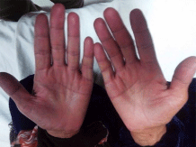
    Figure 1:  The patient’s hands showing bluish coloration at the finger tips
(acrocyanosis).
Work up for polycythemia to exclude secondary cause’s especially renal lymphnangictasia secondary to renal hematoma was initiated. Follow up ultrasound showed organized shrunken hematoma in the left kidney. Serum erythropoietin level was low (2.19 Mu/ml). Testing for mutation on the Janus kinase-2 gene (JAK2) was positive for valine-to-phenylalanine substitution at position 617 (JAK 2 V617F). This established the diagnosis of primary polycythemia Vera according to the revised WHO 2008 criteria of myeloproliferative neoplasms1.Venesection was started and bone marrow biopsy was scheduled.
The result of renal biopsy was obtained at this point. Two cores of renal cortical and corticomedullary tissue were examined. The sections were stained with H&E, periodic acid-Schiff, Masson’s trichrome and Congo red stains. Microscopic examination revealed 23glomeruli with 50% global sclerosis. Most of the tufts showed moderate mesangial sclerosis with frequent segmental sclerotic lesions having luminal hyalinosis and focal capsular adhesions (Figure 2). Moderate mesangial cellular proliferation was noted in two glomeruli (Figure 3), while segmental glomerular basement membrane thickening with peripheral duplication of the basement membranes was noted in 5 glomeruli on light microscopy (Figure 4). Tubulo-interstitium showed mild interstitial fibrosis and tubular atrophy. Arteries showed moderate intimal sclerosis and arterioles showed mild hyalinosis. Immunoperoxidase studies for IgA, IgG, IgM, C3, Kappa and lambda were negative.
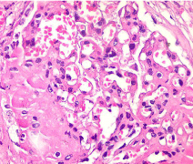
    Figure 2:  Glomeruli moderately enlarged showingsegmental sclerosis &
hyalinosis. (H&E x400).
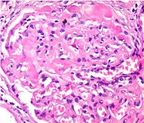
    Figure 3:  Glomeruli are moderately enlarged with Segmental mesangial
proliferation (H&E x400).
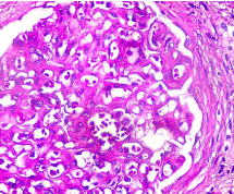
Figure 4: Glomerulus showing basement membrane duplication. (PASx400).
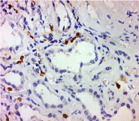
    Figure 5:  Myeloperoxidase staining showing mature and immature
granulocytes within peritubular capillaries .(x200).
Following the clinical diagnosis of PV, further sections were stained for CD61 (megakaryocytes and platelets) and myeloperoxidase (granulocytes). No infiltrating hematopoietic cells were noted in the glomeruli either on light microscopy or by immunohistochemistry. However frequent granulocytes were noted within the peritubular capillaries (Figure 4). Overall features were consistent with Myeloprolifeartive Neoplasms (MPN) related glomerulopathy.
Discussion
Polycythemia Vera (PV) is a relatively rare condition with peak incidence in the age 50-70 years. It is classified under myeloproliferative neoplasms (MPN) according to the WHO classification. Symptoms are insidious in onset with thrombohemorrhagic events and transformation to acute myelogenous leukemia being the major complications.Physical findings in patients with polycythemia Vera (PV) are due to the myeloproliferative process with extramedullary hematopoiesis and splenomegaly present in 75% of patients at the time of diagnosis. Hypertension is a common finding. Pruritus results from increased histamine levels released from increased basophils and mast cells [1,2].
Main differential diagnosis is secondary polycythemia, which in our case could be related to the renal hematoma and subsequent renal lymphamgectasia [3] but this was ruled out by the JAK2 mutation and the low erythropoietin levels.
Renal involvement by MPN is not common. Previous reports included acute renal failure in patients with Essential Thrombocytosis (ET) and tumor lysis syndrome in chronic myelogenous leukemia (CML) [4]. However glomerular involvement with a resulting nephrotic range proteinuria and chronic kidney disease had been rarely reported. Previous reports of glomerulopathy related to MPN have included focal segmental glomerulosclerosis [5], focal segmental glomerulosclerosis with mesangial cellularity [6] and variable degrees of mesangial sclerosis and hypercellularity with features of chronic thrombotic microangiopathy and intra capillary hematopoietic cell infiltration [7,8]. The features encountered in our case resembled those described in the case series reported by Said et al [7]. In their series, infiltrating intraglomerular hematopoietic cells was found in only 36 % of the cases while we couldn’t find any in our case. This could be attributed to the fact that the majority of their cases were Primary Myelofibrosis (PMF) where megakaryocytic proliferation is the main finding. This is not encountered in PV according to the 2008 WHO criteria of myeloproliferative neoplasms.
Conclusion
We report a case of a 60 year old female with primary PV, whose initial presentation was hypertension and nephrotic range proteinuria. This is quite unusual, since glomerular involvement in MPN is typically a late complication that is often un-noticed. Raised awareness and further clinical studies are needed for the early diagnosis and management of this condition.
References
- Tefferi A, Thiele J, Orazi A, Kvasnicka HM, Barbui T, Hanson CA, et al. Proposals and rationale for revision of the World Health Organization diagnostic criteria for polycythemia vera, essential thrombocythemia, and primary myelofibrosis: recommendations from an ad hoc international expert panel. Blood. 2007; 110: 1092–1097.
- Blanc M, Schmutz G, Belzile F, Sabbagh R. Renal lymphangiectasia presenting with hypertension and Polycythemia. Can Urol Assoc J. 2014; 8:163-166.
- Streiff MB, Smith B, Spivak JL. The diagnosis and management of polycythemia vera in the era since the Polycythemia Vera Study Group: a survey of American Society of Hematology members' practice patterns. Blood. 2002; 99: 1144-1149.
- Al-Kali A, Farooq S, Tfayli A. Tumor lysis syndrome after starting treatment with Gleevec in a patient with chronic myelogenous leukemia. J Clin Pharm Ther. 2009; 34: 607-610.
- Au WY, Chan KW, Lui SL, Lam CC, Kwong YL. Focal segmental glomerulosclerosis and mesangial sclerosis associated with myeloproliferative disorders. Am J Kidney Dis. 1999; 34: 889-893.
- Bardy A, Tiple A, Rabant M, Kémény JL, El Karoui K, Hermet M, et al. The myeloproliferative neoplasms-related glomerulopathy. Rev Med Interne. 2014; 35: 222-230.
- Said SM, Leung N, Sethi S, Cornell LD, Fidler ME, Grande JP, et al. Myeloproliferative neoplasms cause glomerulopathy. Kidney Int. 2011; 80: 753-759.
- Paule R, Ponsoye M, Gueutin V, Deray G, Izzedine H. Myeloproliferative neoplasms related glomerulopathy. Rev Med Interne. 2013; 34: 369-372.
Citation: Shehab T and Ismail W. Nephrotic Syndrome as the Initial Presentation of Polycythemia Vera. Austin J Nephrol Hypertens. 2015;2(2): 1035.
Citation: Bucci J and Hansen KE. Should we treat Secondary Hyperparathyroidism in Patients with Pre-Dialysis Chronic Kidney Disease?. Austin J Nephrol Hypertens. 2015; 2(4): 1046. ISSN : 2381-8964