
Research Article
Austin J Neurol Disord Epilepsy. 2016; 3(2): 1023.
Mismatched F-Wave Data with Clinical Findings in a Patient with Cerebrovascular Disease
Suzuki T1,2*, Tani M1,2, Bunno Y1,2, Onigata C2, Uragami S2, Fukumoto Y1, Wakayama I1 and Yoshida S1
1Graduate School of Health Sciences, Graduate School of Kansai University of Health Sciences, Japan
2Clinical Physical Therapy Laboratory, Faculty of Health Sciences, Kansai University of Health Sciences, Japan
*Corresponding author: Toshiaki Suzuki, Graduate School of Health Sciences, Graduate School of Kansai University of Health Sciences, 2-11-1, Wakaba, Kumatori, Sennan, Osaka 590-0482, Japan
Received: May 25, 2016; Accepted: August 08, 2016; Published: August 10, 2016
Abstract
We introduce a 67-year-old male patient with right hemiparesis and cerebral hemorrhage undergoing physical therapy.
He did not move the affected fingers, especially the thumb. According to the clinical findings, MAS (modified Ashworth Scale) of the thenar muscles was 2 and the arm tendon reflex was normal. To investigate the excitability of spinal neural function, we tested the F-wave of the right thenar muscles after stimulating the right median nerve in the wrist at rest.
This patient has hyper tonus but normal tendon reflexes with a normal F/M amplitude ratio. These results suggest that the cause may be the secondary dysfunction because of the shortening of the thenar muscles and not a neurologic factor.
Keywords: F-wave; Muscle tonus; Tendon reflex; Muscle shorting; Cerebrovascular disease
Abbreviations
CVD: Cerebrovascular Disease; MAS: Modified Ashworth Scale
Introduction
F-wave is an index of excitability of spinal neural function. F-waves are recorded from all muscles with the supramaximal stimulation of a motor neurons. The impulse from the supramaximal stimulation of a motor neuron travels to the anterior horn cell and the backfired potential at the anterior horn cell conducts to the recorded muscle. Analysis of the F-wave for excitability of spinal neural function comprises persistence and the F/M amplitude ratio. Persistence is defined by the number of measurable F-wave responses divided by all trials of supramaximal stimulation. In general, persistence reflects the number of anterior horn cells, which the F wave consists of. The F/M amplitude ratio is defined as the mean amplitude of all responses divided by the amplitude of the M-wave. This is the excitability of spinal neural function compared with the excitability of muscle function.
F-waves in patients with cerebrovascular disease (CVD) are thought to be different to those in healthy subjects. In our previous study that investigated the nervous system of hemiplegic patients, excitability of spinal neural function was evaluated using F-wave data of patients with CVD [1]. We reported that the persistence and F/M amplitude ratio in patients with CVD were affected by the grade of muscle tonus, tendon reflex, and voluntary movement.
However, we experienced patients with CVD with mismatched F-wave data and clinical findings. In this report, we introduce atypical case with mismatched F wave data and clinical findings as persistence and F/M amplitude ratio.
Materials and Methods
We introduce a 67-year-old male patient with right hemiparesis and cerebral hemorrhage undergoing physical therapy (20 min, twice a week). Morbidity period from onset is 114 months.
He could move his right arm with movements in the shoulder and elbow joints. Movement in the wrist joint was poor and he did not move the affected fingers, especially the thumb. According to the clinical findings, MAS (modified Ashworth Scale) of the thenar muscles was 2 and the arm tendon reflex was normal.
To investigate the excitability of spinal neural function, we tested the F-wave of the right thenar muscles after stimulating the right median nerve in the wrist at rest.
The subject was comfortably maintained in a supine position with external rotation of both shoulder joints. The skin was prepared with abrasive gel to maintain the impedance below 5KO. The F-waves were recorded using a VIASYS Viking Quest electromyography machine (Natus Medical Inc., CA, USA). We tested the F-wave of the right thenar muscles using a pair of round disks attached to the skin with collodion over the belly and bone of the metacarpal phalangeal joint of the thumb after stimulating the right median nerve at the wrist. The stimulating electrodes comprised a cathode placed over the right median nerve 3cm proximal to the palmar crease of the wrist joint and an anode placed 2cm further. The maximal stimulus was determined by delivering 0.2-ms square-wave pulses of increasing intensity to elicit the largest compound muscle action potentials. Supramaximal shocks (adjusted up to the value of 20% higher than the maximal stimulus) were delivered at 0.5Hz and 90 times for F-wave acquisition. The bandwidth filter ranged from 2Hz to 3kHz.
The persistence and F/M amplitude ratio were analyzed by measurable F-waves. Persistence was defined by the number of measurable F-wave responses divided by 90 trials of supramaximal stimulation. The F/M amplitude ratio was defined as the mean amplitude of all responses divided by the amplitude of the M-wave.
Results
The persistence was 95%. This result was high when compared with normative data (66.8% suzuki et al, 1989 [2]). The F/M amplitude ratio was 1.16%. This result was consistent with normative data (1.81%, suzuki et al, 1989 [2]) (Figure 1) Typical F-wave was showed in Figure 2.
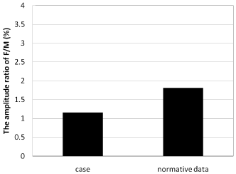
Figure 1: The amplitude ratio of F/M in a patient with CVD.
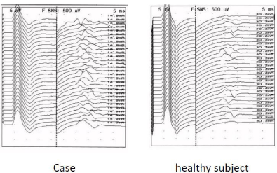
Figure 2: Typical F-wave in a patient with CVD and Healthy subject.
Discussion
We introduced a typical case with mismatched neurological findings, especially those pertaining to the muscle tonus and F-wave results. The neurological findings comprised the MAS of affected thenar muscles and tendon reflex of affected arm. MAS is a score of hyper tonus of affected muscles and is separated into 6 grades (0,1,1+,2,3, and 4). In MAS, we check resistance feeling to stretch testing muscle. It is easy to stretch affected thumb muscle as Score 2.
We hyposis that the resistance factors in MAS were combined spasticity and muscle and skin shorting owing to secondary dysfunction as follows (Figure 3). We tested the tendon reflex of the biceps muscle, triceps muscle, brachioradial muscle, and pronator because tendon reflex of thenar muscles was not tested. Data of tendon reflex of the affected arm was within normal. The grade of tendon reflex is generally thought to express the grade of spasticity in patients with CVD. Hence, the increasing muscle tonus of thenar muscles in this patient was because of muscle shorting owing to secondary dysfunction rather than spasticity. The F/M amplitude ratio from thenar muscle was the same as that in healthy controls in our previous report. From the results for tendon reflex and amplitude ratio of F/M, excitability of spinal neural function was almost normal.
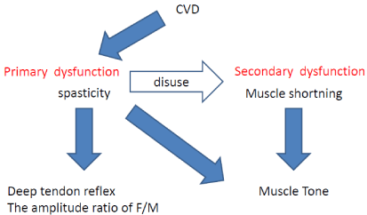
Figure 3: The relationship between spasticity and muscle shortning.
However, we wonder if the persistence was higher although the F/M amplitude ratio was normal; this suggests that the small-typed anterior horn cell was excited. We would like to further investigate this phenomenon in future.
From these results, we suggested that the major cause of hyper tonus of the thenar muscles in this patient was muscle shortening owing to secondary dys function and not spasticity.
Conclusion
This patient has hyper tonus but normal tendon reflexes with a normal F/M amplitude ratio. These results suggest that the cause may be the secondary dysfunction because of the shortening of the thenar muscles and not a neurologic factor.
References
- Suzuki T, Fujiwara T, Takeda I. Characteristics of F-wave During Relaxation in Patients with CVD (In Japanese, Abstract in English). PT Journal. 1993; 27: 277-281.
- Suzuki T, Takeda I, Fujiwara T. Basic research on the F-wave of normal subject at static condition (In Japanese, Abstract in English). J Exerc Physiol. 1989; 4: 17-20.