
Case Report
Austin J Neurol Disord Epilepsy. 2023; 9(1): 1051.
Localized Scleroderma Causes Peripheral Neuropathies and Persistent Elevated Cerebrospinal Fluid Protein: A Case Report
ampliYueman Chen¹; Chenhui Ye²; Yanjun Lin²; Qing Lv³*; Junfen Zhu4; Qinghua Hou²*
1Class 4, 2019 Clinical Medicine Program, Sun Yat-Sen University (Shengzhen), China
2Department of Neurology, Clinical Neuroscience Center, The Seventh Affiliated Hospital, Sun Yat-Sen University, Shenzhen, China
3Department of Rheumatology, The Seventh Affiliated Hospital, Sun Yat-Sen University, Shenzhen, China
4Department of pathology, The Seventh Affiliated Hospital, Sun Yat-Sen University, Shenzhen, China
*Corresponding author: Qinghua Hou Department of Neurology, Clinical Neuroscience Center, The seventh Affiliated Hospital, Sun Yat-sen University, No. 628 Zhenyuan Rd, Guangming, Shenzhen, Guangdong, 518017, China;
Qing Lv, Department of Rheumatology, The Seventh Affiliated Hospital, Sun Yat-Sen University, Shenzhen, China. Tel: +86-755-8120 6812; +86-755-8120 6847 Email: [email protected]; [email protected]
Received: October 31, 2023 Accepted: November 30, 2023 Published: December 07,2023
Abstract
We present a case of peripheral neuropathies and persistent elevated cerebrospinal fluid protein that is attributed to localized scleroderma. This patient’s has been miss diagnosed for more than 14 years, and the diagnosis of localized scleroderma was finally set up by skin biopsy, providing further evidence for the value of skin biopsy in the clinical practice in peripheral neuropathies.
A 62-year-old man was admitted due to volatile tightening sensation on the upper limbs and persistent limb weakness for 14 years. On his first presentation to a local hospital, he was diagnosed as Guillain-Barré Syndrome (GBS) in regard to his symmetry weakness of four limbs and Albuminocytologic dissociation in cerebrospinal fluid (details non-available). After 1 month of standardized treatment of GBS including corticosteroids i.v. and oral corticosteroids subsequential tapper, this gentleman’s limb weakness improved, but his tightening sensation on the upper limbs remained. Nothing of significance was found on the dermatologic examination and serum auto-antibodies assessment at that moment. However, 8 years before the admission, the above symptoms became worse again, and for the second time he was diagnosed as GBS again at a tertiary hospital in Shenzhen. Corticosteroids were once again given for 1 month, together with Intravenous Immunoglobulin (IVIG) therapy, but achieved no effect. Two years prior to the present hospitalization, this patient had begun suffering from a newly-emerged persistent perianal numbness, and a sensation of shoulder heaviness and upper-arm soreness. During the whole course, the patient had not manifested significant disorders in joints, limbs, respiratory and digestive system, nor had he demonstrated Raynaud-phenomenon, skin morphea, or alopecia. There is nothing special in his medical history and family history.
On general physical examination, this gentleman manifested none of the typical characteristics of localized scleroderma or systemic scleroderma, such as sclerotic atrophic patchy alopecia, “mask-face”, skin swells, skin hardens and skin shrinks on the trunk and limbs. Neither as well, no restriction of joint mobility was found on hands and feet. While on neurological examination, this patient was active in mind, clear in articulation and normal in swallowing. No skeletal muscle atrophy was noticed and the muscle strength of four limbs (proximal and distal) were basically normal, and suspicious decrease response in muscle tension/tendon reflex. No subjective sensory deficiencies were detected. However, on electromyography examination, it showed that motor fibers conduction of this gentleman’s bilateral ulnar nerve slows down, and the action potential amplitude of sensory fibers decreases, indicating bilateral motor and sensory fiber damage of this nerve. Latency of skin sympathetic response (SSR) in both hands prolonged, and its amplitude reduced. The SSRs in both feet were non-detectable. Somatosensory Evoked Potential (SEP) examination showed that potential collected at Erb’s point, C7 and cortex on both sides were normal in amplitude, though the wave-interval of left N9-N20 and left N13-N20, and lateral-difference of N9-N20 were prolonged, indicating central-somatosensory pathway damage above C7. The latency, peak-interval and lateral-difference of other waves were all normal. SEP of lower limbs had nothing of significance (Figure 1). No other autoimmune-associated injuries of heart, digestive tract and skeletal-joints was detected on CT/endoscopy examinations. Laboratory investigation indicated positive of anti-scleroderma-70 antibody and anti-nuclear antibody (ANA) (serum titer was 1:100), while other serum assessments were otherwise non-significant, including serological markers of syphilis and HIV infection. Lumber puncture was conducted, and the opening pressure was 140mmH2O, CSF analysis indicated mild elevated cerebrospinal fluid protein (0.848g/l) and normal cell count (2X10^6/L). To unveil the etiology of peripheral neuropathy, we conducted skin biopsies on the medial portions of the left arm and ankle. The skin biopsy showed slight atrophy of the epidermis, hyperplasia of superficial dermal collagen fibers with hyaline degeneration, and infiltration of small lymphocytes around blood vessels (Figure 2), which confirmed as scleroderma by a senior physician (Q.L), and pathology physician (XF.Z).Once the diagnosis was established, immunotherapy with hydroxychloroquine (0.1g t.i.d.) was initiated, and his clinical symptoms mildly improved 2 months later.
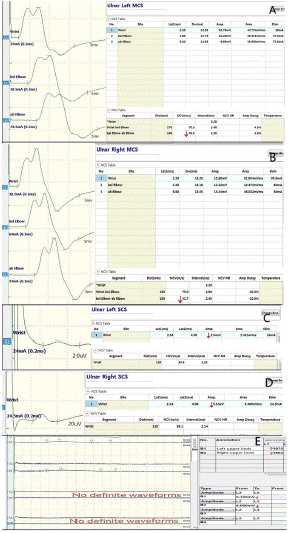
Figure 1: Results of EMG and SEP examination: Ulnar motor nerve conduction velocity on both side decreased (A,B); ulnar sensory nerve action potential amplitude on both sides reduced (C,D); SSR waveforms in four limbs (E): left upper limb (B1), right upper limb (B2), left lower limb (B3), right lower limb (B4). EMG: electromyography; SEP: Somatosensory Evoked Potential; SSR: skin sympathetic response.
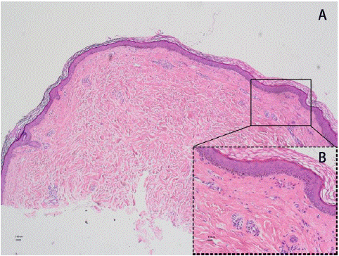
Figure 2: Pathological of skin biopsy (hematoxylin-eosin staining): slight atrophy of the epidermis, hyperplasia of superficial dermal collagen fibers with hyaline degeneration, and infiltration of small lymphocytes around blood vessels. A: 40×, B:200×; Scale bar:500um, 100um.
Discussion
Localized Scleroderma (LS), also known as morphea, is a rare connective tissue disease which is confined to the skin and/or underlying tissues without visceral involvement. The diagnosis of LS mainly relies on the typical clinical manifestation (the linear induration or plaque on trunk, limbs, face and scalp [1]. For atypical, suspicious cases, skin biopsy for histopathological evaluation is the last resort [2]. The histological features of scleroderma fundamentally include the thickening and homogenization of collagen bundles in the dermis and subcutis, accompanying with vascular alterations and inflammatory cell infiltrate. There is no other than these histo-pathological features for morphea. Therefore, differentiating generalized morphea from Systemic Sclerosis (SSc) can be challenging, mostly would have to rely on that patients with morphea do not present with sclerodactyly, Raynaud’s phenomenon, visceral involvement nor nailfold capillary changes, which is typically observed in SSc [3]. The diagnosis of generalized morphea (a subtyple of localized scleroderma) in our case is based on the patient’s typical skin histopathological results and his absence of visceral involvement [4]. As a matter of fact, most atypical subtypes of localized scleroderma, such as nodular or keloidal scleroderma [5], bullous morphea [6], post-irradiation morphea [7] and localized scleroderma presenting with pseudo-ainhum [8], are all usually needed to be confirmed by histopathological biopsy, which emphasizes the importance of histopathological biopsy in the diagnosis of LS.
There are several characteristics of our patient that make it unique to previous cases. First of all, although the course of the disease is as long as 14 years, this patient has not developed typical skin appearance of scleroderma, but only a sensation of skin tightness and the histological changes visible by biopsy, which is extremely rare. Second, though neurologic involvement associated with LS is not uncommon, for example, seizures and headache are quite frequently seen in LS of En coup de sabre (linear scleroderma, ECDS), and it is estimated the involvement of peripheral nervous system manifestations in LS
have a prevalence of 14.4% or higher [9,11]. As far as this case is concerned, based on that the patient's seropositivity for SCL-70 antibody, and the skin biopsy supported localized scleroderma, we consider that the patient's peripheral neuropathy is caused by LS. We make this conclusion based on that we had carefully ruled out the causes of chronical inflammation, mechanical compression, toxicosis, hereditary, metabolism and neoplasm/para-neoplasm, and this association was further supported by the patient’s considerable positive response to targeted immune-therapy. This gentleman’s neuropathy and LS have remained relatively stable in such a long term as long as 14-years, also make this case distinguished from previous cases. Finally, in nerve biopsy study, systemic sclerosis patients with peripheral neuropathy demonstrate perivascular mononuclear cell inflammation with/without vessel wall involvement or fibrinoid necrosis [10], and peri-neural inflammation has also been noticed in LS [11], indicating microvascular mechanism plays an important role in the pathogenesis of scleroderma-associated peripheral neuropathy [12]. Chronic inflammation in the CSF represented as the elevation of intrathecal protein can be observed in some rheumatic diseases involvement of the nervous system, such as systemic lupus erythematosus, sarcoidosis, Behçet's disease and Sjögren's syndrome [13], and scleroderma as well [9]. In the present case, tough the patient’s scleroderma does not develop systemic involvements, nor significant microangiopathic-infiltration as per his skin biopsy results, his sustained-mild- elevation of CSF protein marked this case a very special outlier in LS and autoimmune disease involvement of the central nervous system, which deserves further investigation.
Taken together, skin biopsy helps unveiling a case of localized scleroderma with peripheral nerve system involvement and persistent elevated cerebrospinal fluid protein, which has been miss diagnosed for 14 years.
References
- Careta MF, Romiti R. Localized scleroderma: clinical spectrum and therapeutic update. An Bras Dermatol. 2015; 90: 62-73.
- Papara C, De Luca DA, Bieber K, Vorobyev A, Ludwig RJ. Morphea: the 2023 update. Front Med (Lausanne). 2023; 10: 1108623.
- Ferreli C, Gasparini G, Parodi A, Cozzani E, Rongioletti F, Atzori L. Cutaneous manifestations of Scleroderma and Scleroderma-like disorders: a comprehensive review. Clin Rev Allergy Immunol. 2017; 53: 306-36.
- Kreuter A, Krieg T, Worm M, Wenzel J, Gambichler T, Kuhn A et al. AWMF-Leitlinie Nr. 013/066. Diagnostik und Therapie der zirkumskripten Sklerodermie. J Dtsch Dermatol Ges. 2009; 7: S1-14.
- Bayle P, Bazex J, Marguery MC, Lamant L. Nodules sur sclérodermie en plaque ou morphée [Nodules on localized scleroderma or morphea]. Ann Dermatol Venereol. 2005; 132: 130-2.
- Daoud MS, Su WP, Leiferman KM, Perniciaro C. Bullous morphea: clinical, pathologic, and immunopathologic evaluation of thirteen cases. J Am Acad Dermatol. 1994; 30: 937-43.
- Akay BN, Sanli H, Heper AO. Postirradiation linear morphoea. Clin Exp Dermatol. 2010; 35: e106-8.
- Tajima S, Suzuki Y, Inazumi T. A case of atypical localized scleroderma presenting with pseudoainhum: treatment with tranilast, an anti-fibrotic agent. Acta Derm Venereol. 1996; 76: 162.
- Amaral TN, Peres FA, Lapa AT, Marques-Neto JF, Appenzeller S. Neurologic involvement in scleroderma: a systematic review. Semin Arthritis Rheum. 2013; 43: 335-47.
- Dyck PJ, Hunder GG, Dyck PJ. A case-control and nerve biopsy study of CREST multiple mononeuropathy. Neurology. 1997; 49: 1641-5.
- Succaria F, Kurban M, Kibbi AG, Abbas O. Clinicopathological study of 81 cases of localized and systemic scleroderma. J Eur Acad Dermatol Venereol. 2013; 27: e191-6.
- Fett N, Werth VP. Update on morphea: part I. Epidemiology, clinical presentation, and pathogenesis. J Am Acad Dermatol. 2011; 64: 217-28.
- Reske D, Petereit HF, Heiss WD. Difficulties in the differentiation of chronic inflammatory diseases of the central nervous system--value of cerebrospinal fluid analysis and immunological abnormalities in the diagnosis. Acta Neurol Scand. 2005; 112: 207-13.