
Case Report
Austin Neurosurg Open Access. 2014;1(1): 1003.
Unilateral C1-2 Screw Fixation for Treatment of Atlantoaxial Instability Secondary to Rheumatoid Arthritis - Case Report
Joaquim AF1*, Enrico Ghizoni1, Alexandre Ramos Daoud Yacoub2 and Helder Tedeschi1
1Department of Neurosurgeon, University of Campinas (UNICAMP), Brazil
2Department of Neurosurgery, University of Campinas (UNICAMP), Brazil
*Corresponding author: Joaquim AF, Department of Neurosurgeon, University of Campinas (UNICAMP), Antonio Lapa Street, Number 280, Room 506, Campinas, SP CEP: 13092-621, Brazil
Received: March 10, 2014; Accepted: April 06, 2014; Published: April 08, 2014
Abstract
Background and Importance: Rheumatoid Arthritis (RA) is a systemic chronic inflammatory disease with a high rate of cervical spine involvement, especially at the craniocervical region. The most common presentation of RA in the cervical spine is atlantoaxial subluxation (AAS).
Case: We presented a patient with AAS and severe cervical pain refractory to clinical management who underwent a posterior unilateral C1 lateral mass screw fixation with a laminar C2 screw, using autologous iliac crest bone as a graft for arthrodesis, once the contralateral C1 lateral mass was destroyed by the disease. After two years of follow–up, the patient was without any cervical pain, had no instability in flexion–extension radiographs and also had a C1–2 fusion seen on CT scan.
Conclusion: Unilateral C1–2 screw fixation was a safe and a feasible alternative to treat AAS in patients with RA. It can be a surgical option in patients with unilateral collapse of one atlas lateral mass, avoiding the morbidity of a craniocervical fixation.
Keywords: Rheumatoid arthritis; Atlantoaxial subluxation; Cranial settling; Basilar impression; Vertical subluxation.
Introduction
Rheumatoid Arthritis (RA) is a multi–faceted systemic chronic inflammatory disorder; mainly affect the bone, joints and ligaments, although it may also involve all the other organ systems [1–3]. Its clinical course is quite variable: although it has a chronic nature, acute outbreaks are common, sometimes altered with long periods of quiescent. The proliferative and erosive synovitis progresses to destruction of the particular cartilage, especially in the metatasophalangeal joints, the most common involved joint. Although the cervical spine is not used for scoring radiographic damage in RA, it is a commonly area in the disease course, resulting in spinal instability and neurological impairment in more severe cases [2–6]. Up to 50% of the patients with RA can have some degree of cervical involvement [2]. Of note, in the last two decades, the disease–modifying antirheumatoid drugs (DMARDs) are changing the natural history of this morbid disease, decrease its severity and consequently improving patient's outcome [1,2].
The destructive synovitis progresses to bone erosion and ligamentous laxity, leading to late instability of the spine in a couple of years or even some months. Their clinical and radiological presentations included atlantoaxial subluxation (AAS), the most common form, basilar impression (cranial settling), subaxial subluxation or a combination of them [4,5].
AAS results from laxity of the ligamentous complex that restrains the atlas in the axis, especially the transverse ligament. In AAS, the anterior atlantodental interval (AADI) increases (normal range is lesser than 3 mm), as in our presented case, and the posterior atlantodental interval decreases (PADI), compressing the upper spinal cord [7,8]. Posterior atlantoaxial subluxation is rarer, accounting in less than 10% of the cases with subluxation, generally secondary to fracture of the dens and carry a higher risk of cord injury than AAS. Lateral subluxation can also occur sporadically, resulting in a rotational deformity [2]. We present a case of a patient with RA and AAS that underwent a unilateral C1–2 screw fixation with total neck symptoms resolution for the three years of post–operative follow–up.
Case
A 39–year –old woman presented in our outpatient facility referred from the rheumatology division with a history of refractory cervical pain to analgesics, opioids and even to the use of a rigid cervical collar. She had been treated with DMARDs for the last five years, with a good control of her inflammatory disease and no other clinically evident deformity. Preoperative flexion and extension lateral radiographs showed an increasing anterior atlanto–dental space in flexion compared to extension, suggesting transverse ligamentous laxity (Figure 1A,1B). A small right C1 lateral mass compared to the left one could also be noted (Figure 1C,1D). The patient was neurologically intact. We decided to perform a posterior C1 lateral mass – C2 laminar screw fixation. However, during the procedure, the right C1 lateral mass was collapsed and the bone was extremely fragile, with the C1 screw pulled out just after its insertion. We then opted to leave the left C1 screw and connect it with the right C2 laminar screw, in a unilateral construction. Iliac bone graft was harvested and inserted in the surgical site. Just after surgery and also during the follow–up, the patient had total relief of her cervical pain with visual analogue scale of zero (it was nine points just before surgical treatment), as shown in flexion extension radiographs (Figure 2) and in postoperative CT scan (Figure 2), with a solid bone fusion between C1 and C2.
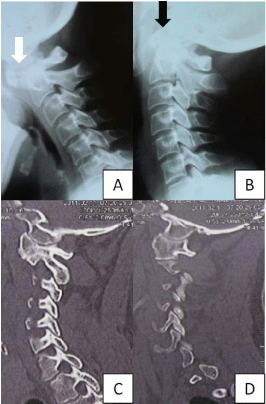
Figure 1: (A) Flexion lateral cervical radiograph showing increase of the anterior atlanto dens interval (white arrow) and 1B) extension lateral cervical radiograph showing decreasing of the atlanto dens interval (black arrow), suggesting ligamentous transverse laxity. In Figure 1C, the right C1 lateral mass is collapsed compared with the left C1 lateral mass in 1D.
Figure 2: (A) Intraoperative picture showing a left C1 lateral mass with a right C2 lamina screw. 2B) Post-operative CT scan showing a left lateral bone bridge connecting C1 and C2. 2C and 2D) Post operative lateral cervical flexion and extension, respectively, radiographs, without change the anterior atlanto dens interval (black arrow).
Discussion
Bone destruction, as well as involvement of the upper cervical spine ligaments, was commonly found in RA. The involvement of the transverse ligament results in atlantoaxial instability, leading to pain and, in non treated cases, neurological impairment due to spinal cord compression [2]. The progressive involvement of the alarligaments, condyles and destruction of the vertebra can result in basilar impression with cranial settling, the most severe presentation of RA in the cervical spine, even though this presentation is reduced nowadays once the severe forms of cervical spine involvement are decreasing with the new DMARDs, as well as the tendency to spare corticosteroids use in these patients [1].
Surgery is an accepted indication in AAS in cases with pain, myelopathy, cord compression or vertebral artery compromise [2].
Many surgical techniques were described in the literature, such as wiring and cable constructions, associated with a high rate of pseudoarthrosis and failure [9]. In the last two decades, the use of screw fixation improved arthrodesis results and patient's outcome, controlling C1–2 motion better than the previous techniques [10–13].Reduction is recommended and can be attempted preoperatively with cervical traction or with direct open reduction and fixation [2]. Occipito–cervical fusions are used in referral centers just for extensive diseases and management of failures [2].
As far as we know, unilateral atlantoaxial fixation was not a reported technique for C1–2 stabilization. In this case, a solid arthrodesis was obtained with a unilateral C1–2 screw fixation. This can be important for sparing occipito–cervical fixation and its higher morbidity in cases where one of the C1 lateral mass is collapsed or destroyed.
References
- Matteson EL. Cervical spine disease in rheumatoid arthritis: how common a finding? How uncommon a problem? Arthritis Rheum. 2003; 48:1775-1778.
- Krauss WE, Bledsoe JM, Clarke MJ, Nottmeier EW, Pichelmann MA. Rheumatoid arthritis of the craniovertebral junction. Neurosurgery. 2010 ;66: 83-95.
- Neva MH, Häkkinen A, Mäkinen H, Hannonen P, Kauppi M, Sokka T. High prevalence of asymptomatic cervical spine subluxation in patients with rheumatoid arthritis waiting for orthopaedic surgery. Ann Rheum Dis. 2006;65: 884-888.
- Zikou AK, Alamanos Y, Argyropoulou MI, Tsampoulas C, Voulgari PV, Efremidis SC, et al. Radiological cervical spine involvement in patients with rheumatoid arthritis: a cross sectional study. J Rheumatol. 2005;32: 801-806.
- Crockard HA, Essigman WK, Stevens JM, Pozo JL, Ransford AO, Kendall BE. Surgical treatment of cervical cord compression in rheumatoid arthritis. Ann Rheum Dis. 1985; 44 :809-816.
- Dvorak J, Grob D, Baumgartner H, Gschwend N, Grauer W, Larsson S. Functional evaluation of the spinal cord by magnetic resonance imaging in patients with rheumatoid arthritis and instability of upper cervical spine. Spine. 1989; 14: 1057-1064.
- O'Brien MF, Casey AT, Crockard A, Pringle J, Stevens JM. Histology of the craniocervical junction in chronic rheumatoid arthritis: a clinicopathologic analysis of 33 operative cases. Spine. 2002; 27: 2245-2254.
- Dreyer SJ, Boden SD. Natural history of rheumatoid arthritis of the cervical spine. Clin Orthop Relat Res. 1999; 366: 98-106.
- Dickman CA, Apostolides PJ, Karahalios DG. Surgical techniques for upper cervical spine decompression and stabilization. Clin Neurosurg. 1997; 44:137-160.
- Goel A, Laheri V. Plate and screw fixation for atlanto-axial subluxation. Acta Neurochir (Wien). 1994; 129: 47-53.
- Goel A, Laheri V. Re: Harms J, Melcher P: posterior C1-C2 fusion with polyaxial screw and rod fixation. Spine. 2002; 27:1589-1590.
- Joaquim AF, Ghizoni E, Rubino PA, Anderle DV, Tedeschi H, Rhoton AL, et al. Lateral Mass Screws Fixation of the Atlas - Surgical Technique and Anatomy. World Neurosurgery. 2010; 74: 359-362.
- Andrei F. Joaquim, Enrico Ghizoni, Helder Tedeschi, Ulysses CausBatista, Alpesh A. Patel,. Axis Instrumentation - Surgical Results. Arq Bras Neuropsiq. 2012; 70: 846-852.