
Case Series
Austin Neurosurg Open Access. 2015; 2(3): 1034.
Two Cases of Traumatic Pseudoaneurysm of Superficial Temporal Artery
Masayuki Goto¹, Keishi Fujita¹*, Tomoya Takada¹, Kazuaki Tsukada¹, Takao Kamezaki¹ and Akira Matsumura²
1Department of Neurosurgery, Ibaraki Seinan Medical Center Hospital, Japan
²Department of Neurosurgery, Institute of Clinical Medicine, University of Tsukuba, Japan
*Corresponding author: Keishi Fujita, Department of Neurosurgery, Ibaraki Seinan Medical Center Hospital, 2190 Sakai, Sashima, Ibaraki 306-0433, Japan
Received: June 19, 2015; Accepted: July 29, 2015; Published: July 30, 2015
Abstract
Traumatic pseudoaneurysms of the Superficial Temporal Artery (STA) are rare vascular lesions that mainly occur after blunt trauma of the temporal region. Such pseudoaneurysms should be diagnosed and treated without any delay, because they can lead to severe headache, facial nerve palsy, arterial bleeding, and bone erosion. The diagnosis of a traumatic pseudoaneurysm of the STA can be established by history of head trauma, physical examination (palpation and bruit), and then be confirmed by imaging studies. The diagnosis and treatment of two traumatic psuedoaneurysm of STA cases are described with good postoperative courses. The diagnosis of traumatic pseudoaneurysm of STA should be excluded during the management of mild blunt head trauma when the area of injury involves the superficial temporal artery location.
Keywords: Trauma; Pseudoaneurysm; Superficial temporal artery
Introduction
It has been reported that traumatic pseudoaneurysms of the Superficial Temporal Artery (STA) account for less than 1% of traumatic aneurysms [1,2]. Most traumatic aneurysms are pseudoaneurysms, consisting of hematoma and fibrous tissue, resulting from injury of all layers of an arterial wall. Traumatic pseudoaneurysms of STA should be diagnosed and treated without delay. Traumatic pseudoaneurysms of STA are usually diagnosed several weeks after blunt trauma to the temporal region, and can cause headache, facial nerve palsy, arterial bleeding and bone erosion [1,3- 6]. In this paper, we present two cases of traumatic pseudoanuerysm of the STA after mild blunt trauma and discuss the treatment of those injuries.
Case Report
Case 1
A 10-year-old child (male) presented in A&E department with a pulsating subcutaneous mass on his left temporal region a few days after a single blow to the left temple with a desk leg. During last nine days, the mass gradually was becoming more painful. At the time of injury, there was no laceration or loss of consciousness. On clinical examination, he was neurologically normal and had a painful pulsating mass without bruit anterosuperior to his left ear. The pulsation disappeared when the subcutaneous artery, which is more proximal than the mass, was compressed. T1-Weighted Image (T1WI) and T2-Weighted Image (T2WI) of Magnetic Resonance Image (MRI) of the patient’s head revealed a subcutaneous isointensity and high-intensity mass, respectively (Figures 1a and 1b). Magnetic Resonance Angiography (MRA) revealed a partial dilatation of the left STA, and Time Of Flight (TOF) showed the lumen of the aneurysm as a lower intensity signal than that of normal cerebral arteries (Figures 1c and 1d). Three-Dimensional Computed Tomographic Arteriography (3D-CTA) revealed a round mass, 17 mm in diameter, off the parietal branch of his left STA (Figure 2). The pulsating subcutaneous mass was a traumatic pseudoaneurysm of the STA. The patient underwent ligation of the proximal and distal ends of the STA and resection of the pseudoaneurysm under sedation and local anesthesia. He recovered without complications during 12 months follow up. Histopathological findings were compatible with pseudoaneurysm (Figure 3).
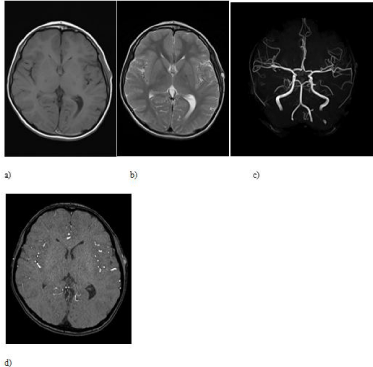
    Figure 1:  a) MRI showing the iso-intensity subcutaneous mass in T1WI; b)
mass in T2WI; c) MRA showing a partial dilatation of the left STA; and d) TOF
showing the lumen of the aneurysm as a lower intensity signal than that of
normal cerebral arteries.
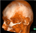
    Figure 2:  3D-CTAG showing a dilatation of the parietal branch of the left STA
with a 17 mm maximum diameter.
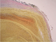
    Figure 3:  Elastica van Gieson stain 4x.
Histopathological examination revealed intra-aneurysmal hematoma
and aneurysmal wall of fibrous connective tissues without internal elastic
membrane.
Case 2
A 45-year-old man complained for a painless and non-pulsating subcutaneous mass in his left temporal region two months after being hit on his left temple by a wooden board that fell from a roof. He presented to A&E department 42 months after the injury due to gradually progressive pulsation of the mass. At the time of injury, there was no laceration or loss of consciousness. On clinical examination, the patient had no neurological symptoms and a painless pulsating mass without bruit located anterosuperior to his left ear. The pulsation disappeared when the subcutaneous artery was compressed. T1WI and T2WI of MRI of his head revealed a subcutaneous iso-intensity mass with a low-intensity portion, and high-intensity mass with low-intensity portion, respectively (Figures 4a and 4b). MRA revealed a partial dilatation of the left STA (Figure 4c), and TOF revealed the residual lumen of the aneurysm had intensity as high as that of normal cerebral arteries (Figure 4d). 3D-CTAG revealed a round mass, 17 mm in diameter, off the trunk of the patient’s left STA (Figure 5). The pulsating subcutaneous mass was diagnosed as a partial thrombosed traumatic pseudoaneurysm of the STA. The patient underwent ligation of the proximal and distal ends of the STA and resection of the pseudoaneurysm under sedation and local anesthesia. He recovered without complication during 6 month follow up. Histopathological findings confirmed the diagnosis of partial thrombosed pseudoaneurysm (Figures 6a and 6b).
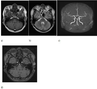
    Figure 4:  a) MRI showing the iso-intensity subcutaneous mass with a flow
void in T1WI; and b) high-intensity subcutaneous mass with a flow void in
T2WI; c) MRA showing a partial dilatation of the left STA; and d) TOF showing
the residual lumen of the aneurysm as high intensity as that of normal
cerebral arteries.
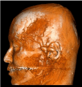
    Figure 5:  3D-CTAG showing a dilatation of the truncus of the left STA with a
17 mm maximum diameter.
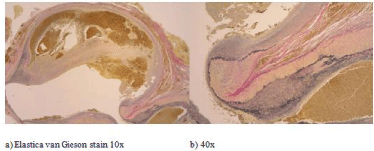
    Figure 6:  a) Histopathological examination revealed intra-aneurysmal
organized thrombus and aneurysmal wall of fibrous connective tissues
without internal elastic membrane and media; b) except at the junction with
the truncus of the STA.
Discussion
The pseudoaneurysms of STA occurred a few days after the blunt trauma in case 1, and two months after in case 2. Traumatic pseudoaneurysms of STA could occur anywhere from a few days to two months post-trauma [1,4,5,7,8]; however Lin et al reported a case that occurred 4 months after trauma [9]. It has been reported that traumatic pseudoaneurysms of the STA tend to occur more frequently in the frontal branch than in the parietal branch due to anatomical characteristics. The frontal branch of the STA is stabilized to the galea without muscle to absorb the impact of trauma in the portion between the temporal muscle and frontal belly. Studies have also found that traumatic pseudoaneurysm of the STA tends to occur in the elderly with arterial sclerosis [6,10,11]. In both our cases, the patients were young. Aneurysmal locations on the muscle and ages of the patients indicated the strength of the trauma impact. Traumatic pseudoaneurysms of STA are not only found as painless pulsating masses, as in our second case, but can also cause headache, as in our first case, facial nerve palsy, arterial bleeding, and bone erosion [1,3,4,5,6]. The bruit of aneurysm can be heard by auscultation in some cases, although not in our cases [3,8,12].
Although the aneurysms described here showed as iso-intensity masses in T1WI, and high-intensity masses in T2WI of MRI, in case 2 the aneurysm included a low-intensity portion in both the T1WI and T2WI; this portion appeared as high-intensity as the normal cerebral arteries in TOF. It has been reported that a large aneurysm shows lower intensity than normal cerebral arteries, as in case 1, because the blood flow in a large aneurysm is slower than that of normal cerebral arteries in TOF [13]. In case 2 of partial thrombosed aneurysm, the residual lumen of the aneurysm was thought to be shown as a flow void in T1WI and T2WI, showing intensity as high as normal cerebral arteries in TOF because the blood flow of the residual lumen was as fast as that of normal cerebral arteries. 3D-CTAG revealed more clearly than MRI and MRA that the parent arteries of the aneurysms in our cases were STAs. Duplex ultrasonic scan, Digital Subtraction Angiography (DSA), MRI and 3D-CTAG have been recommended for differentiating an aneurysm from a hematoma, lipoma, cyst, abcess, angiofibroma, Arteriovenous Fistula (AVF), meningocele and encephalocele [11,14,15], although the traumatic pseudoaneurysm of STA can be diagnosed by the history of head trauma, palpation and auscultation [8].
Traumatic pseudoaneurysm of the STA should be diagnosed and treated immediately, because they can cause headache, facial nerve palsy, arterial bleeding, and bone erosion [1,3-6]. Moreover, postoperative facial nerve palsy has been reported in giant traumatic pseudoneurysm of STA [16]. Ligation resection is recommended for traumatic pseudoaneurysm of the STA because of its prevention effect on rupture and recurrence [14]. Endovascular embolization with coils or embolic materials, and ultrasound-guided embolization with direct aneurysmal puncture for traumatic pseudoaneurysm of STA have also been reported [6,14,15,17]. However, the injection of an embolic material can cause intravascular thrombosis, peripheral ischemia, non-target embolization and allergy; these can be avoided with the endovascular coil embolization technique. Embolization techniques that can diagnose and treat traumatic pseudoaneurysm of the STA at the same time, are inferior to the ligation resection technique from the viewpoint of mass reduction [10,18]. Manual compression for traumatic pseudoaneurysm of STA has been reported to have a 60–90% success rate [15,19]. Although it has been reported that manual compression of the STA proximal to the lesion was effective to induce thrombotic evolution of the aneurysm, a case of arterial bleeding after skin suture and manual compression for pseudoaneurysm of STA following penetrating trauma has also been reported [1,20,21]. Arterial reconstruction of STA has been reported to be useful for preserving blood flow in the skin and preventing wound infection in the case of a small injury of the STA [3]. In both our cases, the diameter of the pseudoaneurysm was 17 mm, and it was thought the territory of the STA was supplied by collateral circulation from the facial artery and the middle meningeal artery [22]. Therefore, we selected the ligation resection, which resulted in good postoperative courses in our cases.
Histopathological examination in case 1 diagnosed the aneurysm as a pseudoaneurysm in which the aneurysmal wall consisted of fibrous connective tissues, inflammatory cells, and small vessels without internal elastic membrane. Histopathological examination in case 2 indicated that the aneurysm, the wall of which lacked an internal elastic membrane, and media, except for the junction with the truncus of STA, was compatible with pseudoaneurysm.
In mild blunt head trauma where the point of impact is on the superficial temporal artery, traumatic pseudoaneurysm of STA should be kept in mind during cases management.
References
- Pourdanesh F, Salehian M, Dehghan P, Dehghani N, Dehghani S. Pseudoaneurysm of the superficial temporal artery following penetrating trauma. J Craniofac Surg. 2013; 24: e334-337.
- Goksu E, Senay E, Alimoğlu E, Aksoy C. Superficial temporal artery pseudoaneurysm: ultrasonographic diagnosis in the ED. Am J Emerg Med. 2009; 27: 627.
- Ayling O, Martin A, Roche-Nagle G. Primary repair of a traumatic superficial temporal artery pseudoaneurysm: case report and literature review. Vasc Endovascular Surg. 2014; 48: 346-348.
- Rancic Z, Pecoraro F, Nigro G, Simon R, Frauenfelder T, Mayer D, et al. Branch ligatures and blood aspiration for post-traumatic superficial temporal artery pseudoaneurysm: surgical technique. Gen Thorac Cardiovasc Surg. 2014; 62: 68-70.
- Kim SW, Jong Kim E, Sung KY, Kim JT, Kim YH. Treatment protocol of traumatic pseudoaneurysm of the superficial temporal artery. J Craniofac Surg. 2013; 24: 295-298.
- Hong JT, Lee SW, Ihn YK, Son BC, Sung JH, Kim IS, et al. Traumatic pseudoaneurysm of the superficial temporal artery treated by endovascular coil embolization. Surg Neurol. 2006; 66: 86-88.
- Li W, Long X, Deng M. Superficial temporal artery pseudoaneurysm: report of a rare case secondary to mandibular condylar fracture. J Craniofac Surg. 2013; 24: e360-361.
- Fukunaga N, Hanaoka M, Masahira N, Tamura T, Oka H, Satoh K, et al. Traumatic pseudoaneurysm of superficial temporal artery. Am J Surg. 2010; 199: e1-2.
- Lin K, Matarasso A, Edelstein DR, Swift RW, Shnayder Y. Superficial temporal artery pseudoaneurysm after face lift. Aesthet Surg J. 2004; 24: 28-32.
- Hasegawa H, Tsutsumi S, Yasumoto Y, Inaba T, Ito M. Traumatic pseudoaneurysm arising from the anterior superficial temporal artery in an infant. Childs Nerv Syst. 2011; 27: 1011-1014.
- Yoshioka F, Hagihara N, Abe T, Kojima K, Watanabe M, Tabuchi K. [Case of an idiopathic dissecting aneurysm of the superficial temporal artery: a case report]. No Shinkei Geka. 2010; 38: 341-345.
- Park IH, Kim HS, Park SK, Kim SW. Traumatic Pseudoaneurysm of the Superficial Temporal Artery Diagnosed by 3-dimensional CT Angiography. J Korean Neurosurg Soc. 2008; 43: 209-211.
- Maeda M. MR Angiography of the Head. Japanese J of Diagnostic Imaging. 2014; 34: 446-456.
- Ahn HS, Cho BM, Oh SM, Park SH. Traumatic pseudoaneurysm of the superficial temporal artery in a child: a case report. Childs Nerv Syst. 2010; 26: 117-120.
- De Vogelaere K. Traumatic aneurysm of the superficial temporal artery: case report. J Trauma. 2004; 57: 399-401.
- Murphy M, Hughes D, Liaquat I, Edmondson R, Bullock P. Giant traumatic pseudoaneurysm of the superficial temporal artery: treatment challenges and case review. Br J Neurosurg. 2006; 20: 159-161.
- Weller CB, Reeder C. Traumatic pseudoaneurysm of the superficial temporal artery: two cases. J Am Osteopath Assoc. 2001; 101: 284-287.
- Weller CB, Reeder C. Traumatic pseudoaneurysm of the superficial temporal artery: two cases. J Am Osteopath Assoc. 2001; 101: 284-287.
- Mann GS, Heran MK. Percutaneous thrombin embolization of a post-traumatic superficial temporal artery pseudoaneurysm. Pediatr Radiol. 2007; 37: 578-580.
- Archer SM, Jones RO. Traumatic pseudoaneurysm of the posterior auricular artery. South Med J. 1992; 85: 47-48.
- Grasso RF, Quattrocchi CC, Crucitti P, Carboni G, Coppola R, Zobel BB. Superficial temporal artery pseudoaneurysm: a conservative approach in a critically ill patient. Cardiovasc Intervent Radiol. 2007; 30: 286-288.
- Quereshy FA, Choi S, Buma B. Traumatic pseudoaneurysm of the superficial temporal artery in a pediatric patient: a case report. J Oral Maxillofac Surg. 2008; 66: 133-135.
- Kim JH, Jung YJ, Chang CH. Superficial temporal artery pseudoaneurysm treated with manual compression alone. J Cerebrovasc Endovasc Neurosurg. 2015; 17: 49-53.