
Case Presentation
Austin Neurosurg Open Access. 2015; 2(3): 1035.
Transfusion-Related Acute Lung Injury Following Lumbar Degenerated Kyphoscoliosis Surgery: A Case Report
Hayato Suzuki¹*, Takashi Kobayashi¹, Naohisa Miyakoshi², Eiji Abe¹, Toshiki Abe¹, Kazuma Kikuchi¹ and Yoichi Shimada²
1Department of Orthopedic Surgery, Akita Kousei Medical Center, Japan
²Department of Orthopedic Surgery, Akita University Graduate School of Medicine, Japan
*Corresponding author: Hayato Suzuki, Department of Orthopedic Surgery, Akita Kousei Medical Center, 1-1-1 Iijima-Nishifukuro, Akita 011-0948, Japan
Received: July 17, 2015; Accepted: August 17, 2015; Published: August 19, 2015
Abstract
Introduction: Transfusion-Related Acute Lung Injury (TRALI) is a syndrome characterized by acute respiratory distress following transfusion. Mortality rates of 5-25% have been reported. Although spine surgery is a risk factor for TRALI, few reports have described TRALI after degenerative kyphoscoliosis surgery. We describe an important case of TRALI following degenerative kyphoscoliosis surgery.
Case Presentation: A 48-year-old Japanese female with degenerative kyphoscoliosis underwent surgical treatment. Extreme lateral interbody fusion from a left-sided retroperitoneal approach and posterior instrumentation was performed. Although 800 g of autologous blood was prepared preoperatively and Cell Saver was used for intraoperative blood salvage, a total of two units of leuko reduced red cell concentrate and two units of fresh frozen plasma were infused. Five hours after transfusion, the patient reported pulmonary distress, and peripheral capillary oxygen saturation was under 60%. Steroid and diuretics were ineffective, and the patient required mechanical ventilation. Mechanical support was continued for 4 days, with gradual recovery to baseline pulmonary function.
Conclusion: A case of TRALI following kyphoscoliosis surgery was successfully treated with mechanical ventilation. We strongly recommend sufficient preoperative preparation of autologous blood, and using Cell Saver for intraoperative blood salvage to minimize autologous transfusion when planning lumbar degenerated kyphoscoliosis surgery.
Keywords: Transfusion-related acute lung injury; Transfusion-associated circulatory overload; Transfusion; Lumbar degenerated kyphoscoliosis; Spinal surgery
Abbreviations
TRALI: Transfusion-Related Acute Lung Injury; RBC: Red Blood Cells; FFP: Fresh Frozen Plasma; WBC: White Blood Cell Count; ESR: Westergren Erythrocyte Sedimentation Rate; CRP: C-Reactive Protein; MRI: Magnetic Resonance Imaging; CT: Computed Tomography; TACO: Transfusion-Associated Circulatory Overload; SSI: Surgical Site Infection; XLIF: Extreme Lateral Interbody Fusion
Introduction
Transfusion-Related Acute Lung Injury (TRALI) is a syndrome characterized by acute respiratory distress following transfusion. Mortality rates of 5-25% have been reported [1-4]. Although spine surgery is a risk factor for TRALI [5,6], few reports have described TRALI after degenerative kyphoscoliosis surgery. We describe a very important case of TRALI following degenerative lumbar kyphoscoliosis surgery.
Case Presentation
A 48-year-old Japanese female with degenerative kyphoscoliosis (Figure 1) underwent anterior spinal fusion from L2 to L5, and posterior spinal fusion from T9 to the pelvis. Preoperatively, 800 g of autologous blood was prepared, and Cell Saver was used for intraoperative blood salvage. Extreme lateral interbody fusion (XLIF; NuVasive Inc., SanDiego, CA, USA) of L2-3, 3-4, and 4-5 from a leftsided retroperitoneal approach and posterior instrumentation from T9 to the pelvis with L5-S lumbar interbody fusion was achieved. The operation lasted 6 h and estimated blood loss during surgery was 1800 g. The patient had no past history of note. Prophylactic antibiotics were given preoperatively, and every 3 h thereafter. Postoperatively, hemoglobin was 6.6 g/dl and hematocrit was 20.1%, and a total of two units of leuko reduced Red Blood Cells (RBC) and two units of Fresh Frozen Plasma (FFP) were infused 1 h after the end of surgery in the intensive care unit. Five hours after transfusion, the patient complained of pulmonary distress, and she became acutely tachycardic (heart rate, 120 beats/min), hypoxic (peripheral capillary oxygen saturation on 3 L oxygen by nasal cannula, 60%) and hypotensive (systolic blood pressure, approx. 70 mmHg). Chest radiography revealed extensive bilateral areas of consolidation in the middle and upper lobes of the lung and bilateral pleural effusions (Figure 2). Because steroids and diuretics did not improve respiratory distress and chest radiography 12 h after surgery revealed extensive bilateral areas of consolidation in the whole lobes (Figure 3), she required mechanical ventilation. Antibiotics and sivelestat sodium hydrate were applied and the patient recovered gradually. Mechanical support was continued for 4 days. The patient subsequently recovered baseline pulmonary function and was discharged in an ambulatory state.
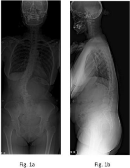
    Figure 1:  Preoperative anterior-posterior (a) and lateral (b) radiographs show
lumbar degenerative kyphoscoliosis.
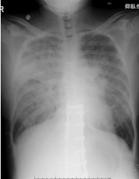
    Figure 2:  Anterior-posterior radiograph at 6 h postoperatively shows
extensive bilateral areas of consolidation in the middle and upper lobes of
the lung.
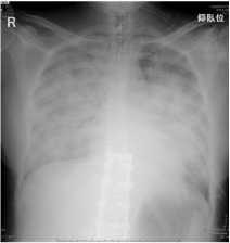
    Figure 3:  Anterior-posterior radiograph at 12 h postoperatively shows
increased consolidation.
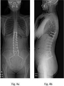
    Figure 4:  Anterior-posterior (a) and lateral (b) radiographs at 2 months
postoperatively show T9-pelvis pedicle screw instrumentation.
Discussion
TRALI is defined as the new onset of acute lung injury occurring within 6 h of blood product transfusion, as indicated by the presence of hypoxemia and bilateral infiltrates on frontal chest radiography, in the absence of circulatory overload [5-7]. TRALI is the number one cause of transfusion-related fatalities reported to the Food and Drug Administration [8]. Mortality rates of 5-25% have been reported [1- 4]. The estimated incidence of TRALI is 1:5000 transfusions [1,2]. Sanchez reported the risk for TRALI increased with each unit of whole blood transfused [9].
Differential diagnoses of acute lung injury after transfusion include Transfusion-Associated Circulatory Overload (TACO), cardiogenic edema, allergic and anaphylactic transfusion reactions, and sepsis due to transfusion of bacterially contaminated blood products [10]. TRALI may be distinguished from TACO and cardiogenic pulmonary edema by the clinical response to diuretics. Although sepsis should be considered when clinical features such as hypotension and fever are noted in a patient after surgery [10], it is very difficult to distinguish from TRALI, because some patients after spine surgery have such symptoms. Anti-bacterial agents should be considered until sufficient recovery of condition is obtained.
Radiographic findings demonstrate bilateral pulmonary infiltrates. Because such findings closely resemble those of fluid overload and/or cardiac failure, differentiation should be based on clinical findings.
Treatment for TRALI depends on the condition of the patient. If the condition is mild, supplemental oxygen alone will be sufficient. However, if the condition of the patient is serious, aggressive respiratory support, supplemental oxygen and mechanical ventilation will be needed [3]. As in our case, corticosteroids and diuretics are not effective [4]. Even with aggressive treatment, the mortality rate from TRALI is 5-25% [4].
Recipient risk factors were chronic alcohol abuse, shock, higher peak airway pressure while being mechanically ventilated, current smoking, positive fluid balance, and spine surgery [5,6]. Transfusion risk factors were receipt of plasma or whole blood from female donors [5,6]. Dunbar described the association between transfusion of maternal blood products and TRALI in pediatric patients undergoing anterior and/or posterior spinal surgery [11]. Patients undergoing spinal surgery frequently demonstrate evidence of acute lung injury [12] and may be at increased risk of TRALI [9]. Spine surgery for degenerative kyphoscoliosis often needs blood transfusions to compensate for marked blood loss. Blood auto transfusion offers numerous advantages over allogenic blood transfusion. Autologous blood donations can reduce the need for homologous transfusions, and strengthen immune ability [13]. We strongly recommend preoperative preparation of autologous blood, and the use of Cell Saver for intraoperative blood salvage.
Many exposures have been applied for degenerative kyphoscoliosis. Posterior correction and instrumentation is the most common. However, for good correction, posterior lumbar interbody fusion or corpectomy are necessary. Interbody fusion or corpectomy can lead to unexpected blood loss. We achieved XLIF and posterior instrumentation. XLIF has several advantages including smaller incisions, decreased operative time and blood loss [14]. Although our patient subsequently needed autologous transfusion, this exposure was expected to reduce blood loss [15]. More technical efforts are needed to reduce intraoperative blood loss.
When the patient complained of pulmonary distress within 6 hours of transfusion, aggressive respiratory support with supplemental oxygen and mechanical ventilation should be considered.
Conclusion
A case of TRALI following kyphoscoliosis surgery was successfully treated with mechanical ventilation. We strongly recommend preoperative preparation of autologous blood, and the use of Cell Saver for intraoperative blood salvage to minimize autologous transfusion.
Acknowledgement
The authors wish to thank Mamiko Kondo and Sachie Miura for their valuable assistance with the editing of this manuscript.
References
- Popovsky MA, Moore SB. Diagnostic and pathogenetic considerations in transfusion-related acute lung injury. Transfusion. 1985; 25: 573-577.
- Popovsky MA. Transfusion-Related Acute Lung Injury: Incidence, Pathogenesis and the Role of Multicomponent Apheresis in Its Prevention. Transfus Med Hemother. 2008; 35: 76-79.
- Silliman CC, Ambruso DR, Boshkov LK. Transfusion-related acute lung injury. Blood. 2005; 105: 2266-2273.
- Silliman CC, Fung YL, Ball JB, Khan SY. Transfusion-related acute lung injury (TRALI): current concepts and misconceptions. Blood Rev. 2009; 23: 245-255.
- Toy P, Lowell C. TRALI--definition, mechanisms, incidence and clinical relevance. Best Pract Res Clin Anaesthesiol. 2007; 21: 183-193.
- Toy P, Gajic O, Bacchetti P, Looney MR, Gropper MA, Hubmayr R, et al. TRALI Study Group: Transfusion-related acute lung injury: incidence and risk factors. Blood. 2012; 119: 1757-1767.
- Kleinman S, Caulfield T, Chan P, Davenport R, McFarland J, McPhedran S, et al. Toward an understanding of transfusion-related acute lung injury: statement of a consensus panel. Transfusion. 2004; 44: 1774-1789.
- Holness L, Knippen MA, Simmons L, Lachenbruch PA. Fatalities caused by TRALI. Transfus Med Rev. 2004; 18: 184-188.
- Sanchez R, Bacchetti P, Toy P. Transfusion-related acute lung injury: a case-control pilot study of risk factors. Am J Clin Pathol. 2007; 128: 128-134.
- Rollins MD, Molofsky AB, Nambiar A, Pandey S, Weiskopf RB, Toy P. Two septic transfusion reactions presenting as transfusion-related acute lung injury from a split plateletpheresis unit. Crit Care Med. 2012; 40: 2488-2491.
- Dunbar N, Cooke M, Diab M, Toy P. Transfusion-related acute lung injury after transfusion of maternal blood: a case-control study. Spine (Phila Pa 1976). 2010; 35: E1322-1327.
- Urban MK, Jules-Elysee KM, Beckman JB, Sivjee K, King T, Kelsey W, et al. Pulmonary injury in patients undergoing complex spine surgery. Spine J. 2005; 5: 269-276.
- Long MY, Liu ZH, Zhu JG. Comparative analysis of autologous blood transfusion and allogeneic blood transfusion in surgical patients. Int J Clin Exp Med. 2014; 7: 2889-2894.
- Arnold PM, Anderson KK, McGuire RA. The lateral transpsoas approach to the lumbar and thoracic spine: A review. Surg Neurol Int. 2012; 3: S198-215.
- Phillips FM, Isaacs RE, Rodgers WB, Khajavi K, Tohmeh AG, Deviren V, et al. Adult degenerative scoliosis treated with XLIF: clinical and radiographical results of a prospective multicenter study with 24-month follow-up. Spine (Phila Pa 1976) 2013; 38: 1853-1861.