
Case Report
Austin Neurosurg Open Access. 2017; 4(1): 1054.
Primary Squamous Papillary-Type Craniopharyngioma in the Cerebellopontine Angle: Report of Two Cases
Miyakoshi A¹*, Kohno M¹, Sora S¹, Sato H¹ and Yokoyama M²
¹Department of Neurosurgery and Stroke Center, Tokyo Metropolitan Police Hospital, Japan
²Department of Diagnostic Pathology, Tokyo Metropolitan Police Hospital, Japan
*Corresponding author: Miyakoshi A, Department of Neurosurgery and Stroke Center, Tokyo Metropolitan Police Hospital, Japan
Received: January 20, 2017; Accepted: February 26, 2017; Published: February 28, 2017
Abstract
We report two rare cases of primary squamous papillary-type craniopharyngioma that developed in the Cerebellopontine (CP) angle. A 54-year-old woman and 26-year-old man were operated on for partially calcified CP angle tumors. One tumor showed is intensity and the other showed hyperintensity on T1-weighted magnetic resonance images. The small nodules of the tumors were the only part requiring contrast enhancement. The tumors were diagnosed neuropathologically as squamous papillary-type craniopharyngiomas. Although the most frequent tumors occurring in the CP angle are schwannomas, meningiomas and epidermoid cysts, the possibility of craniopharyngioma should be considered in cases in which the cyst exhibits hyperintensity in the T1-weighted magnetic resonance imaging scan, inhomogeneous contrast enhancement, and concomitant calcification in the computed tomography scan.
Keywords: Cerebellopontine angle; Ectopic craniopharyngioma; Squamous papillary type; Cerebellopontine angle tumor
Abbreviations
CP: Cerebellopontine; CT: Computed Tomography; MRI: Magnetic Resonance Imaging; MR: Magnetic Resonance; FLAIR: Fluid-Attenuated Inversion-Recovery
Introduction
Craniopharyngioma is a benign epithelial tumor that originates from the Rathke pouch and usually develops around the parasellarregion. By pathology the tumor is divided into two main types, the adamantinomatous type and squamous papillary type. In contrast to the usual parasellar location, primary craniopharyngioma developing in the Cerebellopontine (CP) angle is extremely rare, with only eleven cases reported to date [1-9]. Ten of these cases were of the adamantinomatous type and one was of the squamous papillary type [1-7,9]. Here, we report the neuroradiological findings from two additional cases of squamous papillary-type craniopharyngioma that developed in the CP angle and were isolated from the parasellar region.
Case Presentation
Case 1
A 54-year-old woman presented with hearing impairment and facial numbness on the left side. Additional symptoms included dizziness and a tendency to lean to the left when walking. Neurological examination revealed deafness in the left ear, left facial hyperesthesia, horizontal spontaneous nystagmus to the right and a positive Romberg sign.
A Computed Tomography (CT) scan identified a hyperdense mass with nodular calcified spots on the left CP angle (Figure 1a). Magnetic Resonance Imaging (MRI) revealed an extra-axial tumor, which showed high intensity on T1-weighted Magnetic Resonance (MR) images (Figure 1c) and low intensity in T2-weighted MR images (Figure 1b). The small nodule of the tumor was the only part requiring contrast enhancement (Figure 1 d).
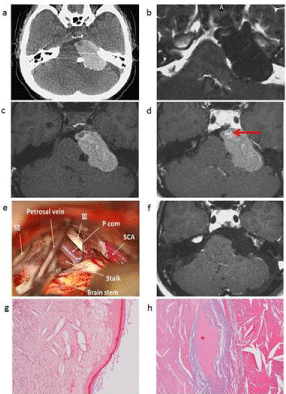
    Figure 1:  A 54-year-old female patient with a squamous papillary-type
craniopharyngioma in the left CP angle. (a) Axial plain CT image shows a
hyperdense mass in the left CP angle with partial calcification. (b) Axial T2-
weighted MRI image reveals a hypointense mass in the left CP angle. (c) The
mass in the left CP angle shows high intensity in the axial plain T1-weighted
MR image. (d) Nodular enhancement was observed in a contrast-enhanced
axial MR image (red arrow). (e) Intraoperative photograph following tumor
removal showing that the pituitary stalk is visible beyond the arachnoid
membrane and does not make contact with the tumor. (f) No apparent
residual tumor in a postoperative axial enhanced MR image. (g, h) The
wall of the cyst consisted of squamous epithelium with a papillary structure.
The tumor contained keratinous material, cholesterin crystal, and a fibrous
inflammatory granuloma.
The patient was therefore operated on via a lateral sub occipital approach. A cystic tumor was identified, with a rust-colored component being visible through the wall of the capsule. Following the opening of the cyst a motor oil-like fluid and semisolid component were drained. The 3rd nerve and pituitary stalk were identified beyond the arachnoid membrane with neither showing any sign of contact with the tumor (Figure 1e).
Postoperative MRI revealed near-total resection of the tumor (Figure 1f). Neuropathology showed that the wall of the cyst was composed of squamous epithelium with a papillary structure (Figure 1g). In contrast, the tumor contained keratinous material, cholesterin crystal and a fibrous inflammatory granuloma (Figure 1h), and was diagnosed as a squamous papillary-type craniopharyngioma. Postoperatively, the patient’s hearing acuity, facial numbness and gait disturbance improved. No sign of recurrence has been observed on follow-up examination for six years.
Case 2
A 26-year-old man presented with facial numbness on the right side, dizziness and a tendency to lean to the left when walking. Neurological examination revealed decreased sensation on the right side of the face. A CT scan of the head showed a hypodense mass with a nodular calcification in the right CP angle (Figure 2a). MRI further identified a cystic extra-axial tumor that partially contained a solid component and was enhanced with gadolinium-containing contrast medium (Figure 2b-2d). The tumor was separate from the sella turcica and pituitary stalk (Figure 2b).
The patient was operated on via a lateral sub occipital and transmastoid-transtentorial approach. Retraction of the cerebellum revealed a cyst with a thin wall that contained a yellow-green fluid. After removal of the tumor we confirmed that it was not attached to the pituitary stalk by intraoperative findings. Additionally, postoperative MRI confirmed near-total removal of the tumor (Figure 2e). Neuropathology revealed that the wall of the cyst was composed of squamous epithelium with a papillary structure (Figure 2f), and that the tumor contained degenerated bone tissue, cholesterin crystal and foreign body-type granuloma (Figure 2g). The final diagnosis of the tumor was a squamous papillary-type craniopharyngioma. Postoperatively, the hypesthesia of the patient’s face improved. No sign of recurrence has been seen on follow-up examination for four years.
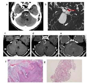
    Figure 2:  A 26-year-old male patient with a squamous papillary-type
craniopharyngioma in the right CP angle. (a) Axial image from a plain CT
scan reveals a hypodense mass with spotty calcification in the right CP
angle. (b) Axial T2-weighted MR image shows a hyperintense cystic mass
containing a solid component in the right CP angle. Tumor is separate from
the pituitary stalk (red arrow). (c) Axial T1-weighted MR image shows that
the mass in the right CP angle is isointense. (d) Partially enhanced tumor in
the right CP angle as seen in a contrast-enhanced axial MR image. (e) No
apparent residual tumor in a postoperative axial enhanced MR image. (f, g)
Cyst wall consisting of squamous epithelium with a papillary structure, and
tumor containingcholesterin crystal and a foreign body-type granuloma.
Discussion
Craniopharyngioma is known to arise around the sella turcica, with the origin thought to be a remnant of the craniopharyngeal duct. Primary craniopharyngioma developing in the Cerebellopontine (CP) angle is extremely rare, with only eleven cases reported to date [1-9]. Ectopic craniopharyngiomas are extremely rare, with only several case reports describing ectopic craniopharyngiomas developing in the CP angle, sphenoid bone [10], third ventricle [11,12], nasopharynx [13,14] and clivus [15]. This pattern raises the possibility that ectopic craniopharyngiomas might develop from migrated squamous epithelial cell remnants originally derived from the craniopharyngeal duct.
Most tumors of the CP angle are schwannomas, meningiomas, and epidermoid cysts [16], with atypical CP angle tumors rarely reported, apart from craniopharyngioma, choroid plexus papilloma, and ganglioglioma [17-20].
Table 1 shows a summary of reported CP angle craniopharyngiomas. Including the two cases reported here, only 13 cases of CP angle Craniopharyngioma have been reported to date [1- 9]. Calcification was identified via CT scan in 6 of the 13 cases.
![]()
    Year
    Author
    Age,    sex
    CT    findings
    MRI    findings
    Pathology


    Density
    Calc
    T1
    T2
    Enhanced


    1990
    Gokalp    HZ et al.
    17, F
    Hypo
    N
    -
    -
    -
    Ad


    2002
    Link ML    et al.
    29, M
    Hyper
    Y
    Mixed
    Mixed
    Heterogeneous
    Ad


    2006
    Aquilina    K et al.
    31, M
    -
    -
    Iso
    -
    Peripherally
    Ad


    2007
    Powers    CJ et al.
    12, F
    -
    -
    Iso
    Hyper
    -
    Ad


    2009
    Yan Y    et al.
    54, F
    Hyper
    Y
    Hypo
    Mixed
    Homogeneous
    Ad


    2011
    Bozbuga    M et al.
    34, M
    Hyper
    Y
    Hyper
    Hypo
    Homogeneous
    Ad


    2012
    Khalatbari    MR et al.
    40, M
    -
    -
    Hypo
    Hyper
    Peripherally
    Ad


    2012
    Khalatbari    MR et al
    22, M
    -
    -
    Mixed
    Mixed
    Heterogeneous
    Ad


    2012
    Khalatbari    MR et al.
    28, M
    -
    -
    Hyper
    Hypo
    Peripherally
    Ad


    2012
    Sharma    M et al.
    26, M
    Hyper
    -
    Hypo
    Hypo
    No
    Sq


    2014
    M S Kim    et al.
    31, M
    Hyper
    Y
    Hyper
    Hypo
    Peripherally
    Ad


    2017
    Present    case 1
    26, M
    Hypo
    Y
    Iso
    Hyper
    Partially
    Sq


    2017
    Present    case 2
    54, F
    Hyper
    Y
    Hyper
    Hypo
    Partially
    Sq


    M: Male; F: Female; Calc: Calcification; Y: Yes; N: No;    Ad: Adamantinomatous type; SqSquamous papillary type; -: not described
Table 1: Summary of previous reports of CP angle craniopharyngioma.
CT scans commonly identify calcification, as in ordinary craniopharyngiomas. The calcification we observed has also been reported in meningiomas, choroid plexus papilloma and ganglioglioma. In contrast, few reports have described calcification in vestibular schwannomas [21]. Four of the 13 cases of CP angle craniopharyngioma showed hyperintensity on the T1-weighted MR image, which reflects the cystic component containing a high concentration of protein in parasellar craniopharyngiomas [22,23]. In contrast, typical CP angle tumors such as schwannoma, meningioma and epidermoid cysts show is intensity or hypointensity on the T1-weighed MR image except for schwannomaor meningiomas with concomitant intratumoral hemorrhage. MRI findings are not uniform in CP angle craniopharyngiomas and show a variety of pattern enhancements (Table 1). In contrast to typical meningiomas and schwannomas, CP angle craniopharyngiomas are not uniformly enhanced. The possibility of craniopharyngioma should therefore be taken into account in cases where the cyst displays hyperintensity in the T1-weighted MRI scan, inhomogeneous contrast enhancement, and concomitant calcification in the CT scan.
Previously reported ectopic craniopharyngiomas were mostly of the adamantinomatous type [1-6,8-15]. Our two cases of squamous papillary-type craniopharyngioma are therefore extremely rare.
Conclusion
Although the most frequent tumors occurring in the CP angle are schwannomas, meningiomas and epidermoid cysts, the possibility of craninopharyngioma should be considered in cases in which the cyst exhibits both hyperintensity in the T1-weighted magnetic resonance imaging scan, with inhomogeneous contrast enhancement, and concomitant calcification in the computed tomography scan.
References
- Aquilina K, O'Brien DF, Farrell MA, Bolger C. Primary cerebellopontine angle craniopharyngioma in a patient with gardner syndrome. Case report and review of the literature. J Neurosurg. 2006; 105: 330-333.
- Bozbuga M, Turan Suslu H, Hicdonmez T, Bayindir C. Primary cerebellopontine angle craniopharyngioma in a patient with Gardner syndrome. J Clin Neurosci. 2011; 18: 300-301.
- Gokalp HZ, Mertol T. Cerebellopontine angle craniopharyngioma. Neurochirurgia (Stuttg). 1990; 33: 20-21.
- Khalatbari MR, Borghei-Razavi H, Samadian M, Moharamzad Y, Schick U. isolated primary craniopharyngioma in the cerebellopontine angle. J Clin Neurosci. 2012; 19: 1516-1519.
- Link MJ, Driscoll CL, Giannini C. Isolated, giant cerebellopontine angle craniopharyngioma in a patient with Gardner syndrome: case report. Neurosurgery. 2002; 51: 221-225.
- Powers CJ, New KC, McLendon RE, Friedman AH, Fuchs HE. Cerebellopontine angle craniopharyngioma: case report and literature review. Pediatr Neurosurg. 2007; 43:158-163.
- Sharma M, Mally R, Velho V, Hrushikesh K. Primary isolated cerebellopontine angle papillary craniopharyngioma. Neurol India. 2012; 60: 438-439.
- Yan Y, Tang WY, Yang G, Zhong D. Isolated cerebellopontine angle craniopharyngioma. J Clin Neurosci. 2009; 16: 1655-1657.
- Kim MS, Kim YS, Lee HK, Lee GJ, Choi CY, Lee CH. Primary intracranial ectopic craniopharyngioma in a patient with probable Gardner’s syndrome. J Neurosurg. 2014; 120: 337-341.
- Cooper PR, Ransohoff J. Craniopharyngioma originating in the sphenoid bone. Case report. J Neurosurg. 1972; 36: 102-106.
- Agrawal R, Misra V, Singla M, Chauhan SC, Singh PA. Intraventricular adamantinomatous craniopharyngioma in a child. Neurol India. 2008; 56: 207-209.
- Davies MJ, King TT, Metcalfe KA, Monson JP. Intraventricular craniopharyngioma: a long-term follow-up of six cases. Br J Neurosurg. 1997; 11: 533-541.
- Graziani N, Donnet A, Bugha TN, Dufour H, Figarella-Branger D, Grisoli F. Ectopic basisphenoidal craniopharyngioma: case report and review of the literature. Neurosurgery. 1994; 34: 346-349.
- Shuman AG, Heth JA, Marentette LJ, Blaivas M, Muraszko KM. Extracranial nasopharyngeal craniopharyngioma: case report. Neurosurgery. 2007; 60: E780-781.
- Kawamata T, Kubo O, Kamikawa S, Hori T. Ectopic clival craniopharyngioma. Acta Neurochir (Wien). 2002; 144: 1221-1224.
- Smirniotopoulos JG, Yue NC, Rushing EJ: Cerebellopontine angle masses: radiologic-pathologic correlation. Radiographics. 1993; 13: 1131-1147.
- Jaiswal AK, Jaiswal S, Sahu RN, Das KB, Jain VK, Behari S. Choroid plexus papilloma in children: Diagnostic and surgical considerations. J Pediatr Neurosci. 2009; 4: 10-16.
- Kwon JW, Kim IO, Cheon JE, Kim WS, Chi JG, Wang KC, et al. Cerebellopontine angle ganglioglioma: MR findings. AJNR Am J Neuroradiol. 2001; 22: 1377-1379.
- Kwon JW, Kim IO, Cheon JE, Kim WS, Chi JG, Wang KC, et al. Cerebellopontine angle ganglioglioma: MR findings. AJNR Am J Neuroradiol. 2001; 22: 1377-1379.
- Milligan BD, Giannini C, Link MJ. Ganglioglioma in the cerebellopontine angle in a child. Case report and review of the literature. J Neurosurg. 2007; 107: 292-296.
- Moller A, Hatam A, Olivecrona H. The differential diagnosis of pontine angle meningioma and acoustic neuroma with computed tomography. Neuroradiology. 1978; 17: 21-23.
- Zhang Y, Yu J, Qu L, Li Y. Calcification of vestibular schwannoma: a case report and literature review. World J Surg Oncol. 2012; 10: 207.
- Samii M, Tatagiba M. Surgical management of craniopharyngiomas a review. Neurol Med Chir (Tokyo). 1997; 37: 141-149.
- Bonneville F, Cattin F, Marsot-Dupuch K, Dormont D, Bonneville JF, Chiras J. T1 signal hyperintensity in the sellar region: spectrum of findings. Radiographics. 2006; 26: 93-113.