
Case Report
Austin J Nucl Med Radiother. 2014;1(1): 1.
Incidental Finding of Marked Bilateral Cerebellar Hypometabolism in an Asymptomatic Patient Imaged with FDG-PET/CT
Liu T* and Mari Aparici C
Department of Nuclear Medicine and Biomedical Imaging, University of California San Francisco, USA
*Corresponding author: Tianye Liu, Department of Nuclear Medicine and Biomedical Imaging, University of California San Francisco, USA
Received: July 02, 2014; Accepted: July 28, 2014; Published: July 30, 2014
Figure 1 an asymptomatic 51-year-old woman underwent an FDG-PET/CT examination for diffuse large B cell lymphoma. 3D MIP image (a), axial FDG image (b) and FDG-PET/CT hybrid image (d) show diffusely decreased FDG uptake in the bilateral cerebellar hemispheres (shown by arrows). This is an abnormal pattern as cerebellum normally shows high FDG uptake on FDG PET scans, such that it could be utilized as an internal reference for brain activity [1]. FDG uptake pattern is normal in the cerebral hemispheres. Possible causes of cerebellar hypometabolism include prior infarction, trauma and hemorrhage. There was no evidence of these entities on corresponding transmission CT (c). Moreover, decreased uptake associated with these abnormalities would be focal. Other diagnostic possibilities include crossed cerebellar diaschisis [2], which would be unilateral; Parkinson's disease [3]; chronic alcoholism [4-5] and antiepileptic medication [6], none of these conditions applied for the patient. Another entity where decreased cerebellar uptake of FDG has been described is olivopontocerebellar atrophy (OPCA) [7], also not applicable to the patient. Diffuse idiopathic cerebellar calcification has also been reported as a possible cause [8], where diffuse coarse calcification was shown throughout the cerebellar hemispheres on CT. Transmission CT of our patient did not show any evidence of calcification(c). An MRI was recommended for further evaluation, which was declined for financial reasons. The patient remained asymptomatic after the FDG-PET/CT examination.
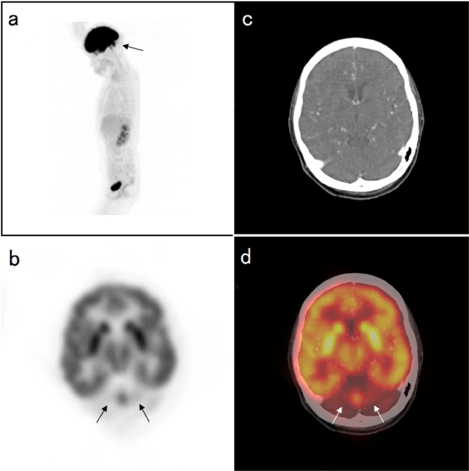
Figure 1 :
References
- Silverman DH, Mosconi L, Ercoli L, Chen W, Small GW. Positron emission tomography scans obtained for the evaluation of cognitive dysfunction. See comment in PubMed Commons below Semin Nucl Med. 2008; 38: 251-261.
- Soria N, Meli F, Blumenkrantz Y, Salas E, Turjanski A. [Crossed cerebellar diaschisis. Study of a patient by magnetic resonance imaging and positron emission tomography]. See comment in PubMed Commons below Rev Neurol. 2013; 56: 425-428.
- Akdemir ÃœÃ, Tokçaer AB, Karakuş A, Kapucu LÃ. Brain 18F-FDG PET imaging in the differential diagnosis of parkinsonism. See comment in PubMed Commons below Clin Nucl Med. 2014; 39: e220-226.
- Adams KM, Gilman S, Koeppe RA, Kluin KJ, Brunberg JA, Dede D, et al. Neuropsychological deficits are correlated with frontal hypometabolism in positron emission tomography studies of older alcoholic patients. See comment in PubMed Commons below Alcohol Clin Exp Res. 1993; 17: 205-210.
- Gilman S, Adams K, Koeppe RA, Berent S, Kluin KJ, Modell JG, et al. Cerebellar and frontal hypometabolism in alcoholic cerebellar degeneration studied with positron emission tomography. See comment in PubMed Commons below Ann Neurol. 1990; 28: 775-785.
- Theodore WH, Fishbein D, Dietz M, Baldwin P. Complex partial seizures: cerebellar metabolism. See comment in PubMed Commons below Epilepsia. 1987; 28: 319-323.
- Gilman S, Markel DS, Koeppe RA, Junck L, Kluin KJ, Gebarski SS, et al. Cerebellar and brainstem hypometabolism in olivopontocerebellar atrophy detected with positron emission tomography. See comment in PubMed Commons below Ann Neurol. 1988; 23: 223-230.
- Nawas MT, Kubal WS, Kuo PH. Decreased cerebellar activity on FDG PET/CT secondary to diffuse idiopathic cerebellar calcification. See comment in PubMed Commons below Clin Nucl Med. 2012; 37: 906-907.