Research Article
Austin J Nucl Med Radiother. 2014;1(1): 4.
Bio-Evaluation of 99mtc Resveratrol in Targeting Chemically Induced Mammary Tumors in Rats
Rozy Kamal1, Vijayta D Chadha1 and Dhawan DK1,2*
1University Institute of Emerging Areas in Science and Technology, India
2Department of Biophysics, Panjab University, India
*Corresponding author: Dhawan DK, Department of Biophysics, Center for Nuclear Medicine (UIEAST), Punjab University, Chandigarh-160014, India
Received: July 10, 2014; Accepted: August 24, 2014; Published: August 27, 2014
Abstract
Objective: The study was aimed at evaluating the efficacy of 99mTc-resveratrol in targeting chemically induced mammary tumours in rats.
Methods: Mammary tumours were induced in a group of female Sprague Dawley (S.D.) rats by giving single intra-peritoneal injection of N-Methyl Nitroso Urea (NMU) (50 mg/kg body weight in normal saline at pH 4-5). Another group of rats did not receive any treatment and served as the normal group. Bio-distribution pattern, expressed in terms of percentage specific uptake values of 99mTc-resveratrol in different organs, at 2 hours and 4 hours post i.v. injection was determined in both normal as well as NMU treated rats. Further, single photon emission computed tomography (SPECT) imaging studies were performed to substantiate the data observed in bio distribution studies.
Results: As observed from the bio distribution and SPECT imaging studies, high uptake of the radiopharmaceutical 99mTc-resveratrol was recorded in liver, spleen and kidneys, following i.v. administration 99mTc-resveratrol to rats. Percent specific uptake in mammary tumours was significantly (p≤0.05) higher than in normal mammary tissue after 2 hours of injection. Increased retention of activity in liver and spleen with significantly lowered uptake in other tissues was observed after 4 hours of injection.
Conclusion: Higher uptake at the tumour site as compared to normal mammary tissue can be viewed as an important finding in this study. However, studies are warranted to increase the uptake of 99mTc-resveratrol at the target site so as to increase its acceptability as a viable mammary cancer imaging agent.
Introduction
The role of resveratrol in chemoprevention and therapy of breast cancer in an animal model is well established [1,2]. The therapeutic role of resveratrol in breast cancer is through its agonist/antagonistic action with estrogens receptors (ER) which are over expressed in breast cancer [3-5]. Moreover, over expression of estrogenic receptors in breast cancer cells has been reported in primary and metastatic stages of cancer. Sixty percent of primary breast cancers and two thirds of advanced ER positive breast cancers are ER positive [6]. These facts make ER, a suitable target for molecular imaging using radio labelled resveratrol. Over-expression of these receptors in mammary cancer cells as compared to normal cells would lead to recognition and increased binding of resveratrol to cancer cells, than to normal cells and thus contribute towards target specificity and localization in cancer tissue. In this way, radio-imaging using radiolabel resveratrol could be used for primary diagnosis of ER positive mammary cancers, and also for evaluating treatment response following radiotherapy and chemotherapy in patients with terminal stages of breast cancer.
Despite numerous reports on cancer specificity of resveratrol in experimental [7] and clinical research, the use of polyphenol family for cancer diagnosis is still unknown and unexplored. Although, a few attempts have been made to radiolabel resveratrol with different SPECT and PET radio nuclides [8,9] but no data is available on radiolabeling with 99mTc. Moreover, data available on targeting of cancerous tissues using radioabeled resveratrol is limited. Therefore, the present study was aimed to assess the efficacy of 99mTc-resveratrol in targeting mammary tumours in experimental animals.
Materials and Methods
Chemicals
N-nitroso-N-methylurea (NMU), and sodium bicarbonate (NaHCO3), n-octanol and stannous chloride dehydrate (SnCl2.2H2O) were purchased from Sigma-Aldrich. Technetium-99m pertechnetate (99mTcO4-) was procured from Post-Graduate Institute of Medical Education and Research (PGIMER) Chandigarh, India. Instant thin layer chromatography-silica gel (ITLC-SG) strips were purchased from MERCK. Trichloroacetic acid (TCA), sodium chloride (NaCl), hydrochloric acid (HCl), sodium di-hydrogen phosphate and disodium hydrogen phosphate were purchased from SRL.
Animals
Thirty female Spargue Dawley (S.D.) rats aged 6-8 weeks and weighing 150-175g were procured from the Central Animal House, Panjab University, and Chandigarh, India. The animals were housed in polypropylene cages under hygienic conditions in the departmental animal house. Before initiating the experiments, the animals were adapted to the laboratory conditions for a week. The rats were maintained on a standard laboratory pelleted feed (Ashirwaad Industries, Tirpari, and Punjab) and water ad libitum throughout the period of experimentation.
Radiolabeling and radiochemical purity
99mTc-resveratrol was prepared by adding 3.7 MBq of 99mTcO4- to a vial containing 100μg of resveratrol (1mg/ml solution in 10% ethanol). To the mixture, 100μg of SnCl2•2H2O [1mg/ml solution in 0.1N HCl] was added and the pH was adjusted to 5-5.5 with 0.05 M NaHCO3. The reaction mixture was vortexes and kept at an ambient temperature for a sufficient time to complete the reaction. Percentage labelling of resveratrol with 99mTc was determined by carrying out chromatographic studies. Briefly, a single spot of preparation was applied on the what man no.1 paper and ITLC-SG strips of appropriate width and length (0.5 x10 cm). Strips were then placed in tubes containing acetone and a mixture of butanol: pyridine: water: glacial acetic acid in the ratio 30:20:20:6 as solvents to measure the amount of free 99mTcO4-, and reduced/hydrolyzed-99mTc (99mTc-RH) fraction, respectively in the preparation. After the solvent touched the earmarked line on the chromatographic paper, the strips were air dried, cut into 0.5 cm long sections and then counted for activity using well-type gamma-sensitive probe (Nucleonix, India).
In vitro stability
In vitro stability of 99mTc-resveratrol with time was evaluated by determining the radiochemical purity of the radio complex as a function of time till 4 hours after the addition of 99mTcO4- to the mixture. To assess the stability of the radio complex in serum, 100μl of the radio complex (3.7MBq) was incubated with 900μl of serum at 37°C up to 4 hours. The samples were then assessed for any dissociated or degraded radio complex at regular time intervals by performing ascending chromatography (What man strip as stationary and 100% acetone as mobile phase).
Plasma protein binding and partition coefficient measurement (log Po/w)
The in vitro plasma protein binding of the complex was estimated in rat plasma by incubating 100 μl of the radio complex with 900 μl of plasma at 37O°C up to 1hours. 1ml of 10% TCA was then added to the complex and the mixture was centrifuged at 2000 rpm for 5 min. Radioactivity was measured in both the precipitate and the supernatant fraction in a well type gamma counter. Protein binding of the complex was expressed as a fraction of radioactivity bound to protein as a % age of total activity.
The log Po/w values of complex was determined under physiological conditions (n-octanol/0.1M phosphate buffered saline (PBS), pH 7.4). 100μl of the preparation containing 3.7MBq of activity was added to a test tube containing 1ml of n-octanol and 0.9 ml of a PBS solution. The tube was vortexes for 1 min and then centrifuged for 5 min at 3,500 rpm. After centrifugation, 500μl of the organic phase was transferred to another tube and was further extracted with 500μl of aqueous phase, as described for the first extraction. After separation of the phases, 50μl aliquots of each phase were taken for radioactivity measurements (in duplicate). The partition coefficient (Po/w) was calculated as the ratio (activity in the n-octanol layer) / (activity in the aqueous layer) and was expressed as log Po/w.
Development of mammary cancer model
Mammary tumours were developed in rats in order to study the efficacy of the radio complex in targeting cancer tissue in vivo. Animals were divided into 2 groups. First group (n=12) i.e. normal group received normal saline once intra-peritonealy (i.p.). The animals in the second group (n=12) were given single intra-peritoneal injection of N-methyl nitroso urea (NMU) in normal saline (1.4% w/v) solution at a dose of 50 mg/kg body weight (pH 4-5). The animals were observed regularly for any palpable tumours for a total duration of 24 weeks. Tumor incidence was recorded in both normal and NMU treated group.
Histology
Histological confirmation of mammary cancer was done by hematoxylin/eosin (H/E) staining. After fixation in 10% formalin, mammary tissue sections were dehydrated using different grades of alcohol and finally imbedded in paraffin wax. The tissues were then cut into thin 5 micron sections and mounted on glass slides to be stained with H/E stain. Stained transverse sections were examined under a light microscope to look for pre-neo plastic /neo plastic changes in NMU treated tissue sections.
Blood kinetics and bio-distribution
Bio evaluation of a radio complex is important as it gives an idea about the in vivo pharmacokinetic behaviour of the radiopharmaceutical and its excretion from the body. Radioabeled conjugate, 99mTc-resveratrol, comprising of 3.7 MBq activity of 99mTc and 100 μg of resveratrol were injected intravenously (i.v.) through the tail vein of the rats. Blood samples were drawn at different time intervals by ocular vein puncture by using sterile capillary, and were counted for radioactivity. Data was expressed as percentage of total injected activity per ml of blood (% specific activity) for n=3.
For bio distribution evaluation, the animals were injected with the radiopharmaceutical and were sacrificed subsequently at desired time point under ether anaesthesia. After sacrificing the animals, organs were carefully removed and isolated to determine the bio distribution characteristics of the tracer. The organ samples were weighed and the corresponding localized radioactivity was measured using an automated well scintillation counter. The percentage of injected dose per gram of tissue (% specific activity) was calculated by comparison with standards representing the injected dose per animal.
SPECT imaging of normal and tumour bearing rats
Radionuclide imaging was performed using a SPECT camera (Siemens) to support bio distribution results. Following intravenous administration of the radio complex (3.7 MBq) to rats, 3 min static images were acquired at different time points after anesthetizing the animals using ether. All the animal experiments were conducted in compliance with the guidelines and approved protocols established by the Institutional Animal Care and Use Committee.
Statistical analysis
Experiment studying each factor was repeated three times and differences in the data were evaluated with one-way analysis of variance (ANOVA) test. Results are reported as mean ± standard deviation (S.D.). The level of significance was set at p ≤ 0.05.
Results
Radio synthesis and characterization of 99mTc-resveratrol
The synthesis of 99mTc labelled resveratrol took 30 min and the labelling efficiency was >85%. The complex was found to be stable up to 4 hours at room temperature. Plasma protein binding was observed in the range of 70-80 % and log o/w value of -0.526 ± 0. 24, was obtained.
Histological evaluation of mammary tumours
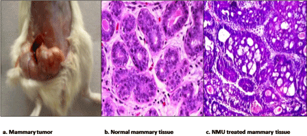
Mammary tumours were obtained in NMU treated rats with a tumour incidence of 60 % whereas no incidence was observed in the normal control group. Histological investigations revealed ductal carcinoma in situ (DCIS) in many glands of NMU treated rats (Figure 1). The mammary tissue from normal control group revealed normal histological appearance without any apparent signs of abnormality.
Figure 1 : Histological confirmation of mammary cancer in NMU treated rats. Morphology and histo architecture of NMU induced mammary tumours, a) A medium size tumour in right abdominal mammary gland. Microscopic images of transverse sections of mammary tissue (20 X) stained with H/E stain b) reveals normal histo-architecture in control group. c) Ductal carcinoma in situ (DCIS) can be observed in mammary tissue from NMU treated rats.
Blood kinetics of 99mTc-resveratrol following intravenous injection to rats.
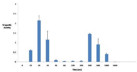
Mean ±S.D. values (n=3) are expressed as percentage of total injected activity per ml of blood (% specific activity).Rise in blood activity is observed at 20 sec and 400 sec post i.v. injection.
Figure 2 :Blood kinetics of 99mTc-resveratrol following intravenous injection to rats.
Mean ±S.D. values (n=3) are expressed as percentage of total injected activity per ml of blood (% specific activity).Rise in blood activity is observed at 20 sec and 400 sec post i.v. injection.
Bio distribution in normal and NMU treated rats bearing mammary tumours
In rats bearing mammary tumours, significantly higher uptake was observed in the mammary tumour at 2 hours after injection of the 99mTc-resveratrol molecular probe (see Table 1) when compared mammary gland uptake in normal rats. Because the molecular probe is cleared primarily through the urinary and digestive systems, the liver and bladder also showed high uptake at 2 and 4 hours. Low % specific uptake values were observed in the stomach and thyroid indicating stability of the probe in vivo. A gradual decrease in radioactivity associated with was all organs except for liver and spleen was observed with time. Further, the uptake pattern of all tissues in NMU treated rats, other than the mammary tissue was almost similar as observed in normal rats.
    Organ
    % Specific    Activity
      Normal rats
    % Specific    Activity
      NMU treated    rats


    2hours
    4 hours
    2hours
    4 hours


    Colon
    0.25    0.12
    0.018±0.002*
    0.22±0.12
    0.023±0.003*


    Heart
    0.232±0.03
    0.044±0.009*
    0.122±0.03
    0.051±0.01*


    Spleen
    4.26±3.4
    4.25±0.24
    4.16±1.4
    4.56±0.14


    Muscle
    0.032±0.02
    0.04±0.01
    0.055±0.02
    0.03±0.001


    Liver
    2.391±0.1
    2.7±0.3
    3.238±0.1
    2.3±0.9


    Kidney
    3.5±1.3
    5.9±1.4
    3.75±1.3
    6.24±1.2


    Bone
    0.28±0.03
    0.082±0.023*
    0.25±0.03
    0.09±0.03*


    Brain
    0.037±0.01
    0.003±0.001*
    0.045±0.001
    0.002±0.0001*


    Small intestine
    0.183±0.05
    0.06±0.035*
    0.135±0.01
    0.04±0.005*


    Lung
    0.35±0.12
    0.05±0.03*
    0.28±0.04
    0.05±0.02*


    Stomach
    0.027±0.02
    0.045±0.013
    0.03±0.01
    0.022±0.01


    Thyroid
    0.45±0.13
    0.11±0.05*
    0.36±0.04
    0.19±0.03*


    Mammary
    0.01±0.001
    0.009±0.001
    0.1±0.02
    0.04±0.01*    # �


    Bladder
    5.43±4.5
    5.2±3.3
    6.53±3.4
    4.7±2.3


    Mammary to Muscle    ratio
    0.31±0.06
    0.225±0.05
    1.85±0.02
    1.31±0.02*#    �
Table 1: Percent specific uptake values in different organs for 99mTc-resveratrol in normal and NMU treated rats.
Mammary cancer imaging using SPECT
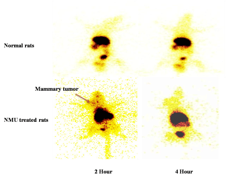
Concomitant with the bio-distribution pattern, the SPECT imaging post 2 hours i.v. administration of 99mTc-resveratrol (Figure 3) confirmed the accumulation of the activity in kidneys, liver and spleen in both normal and tumour bearing rats. After 2 hours post injection, activity observed in the mammary tumour was much higher than background activity, which later appeared diffused at 4 hours.
SPECT imaging of normal and mammary tumor bearing rats following i.v. administration of 99mTc-resveratrol.
Static (3 minutes), SPECT images were obtained at 2hours and 4 hours following intravenous administration of 99mTc-resveratrol (3.7MBq) to pre anesthised (diethyl ether) rats, both normal and those bearing mammary tumors.
Figure 3 :SPECT imaging of normal and mammary tumor bearing rats following i.v. administration of 99mTc-resveratrol.
Static (3 minutes), SPECT images were obtained at 2hours and 4 hours following intravenous administration of 99mTc-resveratrol (3.7MBq) to pre anesthised (diethyl ether) rats, both normal and those bearing mammary tumors.
Discussion
Resveratrol was successfully labelled with 99mTc with good labelling efficiency. Following i.v. injection of 99mTc-resveratrol to normal and NMU treated rats, low % specific uptake values were observed in the stomach and thyroid indicating that the radionuclide (99mTc) did not separate from the ligand (resveratrol) and that the radio complex was stable in vivo. Also, maximum accumulation of the activity in terms of percentage activity per gram of tissue at 2 hours post injection was observed in kidneys followed by spleen and liver indicating both, renal and hepatobilliary route of excretion for the radio-complex. The radio complex seemed to be excreted out mainly via kidneys since high bladder activity was observed after 4 hours post injection. High uptake values in spleen and liver even after 4 hours suggests accumulation of the radiopharmaceutical in these tissues either because of slow excretion via hepato-billiary route or because of reticule-endothelial uptake. Particle aggregation with time due to low water solubility of the complex is understandably the reason for entrapment of these aggregates by the reticule-endothelial cells [10]. However, particle aggregation does not seem to occur until 2 hours post injection since distribution to tissues other than spleen and liver is also observed.
The rate of elimination of the radio-complex from the body is associated to its binding to plasma proteins. High protein binding (70-80%) observed for 99mTc-resveratrol suggests that the rate of elimination of the radiopharmaceutical from the body would be slow. Supporting results were obtained in the blood kinetic profile of the radio complex (Figure 2), which depicts a two-compartment pharmacokinetic model, demonstrating a slow decrease in blood activity after 400 sec (~7 min) post injection. The, abrupt fall in blood activity, observed after 15-20 sec of i.v. administration is due to initial uptake by the central compartment system comprising of liver and other highly perfuse organs [11].
Bio-distribution after 2 hours of administration of radiopharmaceutical, revealed appreciably higher % age specific uptake (p≤0.05) in mammary tumours than in normal mammary tissue. The tumour to muscle ratio in NMU treated rats was significantly higher than normal control values both at 2 hours and 4 hours, thereby indicating the specificity of 99mTc-resveratrol for proliferating cells.
Conclusion
The present study evaluated the tumour localization potential of 99mTc-resveratrol following NMU induced mammary gland carcinogenesis in rats. Higher accumulation of the tracer in cancerous tissue than normal breast tissue is an important finding of the present study. However the accumulation of the complex in the reticulo-endothelial organs, its low water solubility and high protein binding decreases its availability at the tumour site. Therefore, future studies are warranted to increase the uptake of 99mTc-resveratrol at the target site, in order to improve image quality and hence diagnostic efficacy of the radiopharmaceutical.
Acknowledgement
The authors would like to thank the staff of the department of Nuclear Medicine, Oswal Multispecialty Hospital, Ludhiana and Post Graduate Institute for Medical Education and Research (PGIMER) Chandigarh, India for providing necessary material and equipment for the study. The financial support by the Department of Science and Technology (DST), New Delhi is gratefully acknowledged.
References
- Garvin S, Ollinger K, Dabrosin C. Resveratrol induces apoptosis and inhibits angiogenesis in human breast cancer xenografts in vivo. Cancer Lett. 2006; 231: 113-122.
- Provinciali M, Re F, Donnini A, Orlando F, Bartozzi B, Di Stasio G, et al. Effect of resveratrol on the development of spontaneous mammary tumors in HER-2/neu transgenic mice. Int J Cancer. 2005; 115: 36-45.
- Lippman ME, Bolan G. Oestrogen-responsive human breast cancer in long term tissue culture. Nature. 1975; 256: 592-593.
- McGuire WL. Prognostic factors for recurrence and survival in human breast cancer. Breast Cancer Res Treat. 1987; 10: 5-9.
- Hayashi SI, Eguchi H, Tanimoto K, Yoshida T, Omoto Y, Inoue A, et al. The expression and function of estrogen receptor alpha and beta in human breast cancer and its clinical application. Endocr Relat Cancer. 2003; 10: 193-202.
- Hanstein B, Djahansouzi S, Dall P, Beckmann MW, Bender HG. Insights into the molecular biology of the estrogen receptor define novel therapeutic targets for breast cancer. Eur J Endocrinol. 2004; 150: 243-255.
- Chowdhury SA, Kishino K, Satoh R, Hashimoto K, Kikuchi H, Nishikawa H, et al. Tumor-specificity and apoptosis-inducing activity of stilbenes and flavonoids. Anticancer Res. 2005; 25: 2055-2063.
- Kim SK, Lee WS, Han SJ, Kim EJ, Mohammed I. El-Gamal MI, et al. Radiosynthesis and Biodistribution of an 125 I-labeled Resveratrol Derivative Bull. Korean Chem Soc. 2012; 33: 489.
- Gester S, Wuest F, Pawelke B, Bergmann R, Pietzsch J. Synthesis and biodistribution of an 18F-labelled resveratrol derivative for small animal positron emission tomography. Amino Acids. 2005; 29: 415-428.
- Dai WG. In vitro methods to assess drug precipitation. Int J Pharm. 2010; 393: 1-16.
- Winter ME. Basic Clinical Pharmacokinetics. 4th edn. Philadelphia: Lippincott Williams and Wilkins. 2003.


