
Case Report
Austin J Nucl Med Radiother. 2014;1(2): 2.
Scintigraphic and Radiologic Features of Erdheim-Chester Disease: A Case Report
Miguel Hernandez Pampaloni* and Evan Sirc
Department of Radiology and Biomedical Imaging, University of California, USA
*Corresponding author: Miguel Hernandez Pampaloni, Department of Radiology and Biomedical Imaging, University of California, 505 Parnassus Avenue, M-396, San Francisco, CA, 94143, USA
Received: October 30, 2014; Accepted: December 05, 2014; Published: December 08, 2014
Abstract
Erdheim-Chester disease is a rare, non-Langerhans histiocytosis with a highly variable set of systemic manifestations. The disease affects a wide range of ages with a slight male predominance. Radiologically, skeletal abnormalities are most common, including bilateral, symmetric osteosclerosis in the diaphyses of long bones. Imaging of the face can show an extensive retroorbital soft tissue masses. While the anatomic imaging characteristics of Erdheim-Chester have been well-described in previous publications, much less has been reported regarding the scintigraphic manifestations of the disease. This report describes an interesting case with dramatic bone scan and whole-body F-18 FDG PET abnormalities which are correlated with findings on abdominal CT and chest MRI. Erdheim-Chester disease is a rare, non-Langerhans histiocytosis with a highly variable set of systemic manifestations.
Keywords: Scintigraphic; Erdheim-chester disease; Osteosclerosis; Histiocytosis
Case Report
Erdheim-Chester Disease (ECD) is a rare form of non Langerhans' cell histiocytosis. Individuals affected by this disease are typically adults between their 5th and 7th decades of life. The multi systemic form of ECD is associated with significant morbidity, which may arise due to histiocytic infiltration of critical organ systems. The most common presenting symptom of ECD is bone pain. The etiology of ECD is unknown yet thought to be associated with an intense immune response. Bilateral symmetric increased tracer uptake on 99mTc bone scintigraphy affecting the periarticular regions of the long bones is highly suggestive of ECD [1]. However, definite diagnosis of ECD is established only once CD68(+), CD1a(-) histiocytes are identified within a biopsy specimen Clinically, patients may present with bone pain, diabetes insipidus, exophthalmos, neurological signs, and xanthomas, as well as non-specific general symptoms such as fever, weight loss, and weakness. Given its rarity, it is diagnostically elusive and requires a high level of clinical suspicion. Therapeutically, it is of limited alternatives. Currently, interferon-alpha is the most extensively studied agent in the treatment of ECD and serves as the first line of treatment. Treatment with other agents is based on anecdotal case reports and on the basis of biological rationale.
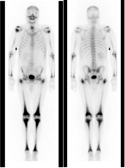
Figure 1 : Anterior and posterior whole body images from a Tc-99m MDP bone scan demonstrating symmetric increased uptake throughout the metapyses and diaphyses of multiple long bones, most prominently about the distal femurs and proximal tibiae.
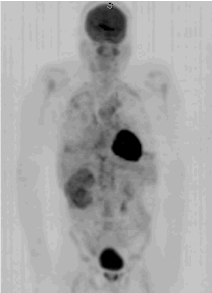
Figure 2: MIP image from a whole body F-18 FDG PET scan from vertex to thighs demonstrating increased bone marrow uptake in the appendicular skeleton, increased uptake in the chest corresponding to periaortic, pericardial, and pleural soft tissue infiltration, and uptake throughout and enlarged right kidney and perirenal soft tissue.
Radiologically, skeletal abnormalities are most common, including bilateral, symmetric osteosclerosis in the diaphyses of long bones. Bilateral symmetric increased tracer uptake on 99mTc bone scintigraphy affecting the periarticular regions of the long bones is highly suggestive of ECD. Imaging of the face can show an extensive retroorbital soft tissue masses. In the chest, pleural and pericardial soft tissue infiltration may be demonstrated, with interstitial lung disease seen in the form of interlobular septal thickening that may mimic hydrostatic pulmonary edema. Abdominal imaging may show renal enlargement with peri-renal soft tissue infiltration, splenomegaly, and retroperitoneal fibrosis. While the anatomic imaging characteristics of Erdheim-Chester have been well-described in previous publications, much less has been reported regarding the scintigraphic manifestations of the disease.
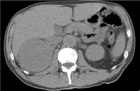
Figure 3: CT scan of the abdomen without contrast-material enhancement demonstrates an asymmetrically enlarged right kidney with perirenal soft tissue infiltration.
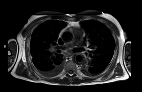
Figure 4: T1-weighted black blood MRI of the chest demonstrating bilateral pleural effusions as well as circumferential soft tissue infiltration surrounding the descending thoracic aorta.
Discussion
This report describes an interesting case with dramatic bone scan and whole-body F-18 FDG PET abnormalities which are correlated with findings on abdominal CT and chest MRI. We describe a patient showing a variety of imaging characteristics that may be interesting to correlate with this disease. The whole-body bone scintigraphy depicts the classic symmetric increased uptake throughout the metapyses and diaphyses of multiple long bones, most prominently about the distal femurs and proximal tibiae [2]. Interestingly whole body F-18 FDG PET scan from vertex to thighs demonstrates a wide variety of abnormal areas of hypermetabolism, which have not been describe in the literature. These include increased bone marrow uptake in the appendicular skeleton, increased uptake in the chest corresponding to periaortic, pericardial, and pleural soft tissue infiltration, and uptake throughout and enlarged right kidney and perirenal soft tissue. These findings seems to suggest that F-18 FDG PET is a valid and useful imaging modality to diagnosis patients with this rare disease, as it can provide relevant information on the skeleton and soft tissues involvement, reflecting the multiorganic aspect of this entity.
References
- Veyssier-Belot C, Cacoub P, Caparros-Lefebvre D, Wechsler J, Brun B, Remy M, et al. Erdheim-Chester disease clinical and radiologic characteristics of 59 cases. Medicine (Baltimore). 1996;75:157-169.
- Dion E, Graef C, Miquel A, Haroche J, Wechsler B, Amoura Z, et al. Bone involvement in Erdheim-Chester disease: imaging findings including periostitis and partial epiphyseal involvement. Radiology. 2006; 238:63.