
Case Report
Ann Nutr Disord & Ther. 2018; 5(1): 1052.
Nail Layering Phenomenon and Its Neutraceutic Therapy
Lee BY*
Department of Pediatrics/Biochemical Genetics, Louisiana State University Health Sciences Center in Shreveport, USA
*Corresponding author: Judith Kimiywe, Kenyatta University, Department of Food, Nutrition and Dietetics, Nairobi, Kenya
Received: February 15, 2018; Accepted: March 07, 2018; Published: March 14, 2018
Abstract
Nail layering phenomenon was common among the slavery laborers. The nail plate separated into two layers due to destruction of the sealing parts, hyponychium and the onychodermal band. The upper layer became very thin, brittle, flat, or even spoon-like, while the lower layer was soft, thick and flattened like a dirty rubber lump inseparable with the finger tissue underneath. The junction of the two separated layers was about 2-4 mm from the finger tip. In advanced cases, it might be about 7mm or more. The upper layer was very vulnerable to daily practice. Once it was accidentally removed, the lower layer revealed as grayish-yellow colored multiple, longitudinal, parallel ridges and grooves.
It was very rare in civil cases. However, an 85-year female was found to have severe nail layering in 10 fingers over 64 years. Her upper layer became very hard and thick, while the lower layer was absent and replaced with an empty cavity in each finger. After parenteral thiamin and VB complex for 7 weeks, the lower soft layer regenerated gradually to fulfill the empty space in all fingers. After 15 weeks of therapy, the finger nail became normal except for the thumbs.
Keywords: Concentration camp syndromes; Intra-nail hemorrhage Malnutrition; Nail-layering; Nationwide Hunger; Thiamin deficiency
Case Presentation
A great variety of nail abnormalities were observed in a labor camp 1958-1962 before and during Nationwide Hunger in China [1,2]. One of them was nail layering, about 75% in occurrence. Under severe malnutrition, nail plate was observed to have layering after the destruction of its normal tiny parts, hyponychium and onychodermal band underneath [3]. The hyponychium is a strip of epithelium beneath the nail plate. It seals the subungual space and allows the nail plate to detach from the nail bed. The onychodermal band seals the nail plate and the hyponychium. These two parts seal the extended free edge of the nail plate and protecting the nail bed. When they were destroyed, the sealed portion of the finger tip and the lower surface of the free edge of nail plate exposed to the environment and lost protection. Then the nail separated into two layers with their junction about 2-4 mm away from the very end of the finger tip as seen in (Figure 1).
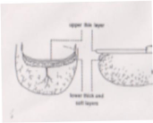
    Figure 1:  Early stage of upper and lower layers separation of the nail. Finger
fissures were commonly seen in slavery laborers. (Permission from Journal
of American College of Nutrition).
The upper layer became very thin, brittle, flattened or even spoonlike (koilonyechia). The lower layer was a soft, flattened mass like dirty rubber lump inseparable from underlying finger tissue. In advanced cases, the junction might reach the transverse middle portion of the nail, about 7mm away from the very end of the finger tip. Upper layer like this was extremely vulnerable to daily wear and tear. Once it was broken by accident, the lower layer underneath revealed as grayishyellow colored multiple, longitudinal, parallel ridges and grooves, not a lump. This should be the epidermis attached to dermis, called matrix crestse (cristae matricis unguis), on the nail bed. Epidermis and particularly its underlying dermis might deeply involve in the pathogenesis of some nail abnormalities, such as surface longitudinal ridges, dark band, and especially the terrible intra-nail hemorrhage [1]. Unfortunately, no picture could be taken and no way to report in that terrible years.
In civil cases, nail layering including finger fissures as in the slavery labors was very rare. However, a severe case was found with thick and hard upper layer but no lower layer, which became an empty space underneath for 64 years. Her unique abnormal nails were normalized during thiamin therapy for her coronary insufficiency as reported in the following.
Case Description
Patient WSF was an 85 years old housewife in Tianjin west. For many years, she had visited multiple top hospitals in Tianjin for her coronary insufficiency with frequent breathlessness, palpitation, chest tightness or pain, leg edema, and general lassitude. Medication included nicergline, isosorbide mononitrite, aspirin, aminophylline and occasionally some others. Clinical signs and symptoms had not been improved but persisted. She then visited this author’s tele-Clinic on October 10, 2016 for cardiac problem.
All her above medicine were totally canceled and only thiamin 300mg plus VB complex one ampule (containing VB1 10mg, VB2 2mg, niacinamide 30mg, VB6 2mg, pantothenic acid 1mg) twice and occasionally once a day were intra-muscularly injected. She became completely free from cardiac signs after 3 months. Very gladly, her unique nail abnormality for 64 years was observed recovering from the 7th week of thiamin therapy. Therefore, nutrition therapy continued for totally 6 months.
When she was 21 years old, every day she worked outside in the cold winter without hand gloves to pull and push a heavy stone roller to press reed-like calamus for weaving commercial products. All her fingers were severely injured with cold. At the beginning, the nail on each finger became very thin, brittle, and flattened or spoon-like. An empty space of the nail size and about 2-3 mm in thickness developed under the brittle thin nail. However, unlike that in slave cases, the upper layer of her 10 nails gradually became very thick and hard without flexibility through 64 years. It was very difficult in manicure as a piece of hard clay. Simultaneously, the empty space between each affected nail and the nail bed persisted also for 64 years. There was no local sign except for uncomfortable feeling and dirty contamination in daily household practice. Hundreds of doctors had never paid any attention on her nails for so many hospitals and years. However, the associate in the tele-Clinic knew according to Lee’s book [2] and took Figures 2 at the same day of her visitation. The empty space could be obviously seen under each finger nail as seen in the three photos. However, the small finger and thumb couldn’t be included within one picture. Figure 3B demonstrated that the empty spaces of the thumbs was heavily contaminated with dirties (Figure 2).
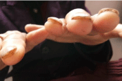
    Figure 2:  The 10 nails of both hands separated from the nail beds with an
empty cavity. The thumb nails were similarly and severely contaminated as
in Figure 3B.
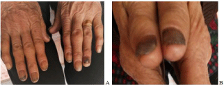
    Figure 3:  A. The lower soft layer regenerated with a preceding dirty portion in
black color after 7 weeks of therapy. B. Lower layer regeneration hadn’t been
observed in the thumb nails.
After 7 weeks of nutrition therapy, cardiac signs greatly improved and the regeneration of the lower layer of the nails started as seen in figures 3A & 3B. Physiologically, the lower layer should generate also from the germinal matrix and the sterile matrix on the nail bed. Germinal matrix locates under the lunula, which is the whitish crescent-shaped base of the nail and close to the nail fold [3-5]. The matrix grew forward inside the empty space under the upper layer and pushed the contaminated content toward the end of the finger tip. The lower layer generation initiated firstly at the small finger nail probably due to its smaller size and thinner plate. Then the middle, index and ring finger nails followed, while the thumb recovered very sluggish and incomplete (Figure 3A&B).
After another 8 weeks of thiamin injection, eight finger nails became normal while the two thumbs nails greatly improved but incomplete as seen in figure 4B. There was a small breaking defect in the right thumb nail tip due to falling down in January 2017. Totally, the patient received parenteral thiamin for six month. Both cardiac signs and the 8 finger nails were normal but the thumb nails improved very sluggish and incomplete (Figure 4A&B).
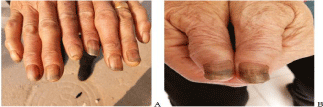
    Figure 4:  A. Eight nails were completely normalized after 15 weeks of
therapy. B. The thumb nails improved greatly but not completely. The small
broken injury at the tip of the right thumb nail was caused by falling down on
the ground.
Discussion
1. This case was quite different from onycholysis [5], in which, there is distal separation of the nail plate from the nail bed. In this case, the nail plate was in its proper location over the nail bed but separated into 2 layers. Unlike milder cases, this patient’s lower layer was absent and replaced by an empty space under each affected nail. After thiamin therapy, the lower layer regenerated. It confirmed that nail layering phenomena observed among the slavery laborers was true and correct. It might be a new clinical entitle and this case might be the first one could be reported with nail layering phenomena because no pictures could be taken in the labor camp.
We called the layers as upper and lower ones before their proper nomenclature available. Her lower layer regenerated in the following order: small finger in first, and then the middle, the index and the ring fingers. Generation of the lower layer of the thumb nails was very sluggish and incomplete probably due to its heavy thickness and the patient old age.
Definitely, the nail sealing parts, hyponychium and onychodermal band, and the nail matrix were severely injured due to cold. Destruction of hyponychium and onychodermal band opened the detrimental way for nail matrix. The germinal matrix (intermediate matrix) became dysfunctional in producing hardened, flat, translucent, non-living, keratin nail cells. Its distal extension, the sterile matrix on the nail bed, totally failed leading to the formation of empty cavity. This confirmed that, physical factor, such as severe low environmental temperature, might also injure the sealing parts and matrix of the nail.
2. Parenteral thiamin was exceedingly effective for nail layering both in slavery laborers and this civil case. The vitamin B complex was used to minimizing the possible imbalance among vitamin B group when thiamin was too much. Based on clinical experiences in the labor camp, vitamin D and vitamin B12 might be only secondary important, however, no clinical study to compare. Interpretation of biochemical mechanism of thiamin therapy is beyond the scope of this paper.
3. Long term parenteral thiamin therapy was very effective for coronary insufficiency. It should be discussed in another paper.
Acknowledgment
The author is a medical volunteer to help patient in the west suburban area of Tianjin City in China through international communication. He is deeply grateful to the associate, Ms. WANG Yanrong, of the tele-Clinic in Tianjin west for her taking the photos.
References
- Lee BY, Hogan DJ, Ursine S, Yanamandra K, Bocchini JA. Personal observation of skin disorders in malnutrition. Clinics in Dermatology. 2006; 24: 222-227.
- Lee BY. New Diseases caused by Hunger and Fatigue. KF Publishing Company Group, USA (Eng & Chn). 2009; 6: 52-53
- Schoon D, Seidel A. Nail Anatomy--Different parts of fingernail. 2014.
- Philips BZ, Gest TR, Schmidt ST, Bhatt RA. Nail Anatomy. Updated. 2013.
- Fawcett RS, Linford S, Stulber DL. Nail abnormalities: Clues to systemic disease. Am Fam Physician. 2004; 69: 1417-1424.