
Case Report
Austin J Obstet Gynecol. 2014;1(5): 2.
A Rare Cause of Developmental Discrepancy in a Dizygotic Twin Pregnancy: Giant Chorangioma
Simavli S1*, Gok S1, Karabulut A1, Oztekin O1 and Akbulut M2
1Pamukkale University Medical School, Department of Obstetrics and Gynecology, Denizli, Turkey
2Pamukkale University Medical School, Department of Pathology, Denizli, Turkey
*Corresponding author: Simavli S, MD, Pamukkale Üniversitesi Kadin Hastaliklari ve Dogum Anabilim Dali Kinikli, 20000 Denizli-Turkey
Received: July 06, 2014; Accepted: September 05, 2014; Published: September 30, 2014
Abstract
Chorioangioma is a benign tumor of the placenta showing similar histologic pattern with hemangiomas. We aimed to present a 9x8x4 cm sized chorioangioma causing twin to twin transfusion like syndrome in a dizygotic twin pregnancy. A 30 weeks old dichorionic-diamniotic twin pregnancy with a 7x5 cm hypo-echoic mass at the placental site of one fetus who revealed a normal development, and the other having intrauterine growth retardation, was admitted to our clinic. Preeclampsia was developed in clinical follow-up at 34th weeks of pregnancy and emergency cesarean section was performed due to fetal distress. Chorioangioma was detected on the placenta of the normal fetus. Pathologic examination of placenta revealed a 9x8x4 cm mass macroscopically with a diagnosis of chorioangioma microscopically. Chorioangiomas may cause twin to twin transfusion like syndrome in dizygotic twins and may result in various spectrum of problems in fetus depending on the size and the texture.
Keywords: Chorioangioma; Twin pregnancy; Intrauterine growth restriction
Case Report
A 23 year old, G1 P0, dichorionic-diamniotic twin pregnancy followed in our clinic until 30 weeks of pregnancy. In that time, sonographic examination revealed intrauterine growth restriction (IUGR) in one fetus and 7x5 cm hypoechoic mass at the placental site of the fetus with normal development. Three weeks of difference was detected between abdominal circumferences (AC) of the fetuses (AC measurements were compatible with 27 and 30 weeks). Amniotic fluid indexes and Doppler indices were within normal limits for both. Patient was followed in outpatient clinics with 10 day intervals. At 34 weeks of gestation, she applied with uterine contractions and 4 cm cervical dilatation. Blood pressure was 160/90 mmHg, and 1+ proteinuria was present in the urine test. Sonographic measurements revealed normal development for the first fetus with a 8x7 cm hypoechoic mass in placenta with slight polyhydramnios, and IUGR for the second one (AC 27 week) with an amniotic fluid index of 8 cm. While umblical arter doppler analysis was normal for the first fetus (S/D:1.93, PI:0.62, RI:0.48), it was abnormal for the second fetus (S/D:3.75, PI:1.16, RI:0.73). She underwent emergency cesarean section due to fetal distress, and 1050 gr female and 2500 gr male babies were born with a 5 minute APGAR scores of 8 and 9 respectively. Pathologic examination of placenta showed a 9x8x4 cm mass macroscopically (Figure 1) with a diagnosis of chorioangioma microscopically (Figure 2).
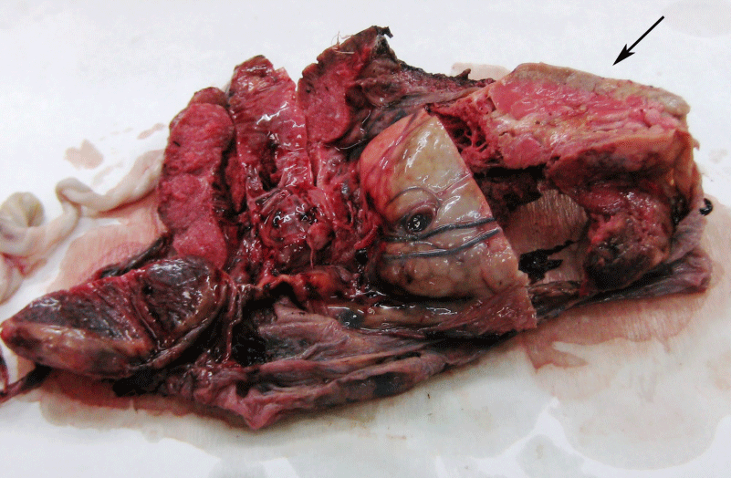
Figure 1:The raised red, round solid lesion seen through the fetal surface is a typical chorioangioma (arrow).
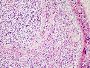
Figure 2: Histology is that of a typical hemangioma, with numerous small sized vessels (H&E,x200).
Discussion/Conclusion
Chorioangioma is a benign tumor of the placenta, and shows similar histological pattern with hemangiomas seen in other body sites [1]. They are originated from chorionic mesenchyme and seen in one in every 100 to 150 placentas [2-4]. They usually small sized and asymptomatic [5,6] Chorioangiomas are thought to be originated from hamartomatous differentiation or hyperplasia of the placental tissue. They usually localized in badly perfused are as of the placenta such as marginal or subchorionic are as and twin pregnancies [7]. It was also localized in marginal site in our case. The ones more than 5 cm in diameter are seen with an incidence of 1/3500-9000 and may be associated with polyhidramnios, preeclampsia, ante partum bleeding, placenta previa, premature birth, thrombocytopenia in mother and asphyxia, disseminated intravascular coagulation (DIC), anemia, IUGR, hydrops, cardiomegaly and neonatal cardiac failure in the fetus [2-5, 8-11]. In our case, besides IUGR both preterm birth and pre-eclampsia were developed at 34th weeks of pregnancy. While the shunts between the vessels in chorioangioma were hold responsible for the cardiac hypertrophy and the failure, infarct occurred in the tumoral tissue may cause DIC [5,12]. In the literature, IUGR was usually reported in fetus with chorioangiomas in twin pregnancies [13]. Small infarcts impairing nutritional exchange and oxygenation of the fetus are mostly accused for development of IUGR. [13-15]. However, in our case, IUGR was seen in the fetus without chorioangioma which cannot be explainable by infarct theory. We supposed that arteriovenous shunts previously described in chorioangiomas [5,12], may result in intense blooding of the placenta and may cause twin to twin transfusion like syndrome with development of IUGR in fetus with normal placenta. Therefore clinical spectrum may vary in twin pregnancies with chorioangiomas depending on the size and structure of the lesion.
In conclusion, chorioangiomas may result in various problems in the fetus depending on the size and the structure of the lesion and the fetus with normal placenta even is under risk in twin pregnancies. These problems must be kept in mind with a close and cautious follow-up of the patient.
References
- Rushton DI. Pathology of placenta. In: Wigglesworth JS, Singer DB, editors. Textbook of Fetal and Perinatal Pathology. First edition. Boston: Blackwell Scientific Publications. 1999: 161-219.
- Demiriz M, Tunca Y, Ozcan A, Celasun B, Finci R. Placental chorioangioma associated with fetal cardiac complication. Acta Obstet Gynecol Scand. 1997; 76: 708-709.
- Kohler HG, Iqbal N, Jenkins DM. Chorionic haemangiomata and abruptio placentae. Case report and review. Br J Obstet Gynaecol. 1976; 83: 667-670.
- Mancuso A, D'Anna R, Corrado F, Cannata ML. Large placental chorioangioma. Acta Obstet Gynecol Scand. 2001; 80: 965-966.
- Mochizuki T, Nishiguchi T, Ito I, Imai M, Isoda H, Masui T, et al. Case report. Antenatal diagnosis of chorioangioma of the placenta: MR features. J Comput Assist Tomogr. 1996; 20: 413-416.
- Mesia AF, Mo P, Ylagan LR. Atypical cellular chorangioma. Arch Pathol Lab Med. 1999; 123: 536-538.
- Ogino S, Redline RW. Villous capillary lesions of the placenta: distinctions between chorangioma, chorangiomatosis, and chorangiosis. Hum Pathol. 2000; 31: 945-954.
- D'Souza D, Olah KS. Infarction of a placental chorioangioma mimicking placental abruption. J Obstet Gynaecol. 1999; 19: 421-422.
- Bauer CR, Fojaco RM, Bancalari E, Fernandez-Rocha L. Microangiopathic hemolytic anemia and thrombocytopenia in a neonate associated with a large placental chorioangioma. Pediatrics. 1978; 62: 574-577.
- Hirata GI, Masaki DI, O'Toole M, Medearis AL, Platt LD. Color flow mapping and Doppler velocimetry in the diagnosis and management of a placental chorioangioma associated with nonimmune fetal hydrops. Obstet Gynecol. 1993; 81: 850-852.
- King CR, Lovrien EW. Chorioangioma of the placenta and intrauterine growth failure. J Pediatr. 1978; 93: 1027-1028.
- Kabukçuoglu S, Öner Ü, Tel N, Ilgici N, Sener T. Placental Chorangiomas. Turk Patoloji Derg 2001; 17: 34-37.
- Salafia CM, Papek EJ. Placenta . In: Damjanov I, Linder J, editors. Anderson's Pathology. Tenth edition. St. Louis: Mosby. 1996; 2310-2353.
- Manning AF. Intrauterine Growth Retardation. Diagnosis. Prognostication and Management Based on Ultrasound Methods. In: Fleisher, Manning, Jeanty, Romero, editors. Sonography in Obstetrics and Gynecology. Fifth edition. Stamford: Appleton and Lange. 1996; 517-37.
- Pardi G, Marconi AM, Cetin I. Pathophysiology of intrauterine growth retardation: role of the placenta. Acta Paediatr Suppl. 1997; 423: 170-172.