1Department of Obstetrics and Gynecology, Texas Tech University Health Sciences Center School of Medicine, USA
*Corresponding author: Kauffman RP, Department of Obstetrics and Gynecology, Texas Tech University Health Sciences Center School of Medicine, 1400 S. Coulter Drive, Amarillo, Texas 79109, USA
Received: June 05, 2014; Accepted: September 15, 2014; Published: November 04, 2014
Citation: Kessler LC and Kauffman RP. Sigmoid Volvulus Presenting During Late Pregnancy: A Report of Two Cases from a Single Institution. Austin J Obstet Gynecol. 2014;1(6): 3. ISSN:2378-1386
Acute bowel obstruction is a rare but serious complication of pregnancy with substantial maternal and fetal morbidity and mortality. Early detection improves maternal and fetal outcomes and a high index of suspicion is necessary since many normal pregnancy-related symptoms may be similar, albeit milder, than those encountered in acute obstruction. Two cases of colon obstruction secondary to rectosigmoid volvulus occurring during the third trimester of pregnancy are presented. Unfortunately, one neonatal death occurred secondary to postoperative hemorrhage, fetoplacental hypoperfusion, and hypoxic encephalopathy. Each case of colon obstruction was readily identified by plain abdominal radiograph. Conventional imaging studies (particularly single-shot abdominal radiographs) do not pose a measurable teratogenic risk to the developing fetus and physicians should not hesitate to order appropriate imaging studies when acute obstruction is considered in the differential diagnosis.
Keywords: Sigmoid volvulus; Bowel obstruction; Pregnancy
Intestinal obstruction during pregnancy is a rare but potentially devastating condition with a reported incidence of 1:1500 to 1:66,431 pregnancies [1,2]. Early intervention and treatment significantly diminishes maternal and fetal morbidity and mortality [2,3]. Symptoms associated with obstruction include vomiting, constipation, abdominal pain, distention, and leukocytosis [4]. Since some or even all of these symptoms may be encountered during normal pregnancy, the recognition of obstruction may be challenging. We present two cases of sigmoid volvulus occurring during late pregnancy from a single institution. One case ended with a neonatal death.
A 21-year old G4P3 presented to Labor and Delivery at 30 gestational weeks complaining of nausea, vomiting, and severe abdominal pain for 24 hours. She denied flatus or bowel movement since the onset of acute pain, although during the course of pregnancy, she had experienced one or two loose bowel movements daily. The pain was colicky and unremitting. She offered no history of prior abdominal surgery. An underlying eating disorder was suspected based on her low prepregnancy BMI (<17 kg/m2), but the patient steadfastly denied behaviors consistent with anorexia nervosa and bulimia.
The patient had been seen four times during the prior month with similar although milder complaints of nausea, vomiting, and poorly localized abdominal pain. Prior examinations and clinical assessments did not suggest an acute abdomen, and no imaging studies were ordered other than routine obstetrical ultrasonography that did not indicate an obstetrical etiology to her complaints. Complete Blood Counts (CBC) and blood chemistries were consistently normal. Each time, her symptoms improved with conservative treatment (antiemetics and intravenous isotonic crystalloid) and the patient was discharged home within a few hours, tolerating a normal diet. The recorded discharge diagnoses included "uterine contractions" and "discomforts of pregnancy".
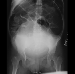
Initial vital signs included temperature 36.9°C, blood pressure 129/86, and pulse 79. Her external Fetal Heart Tone (FHT) tracing was category 1 with no significant uterine activity. Her cervix was dilated to 1cm with 30% effacement. She appeared thin, pale, and acutely ill. A large peristaltic loop of bowel was noted through the abdominal wall skin. No rectal exam was recorded. Ultrasound revealed a singleton fetus, posterior placenta without retroplacental hemorrhage and normal amniotic fluid index. Her biophysical profile score was 10/10. Large dilated loops of maternal bowel were observed at ultrasound. Single view flat plate abdominal X-ray revealed multiple dilated loops of small bowel and dilated colon segment, consistent with large bowel obstruction (Figure 1).
Laboratory data returned with a White Blood Cell (wbc) count of 10,300/μL, hemoglobin 103 g/L, platelets 274,000/μL, potassium 2.7mmol/L, and normal liver enzymes and serum amylase. Urinalysis was significant only for large ketones.
A nasogastric tube was placed and general surgery consultation was obtained. Intraoperative findings were consistent with complete sigmoid volvulus. She underwent an exploratory laparotomy, partial colon resection and temporary diverting colostomy with a Hartmann's pouch. No evidence of bowel necrosis was noted grossly or by histologic analysis.
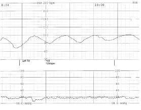
The fetus was monitored by continuous Doppler postoperatively. At the fourth postoperative hour, the fetus developed an acute and persistent sinusoidal tracing (Figure 2). Maternal urine output and blood pressure remained normal, but maternal tachycardia was noted just prior to emergency cesarean delivery for fetal indications. A live female infant was delivered weighing 1340 g with Apgars 1 at one minute, 5 at five minutes, and 5 at ten minutes with a cord arterial pH of 6.92. The infant was admitted to the neonatal ICU.
At the time of cesarean section, a significant intra-abdominal hemoperitoneum was encountered (approximately 1000mL) with arterial bleeding emanating from the proximal colostomy loop. The general surgeon was reconsulted and bleeding was controlled. The mother was resuscitated with packed red cells intraoperatively and transferred to the ICU after delivery. She recovered without incident and eventually underwent successful colon reanastomosis. Unfortunately, the neonate died on day of life 11 due to hypoxic encephalopathy and complications of prematurity.
A 25 year old G3P1A1 presented to the emergency department at 38 weeks' gestation with progressive nausea, abdominal pain, distention, and bilious emesis for five days. The patient's surgical history was significant for prior low vertical cesarean delivery at 32 gestational weeks secondary to severe pre-eclampsia complicated by a central placenta previa.
The patient stated that she had not experienced flatus or a bowel movement for a week except after a self-administered enema two days before. Under normal circumstances, she had daily bowel movements. Intermittent nausea and vomiting for 5 days had given way to intolerance to any oral intake for 24 hours prior to presentation.
The patient was a febrile (37.2°C) and normotensive with a resting pulse of 112. External fetal Doppler revealed a category 1 fetal heart rate tracing. CBC revealed leukocytosis (15,100/μL), hemoglobin 114 g/L, and normal platelet count. Her serum potassium was low (2.9 mmol/L) with a modestly elevated creatinine level (90 μmol/L).
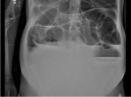
Physical exam demonstrated a markedly distended, tympanic abdomen with absent bowel sounds. No rebound tenderness was present although she had generalized abdominal tenderness. The rectal vault was empty. An upright abdominal radiograph revealed dilated air-filled loops of bowel in the upper and mid-abdomen with multiple air-fluid levels identified. The transverse colon measured 11 centimeters in greatest dimension suggestive of a distal obstruction (Figure 3). A nasogastric tube was placed and general surgery was consulted immediately.
The patient underwent repeat low transverse cesarean delivery, exploratory laparotomy, sigmoidectomy, and primary anastomosis. Repeat cesarean section was necessary to fully access the sigmoid colon. Intraoperative findings included a complete sigmoid volvulus with viable bowel. Post-operatively, both patient and infant did well.
Volvulus of the sigmoid colon occurs when a segment of the rectosigmoid twists on itself causing partial or complete obstruction. The first account of volvulus during pregnancy was published in 1885, and fewer than 100 cases have been reported in the English language literature worldwide since the original report [1]. A handful of newer cases have been reported in the past decade, and it is likely that this phenomenon may be more common than earlier literature reviews have suggested [1,5-7]. The two cases reported herein are the only cases of large bowel obstruction presenting during pregnancy at our institution over the past 15 years (with over 30,000 deliveries). Pregnancy may be an independent risk factor for sigmoid volvulus. Uterine enlargement during pregnancy may upwardly displace and obstruct the sigmoid colon, especially a redundant or elongated colon [8]. The colon twists at the fixation site to the left pelvic sidewall [1]. Constipation, a common complication in pregnancy in association with a large gravid uterus may further promote volvulus development.
The most common causative factors of small and large bowel obstruction in pregnancy include adhesions (58%), volvulus (24%) and intussusception (5%). Less frequently, carcinoma, hernias, and appendicitis might be encountered [2]. Among colon obstructions encountered during pregnancy (excluding the small bowel), sigmoid volvulus is the most common [9], accounting for up to 44% of cases reported in the literature [1,3,9].
Patients presenting with volvulus usually experience abdominal pain, distention, constipation, and nausea. Similar symptoms are also encountered during normal pregnancy; hence, the road to correct diagnosis may be challenging and is frequently delayed. In retrospect, we suspect that the patient in Case #1 had experienced partial or periodic volvulus during the month-long interval before surgical intervention. While rare, a high index of suspicion should remain present in a patient presenting with abdominal pain and emesis. When gastrointestinal obstruction is suspected, there should be no hesitancy to employ abdominal imaging as the risk of delayed diagnosis is far more consequential than the risk of fetal radiation exposure. Simple abdominal radiographs lend minimal exposure to the fetus (approximately 0.1 cGy). Fetal exposure to ionizing radiation dosages <5 cGy is not generally considered teratogenic, but in utero fetal exposure to radiation levels encountered in standard Computerized Tomography (CT) may be associated with a marginal increased risk of childhood leukemia and lymphoma [10-12]. The radiation exposure associated with abdominal CT is usually 5 cGy or less, and withholding intravenous and oral contrast during the study (if feasible) diminishes the radiation exposure required to complete the study [13]. Magnetic Resonance Imaging (MRI) has been employed increasingly in the evaluation of abdominal pain in pregnancy [14,15]. Although radiation is avoided, MRI is expensive and may not be readily available in some quarters. Ultrasonography also may demonstrate bowel dilation and has been successfully used to identify appendiceal enlargement (ie, appendicitis) in pregnancy [16]. In the two cases presented, the necessity to proceed with exploratory surgery was obvious with only a single radiograph.
Historically, maternal mortality rates have been reported as high as 45% due to colon preformation, peritonitis and sepsis with delayed diagnosis or treatment, but with rapid recognition and surgical management, the mortality rate has been reduced to approximately 5% [7]. Fetal mortality rates however have remained relatively consistent at 20-26 percent [17]. This consequential mortality rate is a result of massive maternal sepsis, preterm labor, chorioamnionitis, and/or fetal sepsis; however, none of these conditions was present in Case #1 of this report. Cesarean section was performed within 30 minutes of the time that the fetal sinusoidal rhythm was first observed (a finding suggestive of fetal anemia and/or fetomaternal hemorrhage, strongly correlating with fetal jeopardy) [18], but the fetus died secondary to hypoxic encephalopathy and complications of prematurity despite aggressive resuscitative maneuvers.
Assuming no evidence of fetal compromise, cesarean delivery at the time of surgery should be limited to cases where the uterus must be decompressed to access the site of obstruction or when the pregnancy is at term. Cesarean section was not performed at the initial surgery in Case #1 because the volvulus site was readily accessible and elective delivery would have exposed the 30-week fetus to an array of problems associated with iatrogenic prematurity.
Acute bowel obstruction during pregnancy may be difficult to diagnose promptly given the overlap with common pregnancy-related symptoms. Although rare, a high index of suspicion is warranted. Early diagnosis and treatment substantially diminish maternal and fetal complications including death. Simple radiographic studies (plain abdominal X-rays) are often diagnostic, and appropriate imaging should not be withheld due to concerns about fetal radiation exposure.
Plain abdominal X-ray at 31 weeks gestation demonstrating a large dilated colon loop and a fetus in the maternal pelvis.

Persistent sinusoidal fetal heart rhythm developing shortly after surgery. Intraabdominal bleeding from the colostomy site was identified at emergency cesarean section.

Upright plain abdominal film demonstrating massive dilation of the transverse colon and small bowel air/fluid levels. The pregnant uterus has displaced the abdominal contents cephalad.
