Special Article - Pregnancy Diagnosis
Austin J Obstet Gynecol. 2015;2(2): 1039.
Early Prenatal Diagnosis of Isolated Anal Atresia via Ultrasound and Fetal MRI
Fiedler AG¹ and Ginsberg NA²*
¹Department of Surgery, Harvard Medical School, USA
²Department of Obstetrics and Gynecology, Northwestern University, USA
*Corresponding author: Ginsberg NA, Department of Obstetrics and Gynecology, 30 N. Michigan Avenue, Chicago, Illinois, 60602, USA
Received: March 31, 2015; Accepted: April 12, 2015; Published: April 28, 2015
Abstract
Anal atresia is a rare often, devastating malformation. This abnormality is most commonly associated with Vactrel syndrome and occasionally as an isolated malformation. Prenatal diagnosis is uncommon until late into the third trimester and most frequently diagnosed after birth. We describe a method of earlier detection and confirmation in the second trimester and review diagnosis and management. Early diagnosis is crucial to allow families to make management decisions and plan for delivery.
Keywords: Anal atresia; Prenatal diagnosis; Ultrasound; MRI
Introduction
Anal atresia occurs in approximately 1 in 5000 live births. Anal atresia is a rare finding in isolation, most often presenting in conjunction with Vactrel syndrome, caudal regression syndrome, or Down Syndrome. Prenatal diagnosis of anal atresia is suspected when ultrasound demonstrates dilated loops of bowel, most commonly past 26 weeks of gestation. Fetal MRI has recently been shown to be a useful adjunct to confirming a suspected diagnosis present on ultrasound. Although these modalities can allow the clinician to successfully make this diagnosis prenatally, typically the diagnosis is not discovered until birth. We present the case of a fetus with isolated anal atresia diagnosed on ultrasound and confirmed via fetal MRI at 21 2/7th weeks of gestation.
Case Report
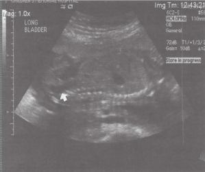
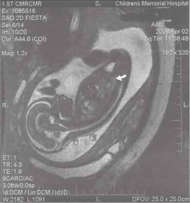
A 30 year old Caucasian woman gravid 1, para 0, abort us 0 with past medical history significant for a cone biopsy. She was on no medication other than prenatal vitamins. She did not smoke, drink alcohol or have and teratogenic exposures. There was no history of pre-existing diabetes and a first trimester random glucose was normal. She underwent routine screening ultrasound at 21 2/7 weeks of gestation. First trimester screening ultrasound was normal. Transabdominal ultrasound demonstrated a dilated fetal rectum to six millimeters with visible haustrations suspicious for anal atresia (Figure 1). The anal ring was not seen. Echocardiographic evaluation of the heart was normal and no other abnormalities were evident. Amniocentesis was performed and revealed a normal 46, XX karyotype. Follow-up evaluation via MRI demonstrated a distended distal sigmoid colon measuring eight to ten millimeters distally, consistent with anal atresia in isolation (Figure 2). No comment was made on MRI about the presence or absence of the anal ring. Management options were discussed. The patient chose to terminate after extensive consultation with a pediatric surgeon. The pregnancy was terminated at 23 weeks. An autopsy of the fetus was performed after the termination, the diagnosis of anal atresia was confirmed and no other abnormalities were identified.

    Figure 1:  Ultrasound at 21 2/7 weeks demonstrating fetal rectal dilation to
six millimeters.

Figure 2: MRI demonstrates a dilated distal sigmoid colon to ten millimeters.
Discussion
Anal atresia is a rare congenital abnormality, present in approximately 1 in 5000 live births [1]. Anal atresia is rarely found in isolation, with prevalence of 1.11 in 10,000 live births in a large cohort of 4.6 million newborns in Europe [2]. Anal atresia is more frequently associated with additional congenital abnormalities such as VACTREL syndrome (vertebral defects, anal atresia, tracheoesophageal fistula with esophageal atresia, radial and renal dysplasia, and limb malformations) and caudal regression syndrome [3-7]. The ability to diagnose anal atresia prenatally is of clinical significance, as prenatal diagnosis allows difficult decisions to be made regarding the continuation of the pregnancy. Furthermore prenatal diagnosis provides time to arrange for delivery in a tertiary care facility that can provide planned immediate surgical attention at birth. Fetal defecation and the neurologic innervations of the colon and rectum provide the physiologic basis for prenatal diagnosis of anal atresia. Fetal defecation illustrated by meconium stained amniotic fluid was previously thought to indicate fetal distress and hypoxia that typically occurred late in the third trimester [7-11]. Current evidence has demonstrated that defecation is potentially a normal process of the fetus [12-15]. Abramovitch and Gray concluded that fetal defecation begins as early as 14 weeks [16]. This concept has been further supported by the detection of high levels of micro-villar intestinal enzymes, such as disaccharidases and intestinal alkaline phosphatases in amniotic fluid between 14 and 22 weeks with a sudden drop in levels following the twenty second gestational week [17]. Yuan has shown via fluorogold retrograde tracing that rats with anorectal malformations have deficient motor innervation of the anal sphincter leading to retained meconium in the fetal period [18]. This explanation provides a physiological basis for the fact that the colon is not prominent ultrasonographically until late second trimester, following adequate neural innervation of the sphincter and subsequent retention of meconium.
Prenatal diagnosis of anal atresia is challenging, with most cases diagnosed at the time of birth. Prenatal diagnosis is established by finding dilated loops of fetal bowel on ultrasound [19]. In a small cohort of twelve patients with documented anal atresia at birth, it was found that a U or V shaped segment of dilated bowel in the lower abdomen or pelvis is particularly suggestive of anal atresia [20]. This diagnosis is generally supported by the presence of additional congenital abnormalities since anal atresia rarely occurs as an isolated anomaly [3]. The youngest fetus diagnosed with sonographic evidence of dilated loops of bowel was 12 weeks of age [21]. This fetus also presented with the additional anomaly of a small perimembranous ventricular septal defect that was visualized sonographically. Harris et. al studied a cohort of patients with sonographically consistent anal atresia where the diagnosis was confirmed either at birth or fetal autopsy. Within the cohort, 11 of 12 patients (92%) with anal atresia demonstrated abnormalities associated with either VACTREL syndrome or caudal regression syndrome [22]. In this cohort, the diagnosis was not suspected until late into the second trimester. The earliest case positively diagnosed was at 26 weeks of gestation [22]. This emphasizes that early diagnosis is difficult to achieve.
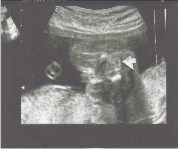
Ultrasound of the anal ring can be used to rule out the presence of anal atresia. This can be seen as early as 15 weeks of gestation; however, at this gestation false negatives are greater. Generally, in the third trimester the ring can reliably be seen (figure 3).

    Figure 3:  The anal complex can be seen between the pelvic bones at the
tip of the arrow. There is a hypoechoic ring, which is the muscular portion
surrounding the hyperechoic mucosa and the central hypoechoic area being
the lumen of the anus. This may be seen from 15 weeks on to term, but may
not be seen well until after 20 weeks of gestation.
Fetal bowel has distinctive characteristics on MRI, with gastrointestinal abnormalities increasingly identified on MRI compared to ultrasonography [23]. Notably, abnormal bowel size, abnormal bowel signal, and normal bowel with abnormal intraabdominal structures are more easily and readily visualized on MRI [24]. MRI is still infrequently used in the prenatal period. As such, formal recommendations and indications for fetal MRI are yet to be established.
Initial management of a patient born with anal atresia involves a thorough perineal inspection to allow the physician to establish the level of the lesion as high, intermediate, or low [25]. Gas in the bladder or meconium in the urine indicates a high anomaly. A low anomaly may be diagnosed with the presence of meconium easily visible through the skin of the perineum. Additionally, finding a well formed gluteal crease and anal region further support the diagnosis of a low lesion. In the absence of a clear diagnosis, the physician should wait 24 hours to allow for the passage of meconium and gas prior to finalizing the diagnosis of anal atresia [26]. Surgical repair begins with immediate sigmoid colostomy in the newborn period [27]. Once the newborn has grown to a sufficient size to tolerate additional surgical procedures, the posterior sagittal anorectoplasty (PSARP) is performed [28]. This procedure allows the surgeon to explore the anatomy of the rectum, surgically correcting any abnormal fistula tracts and creating an anus by connecting the distal rectum to perineal skin. Following PSARP, the patient undergoes anal dilation with hegar dilators until the desired neoanal size is reached. The rationale behind anal dilation following PSARP is the fact that the anus and rectum are surrounded by musculature, remaining closed at rest. Therefore, without post-operative dilation, the anus will tend to heal closed or very narrowly. When the desired neoanus size is reached, the colostomy site is closed [29]. Following repair via PSARP, commonly reported complications are disturbances in motility causing chronic constipation and overflow incontinence [29,30].The degree of chronic constipation experienced by patients post-operatively most closely correlates with the extent of sacral mobilization of the blind pouch during PSARP [31]. Pena and colleagues demonstrated that the rates of chronic constipation are typically elevated in patients undergoing abdominoperineal pull through procedures of intermediate or low abnormalities, with chronic constipation occurring between 18- 61.4% of patients depending on the abnormality [32]. Prenatally, it is impossible to definitively establish the exact level of atresia and therefore estimate the possibility of procedural complications. The risk of anal incontinence and chronic constipation is the primary reason that couples may choose termination. Our case is unique in that the diagnosis of anal atresia was suspected in isolation and confirmed by fetal MRI in a fetus of 21 2/7 weeks of gestation. This case demonstrates the importance of maintaining high clinical suspicion for anal atresia in a fetus with dilated loops of bowel on ultrasound prior to 26 weeks of gestation. Additionally, this case supports the value of MRI in confidently establishing the diagnosis of anal atresia prenatally. MRI provides an accurate distinction between solid and liquid densities, therefore facilitating identification of meconium in the fetal intestine causing colonic dilatation from liquid which is located in the bladder.
References
- Murken JD, Albert A. Genetic counseling in cases of anal and rectal atresia. Prog Pediatric Surgery. 1976; 9:115-118.
- Cuschieri A. Eurocat working group. Descriptive epidemiology of isolated anal anomalies: a survey of 4.6 million births in Europe. American Journal of Medical Genetics. 2001; 103:207-215.
- Quan L, Smith DW. The VATER association: vertebral defects, anal atresia, tracheoesophageal fistula with esophageal atresia, radial dysplasia. Birth Defects. 1972: 8:75-78.
- Terntamy SA, Miller JD. Extending the scope of the VATER association: definition of the VATER syndrome. Journal of Pediatrics. 1974; 85:345-349.
- Nora RH, Nora JJ. A syndrome of multiple congenital anomalies associated with teratogenic exposure. Arch Environmental Health. 1975; 30:17-21.
- Barnes JC, Smith WL. The VATER association. Radiology. 1978; 126:445-449.
- Ladd WE, Gross RF. Congenital malformations of the anus and rectum: report of 162 cases. American Journal of Surgery. 1937; 23:167-183.
- Alger LS, Kisner HJ, Nagey DA. The presence of meconium like substance in second trimester amniotic fluid. American Journal of Obstetrics and Gynecology. 1984; 150:380-384.
- Desmond MM, Moore J, Lindley JE, Brown CB. Meconium staining of the amniotic fluid. A marker of fetal hypoxia. Obstetrics and Gynecology. 1957; 9: 91-103.
- Meis JP, Hall M, Marshall, JR, Hobel CJ. Meconium passage: a new classification for risk assessment during labor. American Journal of Obstetrics and Gynecology. 1978; 131: 509-513.
- Miller PW, Coen RW, Ben irschke K. Dating the time interval from meconium passage to birth. Obstetrics and Gynecology. 1985; 66: 459-463.
- Barss VA, Benacerraf BR, Frigolotto FD. Antenatal sonographic diagnosis of fetal gastrointestinal malformation. Pediatrics. 1985; 46:445-449.
- Bean WJ, Calonge MA, April CN, Geshner J. Anal atresia: a prenatal ultrasound diagnosis. Journal of Clinical Ultrasound. 1978; 6:111-112.
- Grant T, Newman M, Gould R, Schey W, Perry R, Brandt T. Intraluminal colonic calcifications associated with anorectal atresia. Prenatal sonographic detection. Journal of Ultrasound Medicine. 1990; 9: 411-413.
- Mandell J, Lillehei CW, Green M, Benacerraf BR. The prenatal diagnosis of imperforate anus with rectourinary fistula: dilated fetal colon with enterolithiasis. Journal of Pediatric Surgery. 1992; 27:82-84.
- Kimble RM, Trudenger B, Cass D. Fetal defaecation: Is it a normal physiological process? Journal of Paediatrics and Child Health. 2002; 35:116-119.
- Richard A. Mulivor, Donna Cook, Françoise Muller, André Boué, Fred Gilbert, Michael Mennuti et al. Fetal intestinal microvilli in human amniotic fluid. Prenatal Diagnosis. 1987; 6: 429-436.
- Yuan Z. Deficient motor innervations of the sphincter mechanism in fetal rats with anorectal malformation: a quantitative study by fluorogold retrograde tracing. Journal of Pediatric Surgery. 2003; 38: 1383-1383.
- Bean WJ, Calonje MA, April, CN, Geshner J. Anal atresia: a prenatal ultrasound diagnosis. Journal of Clinical Ultrasound. 1978. 6:111-112.
- Harris RD, Nyberg DA, Mack LA, Weinberger E. Anorectal atresia: prenatal sonographic diagnosis. AJR Am Journal Roentgenology. 1987; 149: 395-400.
- Lam Y, Shek T, Tang M. Sonographic features of anal atresia at 12 weeks. Ultrasound Obstetrics and Gynecology. 2002; 19: 523-524.
- Huppert BJ, Brandt KR, Ramin KD, BF. King. Single-shot fast spin-echo MR imaging of the fetus: a pictorial essay. Radiographics. 1999; 19:215-227.https://link.springer.com/article/10.1007%2Fs00261-003-0147-2#page-1
- Veyrac C, Couture A, Saguintaah M, Baud C. MRI of fetal GI tract abnormalities. Abdominal Imaging. 2004; 29:411-420.
- Stephens FD, Smith ED, Paoul NW. Anorectal malformations in children: update 1988 March of Dimes birth defect foundation original series. 2009; 24.
- Pena A, Ashcraft KW, Holder TM. Imperforate anus and cloacal malformations. In: Eds. Pediatric Surgery. 2nd edn. Philadelphia: Saunders, 1993:372-392.
- Guochang L, Yuan J, Geng J, LiT. The treatment of high and intermediate anorectal malformations: One stage or three procedures? Journal of Pediatric Surgery. 2004; 39: 1466-1471.
- Pena A, De Vries P. Posterior sagittal anorectoplasty: important technical considerations and new applications. Journal of Pediatric Surgery. 1982; 17:796-811.
- De Vries P, Pena A. Posterior sagittal anorectoplasty. Journal of Pediatric Surgery. 1982; 17:638-643.
- Holschneider A, Hutson J, Pena A, et. al. Preliminary report on the International Conference for the development of standards for the treatment of anorectal malformations. Journal of Pediatric Surgery. 2005; 40:1521-1526.
- Pena A. Anorectal malformations. Semin Pediatric Surgery. 1995; 4:35-47.