
Case Report
Austin J Obstet Gynecol. 2017; 4(3): 1080.
Prenatal Diagnosis of Gastroschisis at an Imaging Facility in Ghana
Asare HK¹* and Waliwu D²
¹Department of Radiology, Tamale Teaching Hospital, Ghana
²Golden Heart Diagnostic Imaging Center, Ghana
*Corresponding author: Asare HK, Department of Radiology, Tamale Teaching Hospital, Ghana
Received: November 06, 2017; Accepted: November 29, 2017; Published: December 06, 2017
Abstract
Radiographers assuming extended roles in ultrasound practice in Ghana are gradually gaining competence to identify unusual fetal abnormalities during antenatal ultrasound scans. This development is significant to best patient management and pregnancy outcomes. An uncommon fetal defect was recently diagnosed by Ghanaian radiographers at a private imaging center in Tamale, Ghana. Gastroschisis is a birth defect in the anterior abdominal wall allowing portions of the baby`s small and large bowels to herniate outside the fetal body through a small opening in the abdominal walls often located to the right of the umbilicus. This case is presented below.
Keywords: Diagnosis; Gastroschisis; Prenatal ultrasound
Case Presentation
A 27-year-old healthy pregnant woman, gravid 2 para 2, with history of drinking alcohol was referred to a private imaging facility for her first ultrasound scan, to determine gestational age and fetal viability. A systematic ultrasound assessment of the fetus revealed an abdominal wall defect to the right of the umbilical cord insertion with free floating loops of bowel within the amniotic fluid (Figure1). The gestational age was determined as 31 weeks 3 days with estimated fetal weight and Amniotic Fluid Index (AFI) of 2.2kg and 12.2cm respectively. The fetal heart beat was positive. No maternal serum alpha fetoprotein screening test was recorded in the patient`s hospital folder. The woman underwent a successful caesarean section at about 36 weeks.
Discussion
Gastroschisis is the extrusion of abdominal contents directly into the amniotic cavity through a defective anterior body wall. The abnormality is mostly located to the right of the umbilical ring [1,2] and believe to be caused by disruptions in the blood supply to a developing abdominal wall from the omphalomesenteric duct artery at about eighth week of gestation [3]. It is not specifically known what causes the blood supply disruption, but few factors have been associated. For instance, gastroschisis is higher in regions where surface water atrazine levels are elevated especially when conception occurs in the spring, the time when atrazine, the commonly used herbicide, is often applied [4]. Also, several clinical studies have linked aspirin, a United State Food and Drug Administration (FDA) pregnancy category D drug, as an another risk factor [3,5], and according to large a scale study by the California Department of Public Health services, aspirin quadruples the risk of gastroschisis [6]. Similarly, incidences of gastroschisis have been recorded in women who smoke tobacco, use vasoconstrictiveor illicit drugs, consume excessive alcohol during pregnancy, low Body Mass Index (BMI), low socioeconomic status [2,7,8].
A rise in cases of 1 to 6 per 10,000 live births have been reported worldwide recently [7]. The abnormality is suspected when there is obstetric indications such as raised maternal serum Alpha Fetoprotein (AFP), commonly associated with risk of neural tube or abdominal wall defects [1].
Gastroschisis can be diagnosed with prenatal ultrasound scan, as early as first trimester, with an estimated detection rate around 70-72%, especially if transvaginal or high resolution ultrasound is used. Accurate diagnosis of this condition may rise as the use of routine ultrasound becomes more widespread [1,8,9]. This would facilitate the opportunity to counsel the family and to prepare for a safe delivery and optimal postnatal care. Unfortunately, the accuracy of prenatal ultra sound to diagnose abdominal wall defects may be affected by the timing and goals of the scan, fetal position and experience or expertise of the sonographer to differentiate between omphalocele and gastroschisis [10]. Omphalocele is the herniation of a membranous amnion covering abdominal viscera through an enlarged umbilical ring [1,10].
Despite a 97% survival rate of infants born with gastroschisis, it is associated with significant morbidity arising from unpredictably long hospital stay, delay in time to start oral feeding, long term use of total parenteral nutrition, multiple surgical interventions and neonatal complications including sepsis, short bowel syndrome and necrotizing enterocolitis [2,9]. Preterm delivery incidence is 28% compared to 6% pregnancies. Although cesarean section is often the preferred mode of delivery for this condition, it does not convey major merit over vaginal delivery [1].
Sonographic visualization of freely dilated floating loops of bowel within the amniotic fluid originating from the right insertion of the umbilical cord confirms the diagnosis. Polyhydramnios may be evident in high intestinal obstructions while oligohydramnios is present in about 25% of cases [1,2,8].
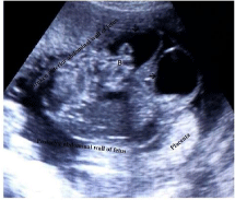
    Figure 1:  Axiral ultrasound view showing sonographic features of
gastroschisis, Free floating bowles (B) in an amniotic fluid (vertical arrow ↓).
Fetus urinary bladder (→horizontal arrow).
Conclusion
Ultrasound is the primary imaging modality in the diagnosis of gastroschisis. The anterior abdominal wall and umbilical cord insertion are easily recognized on ultrasound since it is surrounded by amniotic fluid. However, the diagnosis can be missed if the sonographer or professional performing the scan is not competent in antenatal scanning techniques and image interpretation. An early ultrasound diagnoses facilitates the safe delivery of a fetus with gastroschisis, advanced surgical interventions for correction and highly professional intensive care management to reduce the morbidity and mortality.
Acknowledgement
Appreciation to the patient (pregnant woman) for verbally consenting to publication of the case.
References
- Sree R, Devi SSS, Devi KP, Krupadanam K, Anasuya K. Gastroschisis- a case report. Int J Res Dev Health. 2013; 1: 191-194.
- Page R, Ferraro ZM, Moretti F, Fung KFK. Gastroschisis: antenatal sonographic predictors of adverse neonatal outcome. J Pregnancy. 2014; 12: 1-13.
- Werler MM, Sheehan JE, Mitchell AA. Maternal medication use and risks of gastroschisis and small intestinal atresia. Am J Epidemiol. 2002; 155: 26-31.
- Waller SA, Paul K, Peterson SE, Hitti JE. Agricultural-related chemical exposures, season of conception, and risk of gastroschisis in Washington State. Am J Obstet Gynecol. 2010; 202: 1-6.
- Nakhai-Pour HR, Berard A. Major malformations after first trimester exposure to aspirin and NSAIDs. Expert Rev ClinPharmacol. 2008; 1: 605-616.
- California Department of Health Services. Gastroschisis & Medications. California birth defect monitoring program. 1999; 1-2.
- Durfee SM, Benson CB, Adams SR, Ecker J, House M, Jennings R, et al. Postnatal outcome of fetuses with the prenatal diagnosis of gastroschisis. J Ultrasound Med. 2013; 32: 407-412.
- Brantberg A, BlaasHGK, Salvesen KA, Haugen SE, Eik-nes SH. Surveillance and outcome of fetuses with gastroschisis. Ultrasound obstet Gynecol. 2004; 23: 4-13.
- Kuleva M, Khen-Dunlop N, Dumez Y, Ville Y, Salomo LJ. Is complex gastroschisis predictable by prenatal ultrasound? BJOG. 2012; 119: 102-109.
- Ledbetter DJ. Gastroschisis and omphalocele.SurgClin N Am. 2006; 86: 249- 260.