
Case Report
Austin J Obstet Gynecol. 2018; 5(2): 1095.
Massive Traumatic Vulvar Hematoma in Teenagegirl
Helvacioglu C*, Alay I, Hosgoren M, Bahceci E and Ekin M
University of Health Sciences, Bakirköy, Dr. Sadi Konuk Training and Research Hospital, Clinic of Obstetric and Gynecology, Istanbul, Turkey
*Corresponding author: Caglar Helvacioglu, Department of Obstetrics and Gynecology, Bakirkoy Dr. Sadi Konuk Teaching and Research Hospital, Istanbul, Turkey
Received: January 04, 2018; Accepted: February 12, 2018; Published: February 23, 2018
Abstract
Vulvar hematoma is usually caused by obstetric reasons. Non-obstetric vulvar hematoma is rarely observed. Our case is a 16 year old girl, who admitted to our emergency department with genital swelling which happened after a blunt trauma during a gymnastic routine. In the physical examination a vulvar hematoma of 13 cm was observed on the right labium majus. We prefer surgical intervention in massive vulvar hematomas. Surgical approach reduce morbidity and minimize hospital duration.
Keywords: Vulva; Hematoma; Teenage
Introduction
Vulvar hematomas are usually seen in the obstetric population following repair of episiotomies and birth-related soft tissue injury [1]. However, traumatic on-obstetric vulvar hematomas are rare, but direct vulvar trauma has both short and long term effects physically and psychologically [2]. Coitus, attempts of rape, falling from distance, sportive activities, using foreign objects or aneurisma rupture can be counted as non obstetric reasons [1,2]. Vulvar hematomas are commonly minor and non life threatening; however, non obstetric vulvar hematomas can enlarge massively and can cause hemodynamic instability [3]. Although vulvar hematomas are followed conservatively, some cases require surgery and repairment.
Case Presentation
Our case is a healthy 16 year old girl with no known disease, who admitted to our ER with genital swelling which happened after a blunt trauma during her gymnastics routine. She was presented with symptoms such as swelling and pain, later on added with nausea and dizziness. She had no story of sexual intercourse or sexual trauma. She had no known disease such as bleeding diastases or any history of using anti coagulant drugs. Her vitals which were noted in the ER were; blood pressure was 90/50mmhg and heart rate was 105 beats per minute, respiratual rate was 22 breaths per minute. Macroscopic hematurea was not seen. Her abdomen was soft and non tender.
In our gynecological examination an ecimotic 13x10cm hematoma which was pain full with palpation and covered the right labium majus (Figure 1). She was unable to walk due to the hematoma. There was no evidence of sexual assault, no active bleeding was seen. The hemoglobin level was 11.3g/dl, wbc, plt and INR was in the normal range. There was not any free fluid which indicates intra abdominal bleeding on the Trans abdominal ultrasound. Tran’s perinea ultrasound finding supported the hematoma. Surgery was planned due to the symptoms which were severe pain and not being able to walk. 3cm vertical incision was performed on the right labium majus. The hematoma was drained and the cavity irrigated. There was no bleeding observed after wards. The cavity was sutured with 1.0 vicryl (Figure 2). No urethral, vesical or rectal injury was determined in the examination which was performed under general anesthesia. We insert a Foley catheter in the bladder. The patient was discharged on the first day post operatively without any complications.
2 weeks after the operation, the hematoma was resolved completely and the vulva was symmetrically and normal anatomy (Figure 3).
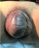
Figure 1:
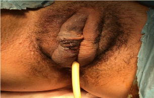
Figure 2:
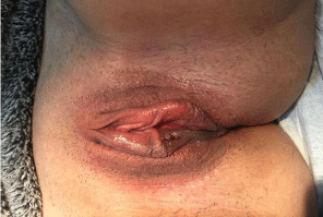
Figure 3:
Discussion
Vulvar hematomas are most frequently reported in postpartum women [3]. The true incidence of non-obstetric vulvar hematoma is unknown [3-4]. Non puerperal injuries have been reported after such diverse insults as straddle injuries (falls, bicycle/tricycle accidents, gokar tracing, cross-country skiing, sleigh riding, mechanical bull riding), physical assaults (kicks, human bites), and vigorous coitus [5].
The vulva consist mostly of loose connective tissue and smooth muscle that is richly supplied by branches of the pudendal artery; a significant branch of the internal liliac artery [6]. The venous drainage consists of labial veins, which are tributaries of the internal pudendal vein and venae comitantes. The injury to labial branches of the internal pudendal artery, which is located in the superficial fascia of the anterior and posterior pelvic triangle, can cause significant vulvar hematomas [7].
Conservative and surgical management are the treatment options for traumatic vulvar hematomas. Treatment criteria differ among hospitals, depending on the preference and experience of the treating physician [4-5]. It is generally accepted that small hematomas without serious symptoms can be managed conservatively with bed rest, compression, ice packing, and analgesia [4]. Large vulva hematomas are best managed with surgical evacuation and primary closure [8]. Benrubi et al. pointed out that conservative treatment prolongs the hospital duration, the need of antibiotics and blood transfusion [5]. Kanai et al. reported better out comes with early surgical intervention [9]. The hematoma should be evacuated and definitive hemostasis obtained [1-10]. Selective arterial embolization is an appropriate alternative to traditional surgical exploration [2-11]. This may be particularly useful in the high-risk operative candidate with prohibitive co morbidities, as the procedure does not require a general anesthetic. However we suggest that on the cases which the hematoma is large enough to inhibit the patient from walking, surgery is a faster and more effective solution.
References
- Virgili A, Bianchi A, Mollica G, Corazza M. Serious hematoma of the vulva from a bicycle accident: a case report. J. Reprod. Med. 2000; 45: 662.
- Kunishima K, Takao H, Kato N, Inoh S, Ohtomo K. Trans arterial embolization of a non puerperal traumatic vulvar hematoma. Radiat. Med. 2008; 26: 168.
- Ernest A, Knapp G. Severe traumatic vulva hematoma in teenage girl. Clinical Case Reports. 2015; 3: 975-978.
- Propst AM, Thorp JM. Traumatic vulvar hematomas: conservative versus surgical management. South Med J. 1998; 91: 144-146.
- Benrubi G, Neuman C, Nuss RC, Thompson RJ. Vulvar and vaginal hematomas: a retro spective study of conservative versus operative management. South Med J. 1987; 80: 991-994.
- Jaraquemada P, Garcia Monaco JMR, Barbosa NE, Ferle L, Iriarte H, Conesa HA. Lower uterine blood supply: extra uterine anastomotic system and its application in surgical devascularization techniques. Acta Obstet. Gynecol. Scand. 2007; 86: 228-234.
- Nelson EL, Parker AN, Dudley DJ. Spontaneous vulvar hematoma during pregnancy: a case report. J. Reprod. Med. 2012; 57: 74-76.
- Hwang KR, Kim SA, Kwon JE, Jeon HW, Choiand JE, So YH. A case of vulvar hematoma with rupture of pseudo aneurysm of pudendalartery. Obstet. Gynecol. Sci. 2014; 57: 168-171.
- Kanai M, Osada R, Maruyama KI. Warning from Nagano: increase of vulvar hematoma and/or lacerated injury caused by snowboarding. J. Trauma. 2001; 50: 328.
- Dash S, Verghese J, Nizami DJ, Awasthi RT, Jaishi S, Sunil M. Severe haematoma of the vulva-a report of two cases and a clinical review. Kathmandu Univ. Med. J. 2006; 4: 228-231.
- Villela J, Garry D, Levine G, Glanz S, Figueroa R Maulik D. Post part umangiographic embolization for vulvo vaginal hematoma: a report of two cases. J. Reprod. Med. 2001; 46: 65-66.