Case Presentation
Austin J Obstet Gynecol. 2018; 5(2): 1096.
Leiomyosarcoma of the Fallopian Tube in a Young Patient
Maria Orsaria¹*, Laura Mariuzzi² and Carla di Loreto³
¹Medical Doctor, Department of Pathology, University Hospital of Udine, Italy
²Assistant Professor, Department of Pathology, University Hospital of Udine, Italy
³Full Professor, Department of Pathology, University Hospital of Udine, Italy
*Corresponding author: Maria Orsaria, Department of Pathology, Azienda Ospedaliero-Universitaria di Udine, Piazzale S. Maria della Misericordia 15, Udine, Italy
Received: January 29, 2018; Accepted: February 16, 2018; Published: February 23, 2018
Abstract
Introduction: Leiomyosarcoma (LMS) of the fallopian tube is a really rare neoplasm. The neoplasms arising in the fallopian tube are infrequent, most of them are malignant and mainly are carcinomas. Pure sarcomas are exceedingly uncommon.
Case Presentation: A 35-year-old woman presented at the Emergency room complaining abdomen lower quadrants pain. The clinical examination was negative, whereas the pelvic imaging techniques showed a solid mass of 7x5cm in the Douglas cavity, into the distal right tube. She underwent complete surgery. Grossly right fallopian tube appeared dilated and the distal part of the tubal lumen was occupied by a solid neoplasm of 7 cm. On immunohistochemistry, tumour cells were positive for desmin, CD34, WT-1, calretinin, Estrogen and Progesterone receptor, while they stained negative for calponin and smooth muscle actin, leading to a diagnosis of fallopian tube leiomyosarcoma, limited to the right tube.
Conclusion: This case is pretty unique considering the young age of the patient, the immature mesenchymal appearance of the tumor and the peculiar expression of immunohistochemical markers (exclusively desmin and CD34).
Keywords: Leiomyosarcoma; Fallopian tube; Young age; CD34
Introduction
The neoplasms arising in the fallopian tube include benign and malignant types, but malignant tumor are more common than benign ones; however both are extremely infrequent, most of them are classified as carcinomas and account for only 0.3% of all cancers of the female genital tract, with occurrences varying between 2.9/ million and 5.7/million [1]. On the other hand, sarcoma of the fallopian tube is exceedingly uncommon, some of them being mixed epithelial-mesenchymal tumors and others pure sarcomas, which may be histologically subtyped if sufficient differentiation is present. Mainly these pure sarcomas are classified as leiomyosarcoma. Here we report a rare case of fallopian tube leiomyosarcoma, limited to the right tube in a young patient, with an immature mesenchymal appearance.
Case Presentation
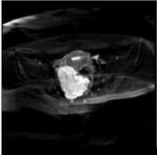
A 35-year-old, gravida 2, para 2, woman presented in March in emergency room with a 1-week history of abdomen lower quadrants pain. No masses were palpable and right lower quadrant was slightly painful. Pelvic ultrasound revealed a right ovarian corpus luteum with an adjacent 47 x 39 mm echocomposite image interpreted as a blood clot. The patient was referred to her Gynecologist. Then she came back to the Emergency room in June complaining again for abdomen lower quadrants pain. No masses were palpable. Ultrasound revealed an ipoechogen 61 x 51 mm mass in the Douglas cavity with a little amount of peritoneal fluid present. She performed also an abdominal Computed Tomography (CT) and a pelvic Magnetic Resonance Imaging (MRI) that showed an oval shape, lobulated, solid mass of 7x5 cm in the Douglas cavity, growing into the distal right tube (Figure 1). No others findings were present on the imaging. Routine preoperative laboratory studies revealed that only the level of C-reactive protein was elevated (32.02mg/L); the levels of CA125, CA19-9, carcinoembryonic antigen, and CA15.3 were within normal limits.

    Figure 1:  Pelvic magnetic resonance imaging (MRI) that showed an oval
shape, lobulated, solid mass of 7x5 cm in the Douglas cavity, growing into
the distal right tube.
No peritoneal dissemination was observed at the laparotomy.
Investigations
Tissues were fixed in buffered formalin, paraffin embedded and routinely processed for histological diagnosis. For immunohistochemistry, the Dako REALTM EnVisionTM Detection System (Glostrup, Denmark), Peroxidase/DAB+, Rabbit/Mouse Code K5007 method was used. The antisera employed are listed in Table 1, together with their source, dilution and antigen retrieval method.
    Antibody
    Clone, source
    Dilution


    Desmin
    D33, Dako
    0.111111111


    CD34
    QBEnd10, Dako
    0.111111111


    WT-1
    6F-H2, Dako
    1:50


    Calretinin
    SP13, Aczon
    0.111111111


    Estrogen Receptor
    SP1, Neomarkers
    0.180555556


    Progesterone Receptor
    636, Dako
    0.111111111


    h-Caldesmon
    h-CD, Dako
    0.111111111


    SMA
    1A4; Dako
    0.180555556


    c-Kit
    Polyclonal, Dako
    0.111111111


    DOG-1
    SP31, Aczon
    0.111111111


    WT1: Wilms’ Tumour 1; SMA: Smooth Muscle Actin; c-Kit: CD117;    DOG-1: Discovered on GIST-1
Table 1: Antibodies employed for immunohistochemistry.
Results
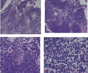
Grossly right fallopian tube appeared dilated with maximum diameter of 7.5cm and a smooth serosal surface; the distal part of the tubal lumen was occupied by a solid neoplasm of approximately 7cm. Histological examination of intraoperative frozen tissue sections of the right adnexal mass resulted in a diagnosis of a ‘‘malignant tumor’’. Accordingly, surgery was performed with total abdominal hysterectomy, bilateral salpingo-oophorectomy, pelvic lymph node sampling, omentectomy, appendicectomy and aspiration of peritoneal fluid. Postoperative histopathological examination of the tumor demonstrated a malignancy characterized by proliferation of atypical middle-sized, focally spindle-shaped neoplastic cells with scant cytoplasm, with high mitotic activity and areas of necrosis and myxoid stroma (Figure 2).

    Figure 2:  Histopathological examination of the tumor demonstrated a
malignancy characterized by proliferation of atypical middle-sized, focally
spindle-shaped neoplastic cells with scant cytoplasm, with high mitotic
activity and areas of necrosis and myxoid stroma (hematoxylin-eosin, original
magnification X40 (a); X100 (b); X200 (c); X 400 (d)).
Immunohistologically, many tumor cells showed a positive reaction to desmin, CD34, WT-1, calretinin, Estrogen and Progesterone receptor (Figure 3), while they stained negative for h-Caldesmon and smooth muscle actin. The tumors thus showed myogenic findings immunohistochemically. The neoplasia was limited to the tube and there was neither direct invasion nor metastatic spread into the other sites. The peritoneal fluid cytology was negative. According to the current surgical staging for tubal cancer, this tumor was staged as FIGO Ia [2]. The patient subsequently received adjuvant chemotherapy.

    Figure 3:  Immunohistologically, many tumor cells showed a positive
reaction to (a) desmin (original magnification X 200) and (b) CD34 (original
magnification X100).
Discussion
Since in 1886, Senger [3] described the first case of pure sarcoma in the fallopian tube and lately, in 1993, Jacoby et al. [4] reported the 34th case of tubal sarcoma and reviewed previous reports, only other few cases are present in the literature [5,6].
Sarcomas of the fallopian tube occur with a median age incidence of 47 years, compared to carcinosarcomas that usually occur in postmenopausal women, with a median age incidence of 60 years [6]. Their presenting symptoms are aspecific, such as pelvic pain and enlargement of the abdomen. Our case was one of the rare cases arising in a young adult patient (35-year-old) and limited to the tube (FIGO Ia), in contrast with the reported cases in the literature that usually show a much higher stage of the disease.
Usually, collecting the description of published case studies, the leiomyosarcoma of fallopian tube are microscopically composed by spindle cells arranged in fascicles with nuclear pleomorphism. They frequently have areas of hemorrhage and necrosis. The mitotic activity was found to be high in most of the reported cases. Our case has a “blue”, almost immature appearance, characterized by a proliferation of spindled or epithelioid cells, with scant eosinophylic or xanthomatous cytoplasm; in the neoplasia there were more cellular areas admixed to more looser ones with a myxoid stroma. This latter appearance could suggest a possible differential diagnosis with an embryonal rhabdomyosarcoma, but there wasn’t a cambium layer and no real rounded or spindled rhabdomyoblasts; moreover the presence of epitheliod cells with vacuolated cytoplasm is not a typical feature of rhabdomyosarcoma. Another possible differential diagnosis of this “immature” appearance was synovial sarcoma that in the tube is pretty exceptional, but the presence of epithelioid cells in absence of a real epithelial component excludes this possibility.
Also immunohistochemistry in this case supported a diagnosis of leiomyosarcoma even if only positivity for desmin was found, with the others smooth muscle markers tested negativity (smooth muscle actin, calponin); in our institute we don’t have routinly h-Caldesmon, that is reported to be usually highly positive in LMS, even if there are some discussion about that specificity in the literature [7,8]. Interestingly our case was also positive for CD34, suggesting a possible Extragastrointestinal Stromal Tumor (EGIST) diagnosis. Foster et al. recently reported a case of a previously diagnosed as tubal leiomyosarcoma that was subsequently reclassified as an EGIST based on molecular evaluation [9]. Anyway our case was negative for c-Kit and DOG-1 by immunohistochemistry and we performed the molecular test by PCR-direct sequencing that confirmed the absence of mutation at exons 9, 11, 13 and 17 of KIT and at exons 12, 14 and 18 of PDGFR. CD34 positivity is usually reported as negative in leiomyosarcoma, but there are rare cases of malignant epithelioid soft tissue tumors, comprising also leiomyosarcomas, positive for this marker [10], or maybe it could be expression of the very immature appearance of our case, that may be composed of progenitor cells, as in analogy with other mesenchymal tissues [11].
We have also sent our case out to a consultant expert in gynaecological pathology that supported our diagnosis.
The prognosis for patients with fallopian tube sarcoma is generally poor; however, it is difficult to evaluate prognostic factors of fallopian tube sarcoma statistically, because of the small number of cases reported. The treatment for tubal sarcomas is primarily surgery, followed by radiation and/or chemotherapy, even if standard adjuvant therapy scheme has not yet been established and they referred usually to uterine leiomyosarcomas [12,13].
Even with radical debulking surgical resection, the early, high rate of local recurrence (most often within the first two years after treatment) and hematogenous metastasis to the lungs, liver and bones compose this tumor’s clinical course. Our case is one of the rare cases of leiomyosarcoma limited to the tube (FIGO Ia); the thorax and abdominal TC didn’t revealed any suspicious image in the lungs, liver and lymph-nodes, after the surgery. At one year follow-up the patient showed no evidence of the disease. We don’t have a long period of follow up of our patient yet, but, in analogy with carcinomas, the limited stage at diagnosis could be the best prognostic factor for our patient. Anyway, for better prognostic evaluation and disease management in such rare cases, it is important to report and compare more cases regarding course of disease and outcome.
Conclusion
A case of leiomyosarcoma of the fallopian tube has been described and the following should be the learning points of this case:
• A preoperative diagnosis of this entity is almost impossible, because clinical symptoms, physical examination and imaging findings are not conclusive and because this is a very rare entity; pathological and immunohistochemical examination of the resected tumour is required for the final diagnosis.
• This rare entity usually occurs with a median age incidence of 47 years, whereas our case was one of the rare cases arising in a young adult patient (35-year-old).
• In the literature the rare cases presented show a high stage of the disease at the time of diagnosis, but our case was limited to the tube (FIGO Ia).
• The histological appearance and growth pattern of fallopian tube leiomyosarcoma are not always so straightforward, infact our case presented as a proliferation of spindled or epithelioid cells, with scant eosinophylic or xanthomatous cytoplasm, giving a “blue” and “immature” appearance to the tumor.
• Immunohistochemically only one or few smooth muscle markers could be expressed, as in this case only desmin showed positivity. Anymore, we can also have expression of some others markers such as CD34, maybe suggestive of the immature appearance of this tumor.
Author Contributions
MO collected the data, conceived, designed and drafted the manuscript, revising it critically for important intellectual content; and gave final approval of the version to be published.
LM and CDL conceived the manuscript and participated in its design, revised it critically for important intellectual content and gave the final approval of the version to be published.
References
- Riska A, Leminen A. Determinants of incidence of primary fallopian tube carcinoma (PTFC). Methods Mol Biol. 2009; 472: 387-396.
- AJCC/UICC TNM, 7th edition, and FIGO 2006 Annual Report.
- Senger E. Ueber ein primaeres Sarkom der Tuben. Centralbl F Gynaek. 1886; 10: 601-604.
- Jacoby AF, Fuller AF, Thor AD. Primary leiomyosarcoma of the fallopian tube. Gynecol Oncol. 1993; 51: 404-407.
- Mariani L, Quattrini M, Galati M, Dionisi B, Piperno G, Modafferi F, et al. Primary leiomyosarcoma of the fallopian tube: a case report. Eur J Gynaecol Oncol. 2005; 26: 33-35.
- Ueda T, Emoto M, Fukuoka M, Miyahara D, Horiuchi S, Tsujioka H, et al. Primary leiomyosarcoma of the fallopian tube. Int J Clin Oncol. 2010; 15: 206-209.
- Rush DS, Tan J, Baergen RN, Soslow RA. h-Caldesmon, a novel smooth muscle-specific antibody, distinguishes between cellular leiomyoma and endometrial stromal sarcoma. Am J Surg Pathol. 2001; 25: 253-258.
- Farah-Klibi F, Ben Hamouda S, Ben Romdhane S, Sfar R, Koubaa A, Ben Jilani S, et al. Immunohistochemical study of endometrial stromal sarcoma and smooth-muscle tumors of the uterus. J Gynecol Obstet Biol Reprod (Paris). 2008; 37: 457-462.
- Foster R, Solano S, Mahoney J, Fuller A, Oliva E, Seiden MV, et al. Reclassification of a tubal leiomyosarcoma as an eGIST by molecular evaluation of c-KIT. Gynecol Oncol. 2006; 101: 363-366.
- Sirgi KE, Wick MR, Swanson PE. B72.3 and CD34 immunoreactivity in malignant epithelioid soft tissue tumors. Adjuncts in the recognition of endothelial neoplasms. Am J Surg Pathol. 1993; 17: 179-185.
- Suga H, Matsumoto D, Eto H, Inoue K, Aoi N, Kato H, et al. Functional implications of CD34 expression in human adipose-derived stem/progenitor cells. Stem Cells Dev. 2009; 18: 1201-1210.
- Hensley ML, Blessing JA, Mannel R, Rose PG. Fixed-dose rate gemcitabine plus docetaxel as first-line therapy for metastatic uterine leiomyosarcoma: A gynecologic oncology group phase II trial. Gynecol Oncol. 2008; 109: 329- 334.
- Kobayashi Y, Tozawa A, Okuma Y, Kiguchi K, Ishizuka B. Complete remission with intraperitoneal cisplatin followed by prolonged oral etoposide in a stage IIIc primary leiomyosarcoma of the fallopian tube patient. J Obstet Gynaecol Res. 2010; 36: 894-897.