
Review Article
Austin Oncol. 2016; 1(3):1011.
Noninvasive Methods for Treatment of Brain Metastases
Wolny-Rokicka E*
Radiotherapy Department, Regional Clinical Hospital in Zielona Gora ul.Zyty, University of Zielona Gora, Poland
*Corresponding author: Edyta Wolny-Rokicka, Radiotherapy Department, Regional Clinical Hospital in Zielona Gora Zyty, University of Zielona Gora, Poland
Received: August 29, 2016; Accepted: October 05, 2016; Published: October 07, 2016
Abstract
Brain metatases occur in 20-40% of patients with cancer. Median survival of patients who develop brain metastases is relatively short - 4 months. Treatment methods such as Whole Brain Radio Therapy (WBRT), Surgery (S) or Radiosurgery (SRS) prolong survival for 3 to 5 months. Treatment options in brain metastases are as follows: symptomatic tratment, surgery, radiotherapy, chemiotherapy and combination of those. Symptomatic treatment of edema and anticonvulsive therapy are used if necessery and depends on neurological patients status. Surgery has been used for single brain metstasis in patients with good performance status. WBRT alone is a treatment of choice for patients with multiple brain metastases, with single brain metastasis not sutable to surgery or radiosurgery, especially those with an active and disseminated systemic disease. SRS is an external irradiation technique to deliver a relatively large radiation dose in a single fraction to an intracranial small target volume.
Chemotherapy alone (CT) is not used unless in clinical trials. Which method to use depends on profile of prognosis factors such as:performance status, patients age,number and volume of metastases.
Keywords: Brain metastases; Radiotherapy; Radiosurgery
Introduction
Brain metastasis is a common manifestation of disseminated malignancy [1,2]. In adults, lung, (36-40%), breast cancer (15-25%) and skin melanoma (5-20%) are the most common sources of brain metastases. Less frequent are colon, rectal, kidney, prostate, testicular, ovarian cancer and sarcoma. Cerebral lesion are mainly located in hemispheres (80%), in cerebellum (15%), in the brain stem (5%), being very rare in the basal ganglia, the pineal gland or hypophysis. Are more common than primary brain tumors [3]. Frequency of disclosure is the result: a more effective treatment of the primary tumor, more effective systemic treatment of disseminated disease (clinical or subclinical), the existence of blood - brain barrier for the majority of cytostatic drugs and the introduction of new imaging techniques. Most patients who develop brain metastases have short time survival. The treatment is used: steroids, surgery, radiation therapy and symptomatic treatment. The choice of treatment depends on the age, general condition of the patient, the number and location of metastatic lesions and the severity of the underlying disease [4]. The last decade has created new possibilities for the treatment of metastatic tumors such as radiosurgery and radiochemotherapy combination treatment with radiation [5].
Treatment Options
Treatment options for brain metastases are: symptomatic and specific therapy directed at the area of the tumor or tumors. To the first treatment should antioedematous and anticonvulsants. Causal treatment includes: surgical resection with or without radiotherapy of the whole brain (WBRT), an independent method of WBRT, radiosurgery (SRS), SRS + WBRT, chemotherapy.
Symptomatic treatment
Using antiedematous drugs is it routine methods. The most popular is glikokortykosteroid. Its mechanism anti oedema is not cleare, although it is probably the action by restoring the continuity of broken capillaries that have been damaged by vasoactive substances secreted by tumor [6]. Most patients start treatment at a dose of 4 to 8 mg dexamethasone daily with a noticeable improvement in neurological status [7,8]. Patients with symptoms such as headache, sleepiness may start treatment with higher doses of dexamethasone (16 mg daily). Patients with small metastatic disease without neurological symptoms do not require treatment with steroids. During brain radiotherapy is used protectively glucocorticosteroids in patients who reveal the acute symptoms of increased swelling of the brain. This results in reducing symptoms and clinical improvement in 75% of patients after just 24 hours after treatment anti-edematous [9]. Consuming of drugs depend on degree of clinical symptoms to reduce side effects in patients who had irradiated brain. After radiotherapy can be reduce doses towards decreasing the risk of occurrence of side effect.
The another group of drugs to reducing brain oedema are osmotic diuretics - mannitol and glycerol; loop diuretics, which include furosemide by rapidly effective diuretic. Anticonvulsant drugs used to control seizures. These drugs (eg, phenytoin, carbamazepine, phenobarbital) through interactions with corticosteroids reduce its activity in the Central Nervous System (CNS), which should be considered when dosing medications.
Surgery
Surgical treatment is used for single metastatic lesions in the brain, taking into account the clinical condition of the patient and tumor localization.The use of intraoperative ultrasonography and magnetic resonance imaging (neuronavigation) allows during surgery more quickly and accurately identify tumor [10]. In locations in centers of speech particularly important is intraoperative mapping. For decreasing of neurological deficit is used mapping. Mapping involves electrical stimulation of the cortex during a treatment of a patient who is woke up under the control of a local anesthetic [11]. It was also shown the benefit of combining surgery with radiation therapy (median survival 9-10 months in compare to WBRT as a independently treatment: 3-6 months) [12,13,14].
Chemotherapy
Chemotherapy can be used in patients with sensitive chemistry tumors (eg. small cell lung cancer, breast cancer, germ cell tumors, gestational trophoblastic disease) and can be an alternative to radiotherapy. Indications and methods of administration of chemotherapy in such cases are selected according to the rules for the corresponding primary tumors.
The response rate after applying alone chemotherapy in small cell lung cancer by Grossi et al. ranged from 21% to 76% [15] and breast cancer by Boogerd et al. from 35% to 60% [16]. That authors suggested that cytostatic treatment should be a first in patients with metastasis and radiotherapy should be used in resistance cases.
Radiotherapy - treatment technique
Whole brain radiotherapy (WBRT): The value of radiation therapy as a treatment of brain metastases were first described by Chao et al., 1954. [17] and the Chu Hilaris in 1961 [18]. They used standard fractionation to total doses from 3000 to 4000r in 3 - 4 weeks. They noticed a benefit in improving the clinical condition and decreased neurological symptoms in 60 to 80% patients and prolong median overvival survival from 6,6 - 8,2 months. Authors as: Hindo et al [19], Shehata et al [20] showed that using high total doses in few fractionated doses is effective as same as standard schema.
WBRT is the treatment of choice for patients who are not candidates for surgery or radiosurgery, especially those with an active disease [12]. The method consists in whole brain irradiation with two coaxial opposed lateral beams. For patients, treatment planning begins with preparing a thermoplastic mask (orfit mask). Is planned to be a homogeneous distribution of the set doses of ionizing radiation in the volume of the entire brain. With the help of the histogram (DVH - Dose Volume Histogram) is check the dose distribution in areas of interest. The used doses are in range 10 Gy in one fraction to 40 Gy in 20 fraction. The most used schemas are 20 Gy in 5 fractions and 30 Gy in 10 fractions. All schemas have comparison result [21,22]. During teleradiotherapy an external beam of high-energy X-rays is generated by a linear accelerator to irradiate and destroy the tumor.
Stereotactic radiotherapy: The concept of stereotactic developed in the early twentieth century and was then examined by Horsley’a and Clark in experimental animals [23].
Only Ernest Spiegl introduced this technique in 1947 for neurosurgery [24] and in 1951 Leksell combined the principles of location stereotactic of ionizing radiation and called this method “Radiosurgery” (SRS) [25]. Initially, Leksell envisioned the development of this method in neurology - ablation selective areas of the brain to treat Parkinson’s disease and chronic pain [26]. However, the increase in clinical experience increased use and efficacy of treatment in primary and secondary brain tumors.
Stereotactic radiosurgery is used most frequently in the treatment of solitary brain metastases. The treatment’s effects of solitary brain metastases are comparable to surgical treatment. This method is based on the strict immobilization of the patient, precise determination of the boundaries of the tumor and using a computer system to the treatment planning and delivery of a high dose of radiation in a well-defined area of the tumor as much as possible sparing healthy tissue. Treatment planning is based on the fusion of a series of CT images and MRI in order to precisely determine the volume of tumor [27]. The stereotactic technique employs an external system of coordinates (usually a stereotactic frame) ensuring maximum precision for the tumor site. The techniques of radiosurgery: static technique (the so-called. conformal radiotherapy CRT) and a dynamic technique (radiosurgery based on the modulation of dose intensity, IMRS - Intensity Modulated Radiosurgery). In CRT - the shape of the radiation beam exposure is constant (static field), while in the dynamic technique- shape of the radiation beam is changed in time of exposure by using a multileaf collimator (dynamic field) [28]. For radiosurgery dose administered once it is most often depending on the diameter of a tumor. The most used doses are 12-24 Gy [29].
Well-defined tumor volume is irradiation as a once (SRS) or in some cases, the treatment may be divided into several sessions or fractions, and the treatment is then called Fractionated Stereotactic Radiotherapy (SRF). In SRS a biological effect is three time greater than the same dose fractionated radiotherapy [30]. Rapid decrease in dose outside the area of interest, reduces the risk of damage to normal nerve tissue in the vicinity of the tumor and critical organs, which in radiosurgery are usually: lenses, eyeballs, a cross between the optic nerves and brain stem (Figure 1).
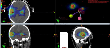
    Figure 1:  Profile of CT scan: transverse, frontal and sagittal. Metastases and
critical organs. Plan of treatment and isodose distribution.
In modern radiosurgery systems, such as the CyberKnife, the tumor can be located without using a stereotactic frame, the reference system being the patient’s anatomical structures (the so-called “internal coordinate system”) (Figure 3).
Volumetric Modulated Arc Therapy (VMAT): RapidArc or volumetric modulated arc therapy (VMAT) is a dynamic technique performed by using a multileaf collimator a variable dose rate and a gantry rotation around the patient. It combines the arc and IMRT techniques and is characterized by a relatively shorter single radiotherapy treatment session compared with other available methods (Figure 2).
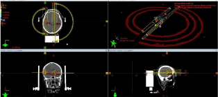
Figure 2: Eclipse system, VMAT technique. Two metastases.
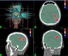
    Figure 3:  Cyber Knife technique. System planning Multi Plan. Two metastasis
Dc 15Gy, 291statical radiation beams.
Summary
The use of methods depend on many prognostic factors as: performans status, age, numbers and volume of tumors. Surgery and SRS are terapeutic methods used in patients with favorable prognostic factors. Whereas the radiotherapy is a treatment method should pay attention to neurological deficits u.e defects of the association that are related to quality of life.
References
- Shiau CY, Sneed PK, Shu HK et al. Radiosurgery for brain metastases: relationship of dose and pattern of enhancement to local control. Int J Radiat Oncol Biol Phys. 1997; 37: 375-383.
- Shehata WM, Hendrikson FR, Hindo WA. Rapid fractionaction technique and re-treatment of cerebral metastases by irradiation. Cancer 1974; 34: 257-261
- Schellinger PD, Meinck HM, Thron A. Diagnostic accuracy of MRI compared to CT in patients with brain metastases. J Neurooncol. 1999; 44: 275-281.
- Fuller BG, Kaplan ID, Adler J, Cox RS, Bagshaw MA. Stereotactic radiosurgery for brain metastases: the importance of adjuvant whole brain irradiation. Int J Radiat Oncol Biol Phys. 1992; 23: 413-418.
- Haie-Meder C, Pellae-Coset B, Laplanche A, Lagrange JL, Tuchais C, Nogues C, et al. Results of a randomized clinical trial comparing two radiation schedules in the palliative treatment of brain metastases. Radiother. Oncol. 1993; 26: 111-116.
- Soffietti R, Ruda R, Mutani R. Management of brain metastases. J Neurol. 2002; 249: 1357-1369.
- Hoskin PJ, Brada M. Radiotherapy for brain metastases.Clin Oncol. 2001; 13: 91-94.
- Schiff D. Single brain metastasis. Curr Treat Options Neurol. 2001; 3: 89-90.
- Cairncross JG, Posner JB. The management of brain metastases. In: Walker MD (ed) Oncology of the Nervous System. Martinus Nijhoff, Boston. 1983; 342-377.
- Patchell RA, Tibbs PA, Walsh JW, Dempsey RJ, Maruyama Y, Kryscio RJ, et al. A randomized trial of surgery in the treatment of single metastases to the brain. N Engl J Med. 1990; 322: 494-500.
- Cairncross JG. Laperriere NJ: Low-grade glioma: To treat or not to treat?. Arch Neurol 1989; 46: 1238.
- Noordijk EM, Vecht CJ, Haaxma-Reiche H, Padberg GW, Voormolen JH, Hoekstra FH et al. The choice of treatment of single brain metastasis should be based on extracranial tumor activity and age. Int J Radiat Oncol Biol Phys. 1994; 29: 711-717.
- Pirotte B. The use of MRI and PET imaging in neuro-oncology. Perspectives in Central Nervous System Malignancies. Conference, Czech Republic, Prague. 2005.
- Mintz AH, Kestle J, Rathbone MP, Gaspar L, Hugenholtz H, Fisher B, et al. A randomized trial to asses the efficacy of surgery in addition to radiotherapy in patients with a single cerebral metastasis. Cancer 1996; 78: 1470-1476.
- Grossi F, Scolaro T, Tixi L, Loprevite M, Ardizzoni A. The role of systemic chemotherapy in the treatment of brain metastases from small – cell lung cancer. Crit Rev Oncol Hematol. 2001; 37: 61-67.
- Boogerd W, Dalesio O, Bais EM, van der Sande JJ. Response of brain metastases from breast cancer to systemic chemotherapy. Cancer. 1992; 69: 972-980.
- Chao JH, Phillips R, Nickson JJ. Roentgen-ray therapy of cerebral metastases. Cancer. 1954; 7: 682-689.
- Chu FCH, Hilaris BB. Value of radiation therapy in the management of intracranial metastases. Cancer 1961;14: 577-581.
- Hindo WA, DeTrana III, FA, Lee MS, et al. Large dose increment irradiation in treatment of cerebral metastases. Cancer. 1970; 26: 138-141
- Shehata MK, Young B, Reid B, Patchell RA, St Clair W, Sims J, et al. Stereotactic radiosurgery of 468 brain metastases ≤2 cm: implications for SRS dose and whole brain radiation therapy. Int J Radiat Oncol Biol Phys. 2004; 1: 87-93.
- Jackson IM, Noren G. Gamma knife radiosurgery for pituitary tumors. Best Practice and Research in Clinical Endocrinology and metabolism. 1999; 13: 461-469.
- Sause WT, Crowley JJ, Morantz R, Rotman M, Mowry PA, Bouzaglou A, et al. Solitary brain metastasis: results of an RTOG/SWOG protocol evaluation surgery + RT vs. RT alone. Am J Clin Oncol. 1990; 13: 427-432.
- Horsley V, Clarke RH. The structure and functions of the cerebellum examined by a new method. Brain. 1908; 31: 45-124.
- Spiegel EA, Wycis HT, Marks M, Lee AJ. Stereotaxic apparatus for operations on the human brain. Science. 1947; 106: 349-350.
- Leksell L. The stereotaxic method and radiosurgery of the brain. Acta Chir. Scand. 1951; 102: 316-319.
- Leksell L. Stereotactic radiosurgery. J.Neurol Neurosurg Psychiatr.1983; 46: 797-803.
- Graus F, Walker RW, Allen JC. Brain metastases in chlildren. J Pediatr. 1983; 103: 558-561.
- Ślosarek K, Składowski K, Rembielak A et al. Intensity Modulated Radiation Therapy (IMRT) – description of the technique. Nowotwory J Oncol. 2002; 52: 614-618.
- Shaw E. Single dose radiosurgical treatment of recurrent previously irradiated primary tumors brain metastases: final report of RTOG protocol 90-05. Int J Radiat Oncol Biol Phys. 2000; 47: 291-298.
- Steiner L. Stereotactic radiosurgery with the Cobalt 60 Gamma Unit in the surgical treatment of intracranial tumors and arteriovenous malformations in operative neurosurgical techniques. Grune and Staton. 1985; 515.