
Review Article
J Ophthalmol & Vis Sci. 2016; 1(1): 1009.
Role of Optical Coherence Tomography in Macular Disorders
Pandey AN*, Raina AV and Sharma PD
Department of Ophthalmology, Govt. Medical College, Uttarakhand
*Corresponding author: Pandey AN, Department of Ophthalmology, Govt. Medical College, Srinagar Garhwal, Uttarakhand
Received: September 07, 2016; Accepted: October 10, 2016; Published: October 19, 2016
Abstract
Optical Coherence Tomography (OCT) is an emerging technology for performing high-resolution cross-sectional imaging. OCT is analogous to ultrasound imaging, except that it uses light instead of sound. OCT can provide cross-sectional images of tissue structure on the micron scale in situ and in real time.
High resolution cross-sectional imaging of the retina is useful for identifying, monitoring and quantitatively assessing macular diseases. Optical Coherence Tomography (OCT) uses low coherence or white light interferometer to perform high resolution measurements and imaging. Optical Coherence Tomography (OCT) has a great potential as a diagnostic and research tool. However it has its own limitations in the presence of media opacities like vitreous hemorrhage, corneal edema and dense cataract. Hence, an attempt has been made to study the role of Optical coherence tomography in diagnosis of macular disorders.
Keywords: Optical coherence tomography; Macular disorders; HMD
Introduction
The macula in retina is responsible for sharp, clear central vision and the ability to perceive color. The tightly packed photoreceptor cells in the macula control all of the eye’s central vision and are responsible for the ability to read, drive a car, watch television, see faces and distinguish detail. There are two types of photoreceptor cells in the retina, rods and cones. The rods provide vision at low light levels, while the cones provide sharp vision and discrimination.
High resolution cross-sectional imaging of the retina is useful for identifying, monitoring and quantitatively assessing macular diseases. Image resolutions of 1 to 15 μm can be achieved one to two orders of magnitude higher than conventional ultrasound. Cross-sectional images of the retina are obtained at the resolution of 1-10 micron. Optical Coherence Tomography (OCT) uses low coherence or white light interferometer to perform high resolution measurements and imaging. The high resolving power (10 um-Time Domain, 5 um- Spectral Domain) provides excellent detail for evaluating the vitreoretinal interface, neurosensory retinal morphology and the RPEchoroid complex. The infra-red light beam has a wave length of 820 nm. OCT has made it possible to understand better the vitreoretinal relationship at the macula. Optical Coherence Tomography (OCT) has a great potential as a diagnostic and research tool. However it has its own limitations in the presence of media opacities like vitreous hemorrhage, corneal edema and dense cataract, which attenuate the incident and reflected light. Also, it cannot assess function and can only provide details about structural alterations. Hence, an attempt has been made to study the role of Optical coherence tomography in diagnosis of macular disorders [1,2].
Literature
Anatomy of the macula
The macula lutea is an oval, yellowish area of the center of the posterior part of the retina. It measures about 5.5 mm in diameter and lies about 3mm to the lateral side of the optic disc.
The yellowish coloration of the macula is caused by a yellow carotenoid pigment, xanthophylls, which is present in the retinal layers from the outer nuclear layer inward.
Parts of the macula [1-4]
- Umbo
- Foveola
- Foveal avascular zone
- Fovea
- Parafoveal area
- Peri foveal area
Umbo: It is a tiny depression in the centre of the Foveola which corresponds to the ophthalmoscopically visible foveolar reflex.
Foveola (350 microns): It is the small central region in which the thickness of the retina is reduced so as to contain only photoreceptors, glial cells and muller cells.
Foveal avascular zone (800 microns): It is located inside the fovea but outside the foveola.
Fovea (1500 microns): It is a small depression where the retina is reduced to about half its normal thickness moving towards the centre of the retina. The inner nuclear layer is reduced to a double row of cells at the edge of the fovea.
The foveal sides of the depression are called the clivus; the floor of the depression is the foveola. There are no blood vessels overlying the fovea and no rod cells in the floor of the fovea. It is here that there is the highest concentration of cones is found.
Parafoveal area (500 microns): It is characterized by the densest accumulation of the nerve cells in the entire retina especially ganglion cells and the inner nuclear layer. The outer boundary is the point where the ganglion cell layer has four rows of the nuclei [3,4].
Peri foveal area (1500microns): It ends where the ganglion cells are reduced to a single layer [4,5].
Histology of the macula
The centre of the macula is the fovea containing the following layers
- Internal limiting membrane
- Outer plexiform layer
- Outer nuclear layer
- Layer of cones
- Retinal pigment epithelium
Among the 301 patients, 428 images were studied, reviewed and diabetic macular edema, CSR, ARMD and occlusive diseases were diagnosed using SD-OCT, concluded that SD-OCT was useful in complementing the clinician’s assessment of macular diseases [6,7].
Among the 4 eyes in 4 patients of macular degeneration by Optical Coherence Tomography (OCT), series comprised one eye of Alport syndrome and 3 eyes of cone or cone-rod dystrophy [1]. In Alport syndrome, OCT showed thinning of sensory retina and high-reflective layer corresponding to retinal pigment epithelium and choriocapillaris. In 3 eyes of cone or cone-rod dystrophy, OCT showed absence of low-reflective layer corresponding to the photoreceptors. The foveal thickness in the 4 eyes ranged from 25 to 73 micron, average 56.0±21.4 microns. It averaged 136.3±12.3 microns in normal eyes. The difference was significant (p<0.05). The diameter of macular depression averaged 1,452.8±149.3 microns in the 4 eyes and 1,258.5±47.1 microns in normal eyes. The difference was significant (p<0.05) [1].
OCT Scan protocols in macular disease [8,9]
The protocols that are helpful in macular diseases are the following:
- Line scan: The line scan gives an option of acquiring multiple line scan without retaining to main window. Both the length and angle can be changed according to resolution.
- Radial lines
- Macular thickness map
- Fast macular thickness map
- Raster line
- Repeat
Principle [9,10]
Optical Coherence Tomography (OCT) is an emerging optical imaging method, which allows the acquisition of cross-sections in a non-destructive and contactless manner. Now a day, OCT systems provide axial resolutions ranging from 1-10 μm and allow real time in situ and in vivo imaging.
Optical coherence tomography is based on the phenomenon of white light interferometry and very sensitive to small changes in the refractive index of the sample. Therefore it delivers complementary information to other imaging techniques like X-ray Computed Tomography (CT) or Magnetic Resonance Imaging (MRI). Near infrared light waves are partially reflected at different depths within the sample and their arrival times at a detector are interferometrically compared with a reference wave. The detected signal contains information on the position of the scatters within the sample, on their reflectivity, velocity and polarisation properties. Cross-sectional images can be reconstructed by collecting depth scans at different adjacent positions [10].
Various types of OCT
- Time domain OCT [9-11]
- Frequency Domain OCT (FD-OCT) [12,13]
- Spatially Encoded Frequency Domain OCT (Spectral Domain or Fourier Domain OCT)
- Time encoded frequency domain OCT (also swept source OCT)
- Optical coherence tomography
- Diabetic macular edema [7,13]
Optical Coherence Tomography (OCT) allows characterization of the profile and the intra-retinal structural changes of many macular disorders [13].
This has five distinct patterns on OCT (Figures 1-4)
- Sponge-like retinal thickness
- Cystoid macular edema
- Subfoveal serous detachment
- Vitreomacular traction
- Taut posterior hyaloid
- Early macular holes
- Presence of PVD
- Size of macular holes
- Closure pattern following Vitrectomy [6]
OCT is very useful in defining treatment options and for monitoring response to intervention [14].
Macular hole: OCT is extremely used for assessing the following:
ARMD: OCT has become very popular in ARMD because of early disease detection, monitoring of therapy and early evidence of reactivation [9].
ERM: These are clearly visible on the vitreoretinal interface on OCT [11-13].
CSR: OCT allows detection of early CSR [12,13] (Figure 3).
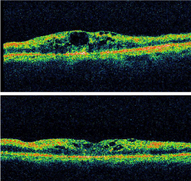
Figure 1: A) CME before treatment, B) CME after treatment.
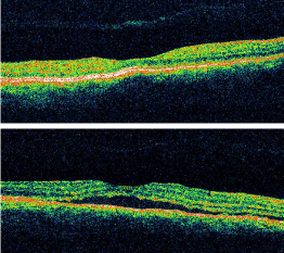
Figure 3: A) CSR before treatment, B) CSR after treatment.
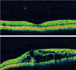
    Figure 4:  A) Macular edema before treatment, B) Macular edema after
treatment.
Macular Disorders
Age-related macular degeneration [14,15]
Age-Related Macular Degeneration (ARMD), also called senile macular degeneration, is a bilateral disease. It is a leading cause of blindness in developed countries, in population above the age of 65 years.
It is of two types
- Non-neovascular (Dry)
- Neovascular (Wet or exudative)
- Serous PED
- Fibro vascular PED
- Hemorrhagic PED
- Drusenoid PED
- Kato C, Mori T, Herai T, Liu Y, Tanifuji N. Optical coherence tomographic findings in atrophic macular disorders. Japanese journal of clinical ophthalmology. 2000; 54: 1429-1433.
- Gupta V, Gupta A, Ram M. Atlas of Optical Coherence Tomography of macular diseases. Jaypee. 2004.
- Haouchine B, Massin P, Tadsyoni R, Erginay A, Gaudric A. Diagnosis of macular pseudoholes and lamellar macular holes by optical coherence tomography. American Journal of Ophthalmol. 2004; 138: 732-739.
- Hee MR, Puliafito CA, Duker JS, Reichel E, Coker JG, Wilkins JR, et al. Topography of diabetic macular edema with optical coherence tomography, Ophthalmology. 1998; 105: 360-370.
- Hee MR, Puliafito CA, Wong C, Reichel E, Duker JS, Schuman JS, et al. Optical coherence tomography of central serous chorioretinopathy. AM J Ophthalmol. 1995; 120: 65-74.
- Johnson MW. Improvements in understanding and treatment of macular holes. Curr Opin Ophthalmol. 2002; 13: 152-160.
- Massin P, Duguid G, Erginay A, Haouchine B, Gaudric A. Optical Coherence Tomography for evaluating diabetic macular edema before and after Vitrectomy. American Journal of Ophthalmol. 2003; 135: 169-177.
- Natarajan S, Sharma S, Hitendra B. Diagnostics in macular disorders. Jaypee. 2005.
- Rogers AH, Martidis A, Greenberg PA, Puliafito CA. optical coherence tomography findings following photo dynamic therapy of choroidal neovascularisation. Am J Ophthalmol. 2002; 134: 566-576.
- Singh M, Chee CK. Spectral domain optical coherence tomography imaging of retinal diseases in Singapore. Ophthalmic surgery Lasers and Imaging. 2009; 40: 336-341.
- Wilkins JR, Puliafita CA, Hee MR, Duker JS, Reichel E, Coker JG, et al. Characterization epiretinal membranes using optical coherence tomography. Ophthalmology. 1996; 103: 2142-2151.
- Fercher AF. “Ophthalmic interferometry,” Proceedings of the International Conference on Optics in Life Sciences, Garmisch-Partenkirchen, Germany. 1990.
- Huang D, Swanson EA, Lin CP, Schuman JS, Stinson WG, Chang W, et al. Optical coherence tomography. Science. 1991; 254: 1178-1181.
- Zysk AM, Nguyen FT, Oldenburg AL, Marks DL, Boppart SA. Optical coherence tomography: a review of clinical development from bench to bedside. Journal of biomedical optics. 2007; 12: 51403.
- Fercher AF, Hitzenberger CK, Drexler W, Kamp G, Sattmann H. In Vivo Optical Coherence Tomography. Am J Ophthalmol. 1993; 116: 113-114.
Non-neovascular ARMD
Drusen: Drusen can be hard drusen or soft drusen.
Hard drusen in OCT: On OCT they are seen as focal elevation of the RPE with mildly thinned overlying retina. The elevated RPE does not shadow the reflection.
Soft drusen in OCT: images show small modulation in the contour of the retinal pigment epithelium with lack of shadowing below the contour changes, similar to serous RPE detachment. However, unlike a PED, soft drusen have shallow margins and mild back scatter into the choroid.
Geographic atrophy: OCT shows a thinned out overlying retina and decreased reflectivity of RPE with enhanced choroidal layer.
Neovascular ARMD (exudative)
OCT shows retinal thickening with mild back scattering from outer retinal layers, with disruption in the RPE in an area of moderate reflectivity with fusiform thickening of the reflective band corresponding to the CNVM. The lesions merge with RPE choriocappileries layer [12,13].
Occult CNVM: OCT shows an enhanced optical scatter signal at the choroidal level extending into sub-RPE space which is thicker than the adjacent reflection from the pigmented epithelium and choriocappileries, consistent with a neovascular membrane. The retina overlying the lesion appears thickened and elevated, with reduced reflectivity, suggesting fluid accumulation (Figure 2).
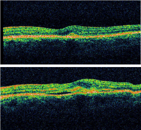
Figure 2: A) CNVM before treatment, B) CNVM after treatment.
Retinal pigment epithelium detachment (PED) [10,15]
PED can be differentiated into the following types
Serous PED: Serous PED-clinically seen as an area of RPE detachment.
Fibro vascular PED: OCT shows elevated RPE, along with mild to moderate back scatter persisting beneath the RPE, consistent with the Fibro vascular PED. It may have associated overlying neurosensory detachment or overlying cystoid macular edema.
Hemorrhagic PED: OCT through the hemorrhagic PED shows elevated RPE with moderate back scatter, which attenuates quickly with depth below the RPE. This back scatter, appearing immediately below the raised RPE is consistent with the hemorrhagic. The RPE band may be indistinguishable from hemorrhagic. There may be associated neurosensory elevation and cystoid changes in the overlying retina.
Drusenoid PED: OCT defines a RPE elevation with optical back scatter below the RPE, with small modulations in the RPE contour with moderate optical reflectivity below the lesion.
Feeder vessels
OCT may reveal areas of increased back scatter corresponding to the scarring membrane, with associated thickening of RPE choriocapillaries layer.
Loculated fluid
OCT shows a well defined region of hyper-reflectivity at the level of the RPE-choriocapillaries, consistent with the CNVM. The neurosensory retina above the membrane shows thickening as well as focal areas of reduced optically, corresponding with the fluid.
RPE rip
OCT through the lesion shows the elevated neurosensory retina and RPE which shadows the reflections from the choroid below. In the area of RPE rip two highly reflective layers are seen below the sensory retina corresponding with a folded double layer of pigment epithelium, with attenuation of reflections below the layer. In the area corresponding with absent RPE, enhanced reflectivity and optical penetration of the choroid is noted.
Disciform scar
OCT reveals a focal area of enhanced back scatter corresponding to the chorioretinal scar. The overlying neurosensory retina shows thinning throughout [8].
Central Serous Chorioretinopathy (CSCR) [5,11]
OCT shows on elevated neurosensory retina with an optically clear space underneath and a reflective layer corresponding to the RPE and choriocapillaries underneath it, corresponding to the CSR. With the advent of OCT the existence of PED with CSR has been shown to be much more than thought previously. In cases of associated PED, below the elevated neurosensory retina lies optically clear space below which highly reflective dome shaped elevation with attenuated shadows below it suggest the presence of PED. There may be hyper reflective spots in the optically clear space below the neurosensory retina corresponding with the sub retinal exudation and fibrin seen in resolving or chronic CSCR. CSCR may also have associated Choroidal Neovascular Membrane (CNVM). In such situations, there will be a delineated hyper fluorescence on angiogram corresponding with the CNVM. OCT will show high reflectivity at the level of RPE choriocapillaries suggestive of a CNVM.
Epiretinal Membrane (ERM) [8,12]
OCT is very useful tool in grading the ERM and providing information about the underlying retinal traction and secondary changes in the retina. OCT reveals a hyper-reflective membrane at the vitreoretinal interface, adherent to the underlying retina, with distortion of the inner retinal layer. The traction on the underlying retina is seen as folds in the inner retinal layer. On OCT, ERMs are seen as clearly separable where clear space is visible between the epiretinal membrane and inner retinal surface and global ERM where no area of separation is seen between the ERM and inner retinal surface. An increased thickness of retina with optically empty spaces in retinal layers along with reduced back scattering from outer retinal layers will indicate Cystoid macular edema, a lamellar macular hole may be associated with ERM. A macular pseudo hole formed by the ERM needs to be differentiated from a true macular hole. A macular pseudo hole will show an abnormally steep foveal contour resembling a partial thickness macular hole, but the outer retinal layer is seen intact throughout the image ruling out a full thickness macular hole. It is important to distinguish an ERM from a detached posterior hyaloid as both appear as a reflective band anterior to the retina on OCT. The posterior hyaloid has a weak, patchy reflection that is inner than an ERM (Figure 5).
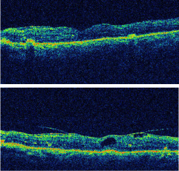
Figure 5: A) ERM before treatment, B) ERM after treatment.
Macular Hole [14,15]
In OCT the following are seen:
Stage IA of the macular hole is seen as foveal pseudo cyst in the retinal layers.
Stage IB is seen as partial disruption of the outer retinal layers. The posterior vitreous seen attached to its edges. It may develop to a lamellar macular hole.
Stage II reveals as a full thickness macular dehiscence with attached posterior vitreous. The surrounding retina may show thickening and cystic changes.
Stage III reveals a large macular hole with attached posterior vitreous.
Stage IV reveals a macular hole larger than 400 μ in diameter, with a detached posterior hyaloid. There may be an associated optically empty space below the neurosensory retina at the edges of the dehiscence suggestive of sub retinal fluid, along with thickening and cystic changes in the retinal layer [8] (Figure 6).
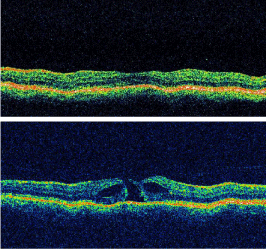
Figure 6: A) Macular hole before treatment, B) Macular hole after treatment.
Heredomacular Degeneration (HMD)
Best’s disease
OCT shows a characteristic dome shaped lifting of retina including the RPE layer, with a high reflective deposit in the sub RPE space, corresponding to the accumulated lipofuschin. This is in contrast to the presentation of a serous PED.
Foveal schisis
OCT is diagnostic as it reveals an elevation of the foveal pit with peri foveal splitting of the neurosensory retinal layers.
Stargardt’s disease
OCT reveals thinned out neurosensory retina along with increased reflectivity of the RPE choriocapillaries layer.
Retinitis pigmentosa
OCT reveals a thinned out neurosensory retina; associated CME may be seen [8].
Conclusion
OCT can certainly play a vital role along with the clinical examination in the diagnosis and evaluation of macular disorders. OCT can be included as a part of evaluation of any disorder affecting the macula which can be performed and interpreted by an ophthalmologist. It is useful for both screening and monitoring macular diseases and for deciding intervention. It is useful for assessing response to intervention such as laser photo coagulation, intravitreal injections and surgery.
References