
Case Report
Austin J Orthopade & Rheumatol. 2014;1(1): 4.
Brachial Plexus Injury Accompanying Glenohumeral Instability Case Report and Literature Review
Zumwalt M1* and Wooldridge A1
1Department of Orthopaedic Surgery and Rehabilitation, Texas Tech University Health Sciences Center, USA
*Corresponding author: Zumwalt M, Department of Orthopaedic Surgery and Rehabilitation, Texas Tech University Health Sciences Center, 3601 4th Street, MS 9436 Lubbock, TX 79430, USA, Tel: 806-743-2465; Fax: 806-743-1305; Email: [email protected]
Received: August 04, 2014; Accepted: October 22, 2014; Published: October 27, 2014
Abstract
Glenohumeral instability is a frequent condition after traumatic shoulder dislocation. Concomitant peripheral nerve injury is common and typically involves the axillary nerve. We present an unusual case of persistent suprascapular and long thoracic nerve injury, with resultant scapulothoracic dyskinesia contributing to glenohumeral instability. The upper extremity neuropraxia was followed with “benign neglect” for spontaneous resolution, prior to the treatment decision of performing anterior capsulorrhaphy alone versus the combined procedure with latissmus dorsi transfer and/or neurosurgical intervention.
Keywords: Brachial plexus injury; Glenohumeral instability; Shoulder dislocation; Scapulothoracic dyskinesia
Introduction
Shoulder dislocations typically occur due to major blunt trauma, but can also be due to minor trauma [1-3]. In most instances the forces imparted are indirect rather than direct impact [4]. Approximately 95% of shoulders dislocate anteriorly [2,5]. Subsequent glenohumeral instability is due to injury to the labrum, capsule, and nerve damage affecting the scapulothoracic articulation [4,6]. While peripheral nerve injury is common, brachial plexus injury is rare and can contribute to persistent glenohumeral instability due to scapulothoracic dyskinesia [3,7,8]. In this paper, we report an unusual case of an 18-year-old female with anterior shoulder instability secondary to recurrent dislocations, and concomitant suprascapular plus long thoracic nerve neuropraxia contributing to scapulothoracic instability.
Case Presentation
18 year-old left-hand dominant female sustained a right anterior shoulder dislocation after being tackled five years prior. Two years later, she sustained a second dislocation during an overhead basketball pass. In the past year, she lifted a heavy backpack when her right shoulder dislocated anteriorly again. Since her most recent closed reduction, she has had continued shoulder pain and gross deformity about the shoulder girdle. Feelings of instability and painful subluxation in her right shoulder have interfered with her activities of daily living and prevented her from participating in overhead sports. She has undergone physical therapy rehab exercises without complete resolution of her symptoms. She was most concerned about the arm “hanging down, slipping in and out, and her shoulder blade sticking up.”
History
18 year-old left-hand dominant female sustained a right anterior shoulder dislocation after being tackled five years prior. Two years later, she sustained a second dislocation during an overhead basketball pass. In the past year, she lifted a heavy backpack when her right shoulder dislocated anteriorly again. Since her most recent closed reduction, she has had continued shoulder pain and gross deformity about the shoulder girdle. Feelings of instability and painful subluxation in her right shoulder have interfered with her activities of daily living and prevented her from participating in overhead sports. She has undergone physical therapy rehab exercises without complete resolution of her symptoms. She was most concerned about the arm “hanging down, slipping in and out, and her shoulder blade sticking up.”
Physical examination
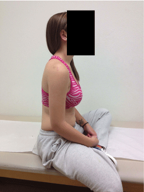
Upper extremities with bilateral ligamentous laxity (mainly elbow hyperextension and inferior sulcus sign); right shoulder girdle with mild atrophy of both spinati; tender over the trapezius, anterior joint line, sternocleidomastoid and paracervical musculature; active total elevation 175 degrees; decreased internal plus increased external rotation as compared to the other side to mid-high thoracic levels; 4+/5 drop arm test; superomedial protraction of the right scapula and winging with push-up; negative Spurling’s, positive inferior sulcus and anterior apprehension signs, equivocal O’Brien’s, negative relocation test and posterior apprehension sign; anterior subluxation with the arm adducted and relocation with 90 degree elevation (Figure 1-3).
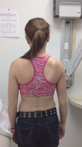
Figure 1: Scapular prominence on right with arm adducted as side (Pre-op).
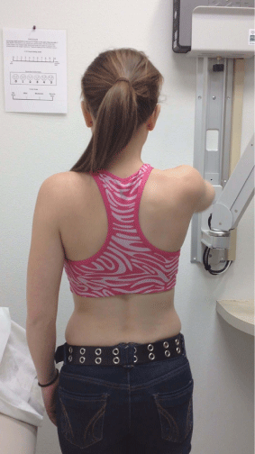
Figure 2: Reduction of shoulder joint with arm elevation to 90 degrees (Preop).
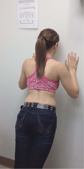
Figure 3: Right–sided scapular winging with push–up position (Pre-op).
Tests and Results
Radiographs of right shoulder
Grashey AP, Scapular Y, and axillary lateral views reveal medialization of the scapula and anterioinferior subluxation of the humeral head (Figure 4-6).
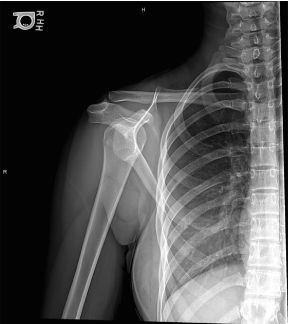
Figure 4: AP view of right shoulder with apparent humeral head subluxation.
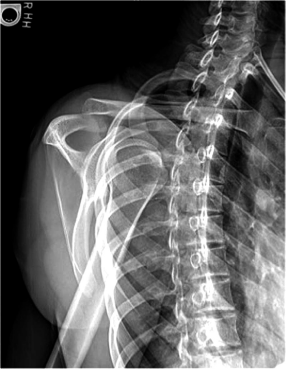
Figure 5: Y–lateral of right shoulder with anterior glenohumeral dislocation.
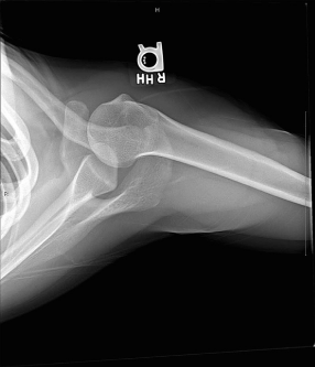
Figure 6: Axillary lateral right glenohumeral joint with anterior subluxation.
MR arthrogram of right shoulder
Partial tears of the supraspinatus and subscapularis. Redundant inferior capsule with signal changes involving the middle and inferior glenohumeral ligaments, along with the anterior glenoid labrum. Humeral head Hill-Sachs lesion (Figure 7-8).
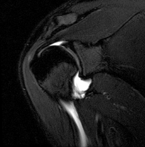
Figure 7: Coronal MRI of redundant inferior capsule and signal changes involving glenohumeral ligaments and glenoid labrum.
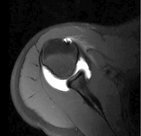
Figure 8: Axial MRI demonstrating Hill-Sachs lesion.
Electrodiagnostic testing (NCS/EMG) of right upper extremity 5 months post recent injury
Incomplete lesion of the right brachial plexus upper trunk involving primarily the long thoracic and suprascapular nerves with neural continuity consistent with stretch injury.
Repeat NCS/EMG 3.5 months later
Resolving brachial plexus upper trunk stretch injury.
Diagnosis
Right suprascapular and long thoracic nerve neuropraxia associated with scapulothoracic dyskinesia.
Right shoulder anterior glenohumeral instability.
Treatment and Outcomes
- Brachial plexus injury treated with avoidance of further traction/pressure.
- Scapulothoracic dyskinesia managed conservatively with TLO bracing.
- Multidirectional instability treated with physical therapy and shoulder stabilizing brace. After resolution of neurologic symptoms (1 year from most recent traumatic episode), patient underwent arthroscopic shoulder capsulolabral reconstruction/plication.
- At 5 months follow-up, patient is symptom free regarding the right shoulder. She is doing push-ups and playing basketball again; complains of myofascial type symptoms involving paracervical and trapezius musculature only, and is pleased with the cosmetic appearance of her shoulder blade; completed formal physical therapy and doing rehab exercises on her own: ATE 175 degrees, abduction 175 degrees, IR to the mid thoracic level, ER to the C7/T1 junction; negative anterior apprehension sign/Obrien’s test; 5/5 rotator cuff strength (Figure 9-11).
- Kosiyatrakul A, Jitprapaikulsarn S, Durand S, Oberlin C. Recovery of brachial plexus injury after shoulder dislocation. Injury. 2009; 40: 1327-1329.
- Paxinos A, Walton J, Tzannes A, Callanan M, Hayes K, Murrell GA. Advances in the management of traumatic anterior and atraumatic multidirectional shoulder instability. Sports Med. 2001; 31: 819-828.
- Shears E, Sunderamoorthy D, Ali SA. Brachial plexus injury after anterior shoulder dislocation: a case report. Acta Orthop Belg. 2005; 71: 489-490.
- Mehta MP, Kottamasu SR. Anterior dislocation of the shoulders with bilateral brachial plexus injury. Ann Emerg Med. 1989; 18: 589-591.
- Good CR, MacGillivray JD. Traumatic shoulder dislocation in the adolescent athlete: advances in surgical treatment. Curr Opin Pediatr. 2005; 17: 25-29.
- Martin RM, Fish DE. Scapular winging: anatomical review, diagnosis, and treatments. Curr Rev Musculoskelet Med. 2008; 1: 1-11.
- Warner JJ, Navarro RA. Serratus anterior dysfunction. Recognition and treatment. Clin Orthop Relat Res. 1998; : 139-148.
- Wiater JM, Flatow EL. Long thoracic nerve injury. Clin Orthop Relat Res. 1999; : 17-27.
- Hafizuddin A AH. Dislocation of Should Joint. KYAMC Journal. 2011; 2: 140-144.
- Yeap JS, Lee DJ, Fazir M, Kareem BA, Yeap JK. Nerve injuries in anterior shoulder dislocations. Med J Malaysia. 2004; 59: 450-454.
- Streubel PN, Krych AJ, Simone JP, Dahm DL, Sperling JW, Steinmann SP, et al. Anterior glenohumeral instability: a pathology-based surgical treatment strategy. J Am Acad Orthop Surg. 2014; 22: 283-294.
- Nikolaou VS, Pilichou A, Staramos D, Chronopoulos E, Korres D, Efstathopoulos N. Axillary artery and brachial plexus injury after anterior shoulder dislocation: Report of a case and review of the literature. Eur J Orthop Surg Traumatol. 2008; 18: 595-598.
- Cutts S, Prempeh M, Drew S. Anterior shoulder dislocation. Ann R Coll Surg Engl. 2009; 91: 2-7.
- Bedi A, Ryu RK. The treatment of primary anterior shoulder dislocations. Instr Course Lect. 2009; 58: 293-304.
- Novak CB, Mackinnon SE. Surgical treatment of a long thoracic nerve palsy. Ann Thorac Surg. 2002; 73: 1643-1645.
- Zoltan JD. Injury to the suprascapular nerve associated with anterior dislocation of the shoulder: case report and review of the literature. J Trauma. 1979; 19: 203-206.
- Kömürcü M, Ulas ÜH, Özdemir T, Koca K, Atesalp AS. Anterior shoulder dislocation with brachial plexus injury (Case report and review of the literature). Gulhane Med J 2002; 44: 453-456.
- Safran MR. Nerve injury about the shoulder in athletes, part 1: suprascapular nerve and axillary nerve. Am J Sports Med. 2004; 32: 803-819.
- Gupta V, Posner B. Trauma to the long thoracic nerve and associated scapula winging in a low-velocity rear-end automobile collision: case report. J Trauma. 2004; 57: 402-403.
- Lorei MP, Hershman EB. Peripheral nerve injuries in athletes. Treatment and prevention. Sports Med. 1993; 16: 130-147.
- Safran MR. Nerve injury about the shoulder in athletes, part 2: long thoracic nerve, spinal accessory nerve, burners/stingers, thoracic outlet syndrome. Am J Sports Med. 2004; 32: 1063-1076.
Normal contour of right scapula with arm relaxed at side (Post-op).
Figure 9:Normal contour of right scapula with arm relaxed at side (Post-op).
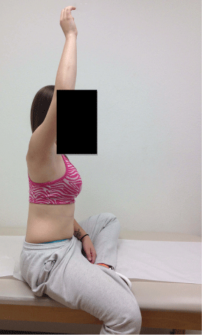
Figure 10: Almost full active elevation of right shoulder (Post-op).
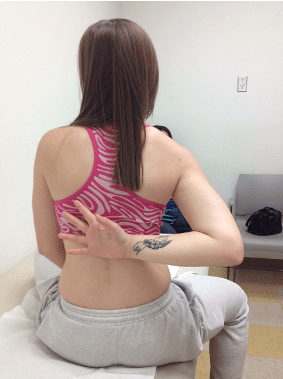
Figure 11: Almost full active elevation of right shoulder (Post-op).
Discussion
The shoulder has an inherent unstable anatomical configuration to allow for a wide range of motion. Therefore, the shoulder is the most frequently dislocated joint [2,5,9-13]. Traumatic shoulder dislocations occur almost three times more often in males versus females, and close to 50% of cases occurring between the ages of 15-30 years old [11,13]. The risk for recurrent dislocation is higher in males and in young patients with incidence ranging from 80-95% [2,5,12- 14]. Constitutional laxity and/or scapulothoracic dyskinesia can both contribute to ongoing instability [7,8].
Causes of shoulder dislocation are variable, stemming from relatively atraumatic to more violent forces such as motor vehicle accident. The arm is typically in a position of hyperextension, external rotation, and abduction when the injury occurs [1,4,9,13]. Since traction on the shoulder usually accompanies the traumatic episode, the incidence of peripheral nerve injury is common, occurring in up to 45 % of cases [1,3,6,15-19]. The occurrence of neurologic trauma ranges from 5-75% as confirmed by electrodiagnostic studies, and clinical diagnosis ranging from 3-21% [4,10,16]. Up to 15% of neural injury occurs in acute and more than 5% in recurrent shoulder dislocations [12]. The axillary nerve is injured most frequently, followed by other peripheral nerves including the musculocutaneous, median, radial, and/or ulnar nerves [10,12,16,17]. Fortunately, the majorities of nerve injuries are transient neuropraxias and improves following shoulder reduction [12].
Brachial plexus injury is rarely associated with anterior glenohumeral dislocation, and can take from 8 to 18 months for complete resolution [1,3,12,20,21]. There are very few reports from European literature describing other more proximal injuries at the cord level. The majority of brachial plexus lesions involve postganglionic and/or infraclavicular sites [1]. The injury pattern is dependent upon the arm position at time of injury. Abduction/ internal rotation applies tension to all cords of the brachial plexus; elbow and wrist extension causes distraction to the medial cord; elbow flexion stretches the posterior plus medial cords [3,13]. Only some isolated cases of suprascapular nerve involvement have been reported in association with shoulder dislocation. Injury to this nerve has a low chance of recovery since it has to withstand tensile forces near the point of attachment close to the spinal cord. Even then, supraclavicular lesions recover better than the axillary nerve but not as well as infraclavicular lesions [4].
Conclusions
This unusual case of a mixed brachial plexus injury accompanying anterior shoulder dislocation in a young female from low energy mechanism emphasizes the importance of assessing for simultaneous nerve injury at the time of dislocation. Treatment decisions were guided by the resolution of the brachial plexus neuropraxia. Without resolution of the neuropraxia, the patient would have required additional muscle transfer and/or neurosurgical intervention. While static stability can be restored by capsulorrhaphy, dynamic instability must also be correct by addressing the scapulothoracic component. In cases of concomitant nerve injury, patients must be counseled on the length of healing and expectations about complete recovery.
Acknowledgment
I would like to thank the following people for their technical expertise in assisting me with the completion of this article: Ms. Beverly Reed, Ms. Margaret Vugrin, and Mr. Charles Henderson. Without their tireless dedication and on-going support, the task would have been much more arduous MZ.
References