
Case Report
Austin J Orthopade & Rheumatol. 2014;1(2): 2.
Isolated Upper Thoracic Chance Fracture with Neurological Deficit, Without Bone Injury, Case Report
Meriç Unal1*, Ahmet Cemil Turan2 and Omer Akçali3
1IspartaSifa Hospital Orthopaedics and Traumatology, Isparta/Turkey
2Izmir Medical Park Hospital Orthopaedics and Traumatology, Izmir/Turkey
3DokuzEylül University Faculty of Medicine Orthopaedics and Traumatology, Izmir/Turkey
*Corresponding author: Meric Unal, Isparta Sifa Hospital, Department of Orthopaedics and Traumatology, Isparta/Turkey
Received: October 10, 2014; Accepted: October 27, 2014; Published: November 04, 2014
Introduction
Chance fracture is known as horizontal splitting of vertebral corpus and neural arc. Mostly seen mechanism is hyperflexion of vertebra [1,2,3,4]. Horizontal splitting includes vertebral corpus, intervertebral discs or both [2]. Mostly seen at thoracolumbar region [3]. Upper thoracic chance fracture is very rare [1]. Injury mechanism is usually flexion-distraction injury and usually seen as seat belt injury at the time of car crush [1,3,4].
In this case report, patient had spinal cord injury caused by traffic accident outside car. There were no bone injury detected at direct radiography, computed tomography (CT) and magnetic resonance imaging (MRI) but there was T1-T2 intervertebral disc horizontal splitting and interspinous and supraspinous ligament separation at the same level.
Case
70 year old male examined after car crush. At neurological examination, total loss of bilateral lower extremity muscle strength and hypoestesia under second thoracic vertebra. There was no anal tonus and sense. Bulbocavernous reflex was not detected. Abdominal skin reflex also could not detect bilaterally. Bilateral upper extremity examination was normal. There were no other problems at systemic examination.
Direct radiography, includes all vertebras was taken (Figure 1,2). Lateral radiography at swimmer position was also taken for exact determination of cervicothoracic junction. There were no abnormalities found at direct radiographic examination. At the thin slice CT examination, there were no bony lesions found (Figure 3).
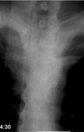
Figure 1: Upper Thoracic AnteroposteriorDirect Radiography (No Bony Lesion).
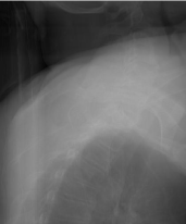
Figure 2: Upper Thoracic Lateral Direct Radiography (No Bony Lesion).
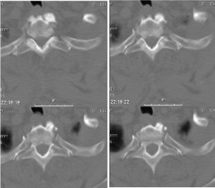
Figure 3: Axial CT scan of Thoracic-2 Vertebra (No Bony Lesion).
Because of the unexplained neurological condition, MRI was performed (Figure 4). T2 fat suppression slices showed injury at T1-T2 intervertebral disc space includes T2 vertebra upper end plate and also injury at supraspinous and interspinous ligaments at the level of T1 and T2 vertebra (Figure 5). At spinal cord examination, on MRI, hyperintense appearance found at T2 weighted slices that are compatible with edema or hematoma. Sternum, ribs and other vertebras were found normal. Lung examination and radiography were also normal.
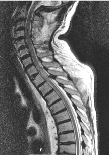
Figure 4: Sagittal View of Thoracic MRI.
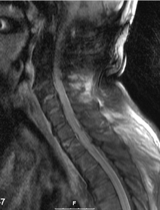
Figure 5: Sagittal View of Thoracic MRI (Fat Supression).
30 mg/kg methylprednisolone was given intravenous bolus. 5.4 mg/kg/day methylprednisolone intravenous infusion was started for maintenance therapy. Injury was accepted stable and start to treat conservatively. On the second day of the injury sudden onset respiratory distress and cardiac arrest developed and patient was intubated. Food particles found inside the intubation tube, so food aspiration was thought. Patient did not respond to cardiopulmonary resuscitation for 45 minutes and accepted as exit us. Family did not accept autopsy.
Discussion
Chance fracture defines a group of injuries results as horizontal splitting of vertebral corpuses and neural arc [1,2,3,4]. Horizontal splitting can also seen at intervertebral discs [2]. Injury mechanism is usually hyperflexion of vertebra over a fixed point, because of this mechanism, injury is defined as seat belt injury [1,3]. Accepted mechanism is flexion-distraction injury [3].
This type of injury is almost always seen at thoracolumbar region [2]. It is very rare at thoracic region because of the protective effect of the ribs [3]. Chance fractures are generally associated with high energy traumas so associated internal organ injuries usually seen [2].
In our case, there were no direct radiography and CT findings that explain the neurological status. At MRI; horizontal splitting was found either T1-T2 intervertebral discs or interspinous and supraspinous ligaments. Spinal cord edema was also found at MRI. Case is considered as chance fracture at a very unusual localization.
In the case of cervical and lumbar degeneration and associated degenerative fusion, thoracic region become more susceptible for injuries. Chance type injury without bony lesion at the upper thoracic region in the absence of high energy hyperflexion injury can be developed more than expected. It is always considered in older patients with cervical and lumbar degeneration.
References
- Dhall SS, Tumialán LM, Mummaneni PV. Chance fracture of the second thoracic vertebra: case illustration. J Trauma. 2006; 60: 922.
- Groves CJ, Cassar-Pullicino VN, Tins BJ, Tyrrell PN, McCall IW. Chance-type flexion-distraction injuries in the thoracolumbar spine: MR imagingcharacteristics. Radiology. 2005; 236: 601-608.
- Davis JM, Beall DP, Lastine C, Sweet C, Wolff J, Wu D. Chance fracture of the upper thoracic spine. AJR Am J Roentgenol. 2004; 183: 1475-1478.
- Todkar M. Case report: unusual mechanism of chance fracture in an adult male. MedGenMed. 2005; 7: 26.