
Case Report
Austin J Orthopade & Rheumatol. 2016; 3(2): 1031.
Chronic Clavicular Malunion Treated with Corrective Osteotomy
Sally Corey, Shaw KA* and Terry Mueller
Department of Orthopaedic Surgery, Dwight D. Eisenhower Army Medical Center, USA
*Corresponding author: K. Aaron Shaw, Department of Orthopaedic Surgery, Dwight D. Eisenhower Army Medical Center, Georgia, USA
Received: June 04, 2016; Accepted: July 20, 2016; Published: July 21, 2016
Abstract
The standard of care for the majority of mid-shaft clavicle fractures is nonoperative management, especially in the pediatric and adolescent population. Although a degree of malunion can be expected in nearly every displaced midshaft clavicle fracture treated non-operatively, most are asymptomatic. Recent emphasis on patient-reported outcome measures reveal symptomatic clavicular malunions are more prevalent than previously believed. We report a case of a chronic clavicular malunion following multiple clavicular fractures sustained during childhood, treated with a corrective osteotomy nineteen years following injury.
Keywords: Clavicle fracture; Malunion; Corrective osteotomy
Introduction
Clavicle fractures are common, representing approximately 5% of all fractures, the majority occurring in the mid-shaft region [1]. In adults, indications for operative intervention include shortening of >1.5cm, skin tenting, neurovascular compromise, and open fractures [2], although these remain controversial [3]. Most fractures treated operatively will have satisfactory results; however, one in four will require a reoperation [4]. In patients who do not receive initial operative intervention, some degree of malunion does occur and can become symptomatic [5]. Prevention of symptomatic malunion with surgical intervention remains a controversial topic [6].
In this article, we report the case of a chronic clavicular malunion following multiple clavicle fractures sustained in childhood, resulting in shoulder dysfunction, as well as a delibitating cosmetic deformity in adulthood. She underwent a corrective osteotomy with resultant improvement in shoulder function 19 years following the most recent injury. The patient provided consent for print and electronic publication of this case report.
Case Presentation
A 34-year-old right hand-dominant female was evaluated in the orthopaedic clinic for right shoulder pain and dysfunction. She reported a history of 5 fractures to the right clavicle since the age of 9, with the most recent occurring at the age of 15. These injuries resulted in a noticeable deformity about the shoulder and clavicle. She had been treated with non-operative management for all fractures, consisting of sling immobilization followed by progressive return to activity. The patient experienced persistent pain and right shoulder dysfunction which progressed throughout adulthood to the extent that she would not use her right upper extremity. Her shoulder pain was activity related, but absent with rest. She had no complaints of distal neurovascular aberration or thoracic outlet syndrome. She was particularly concerned with the cosmetic deformity of the shoulder and had sought care from multiple orthopaedic surgeons due to her shoulder dysfunction but was repeatedly counseled against surgical intervention.
On physical exam, there was an obvious deformity to her clavicle, with the shoulder held protracted. Asymmetry in the height of her shoulders is present with inferior displacement of the affected shoulder. The shoulder and clavicle was nontender and range of motion consisted of forward flexion to 150 degrees and abduction to 110 degrees. Radiographs of the clavicle and shoulder demonstrated a malunion of the right clavicle (Figure 1A). Bilateral views of the acromioclavicular joints showed 1.3 cm of shortening in comparison to the contralateral clavicle and Computed Tomography (CT) demonstrated 80 degrees of apex posterosuperior angulation (Figure 1B).
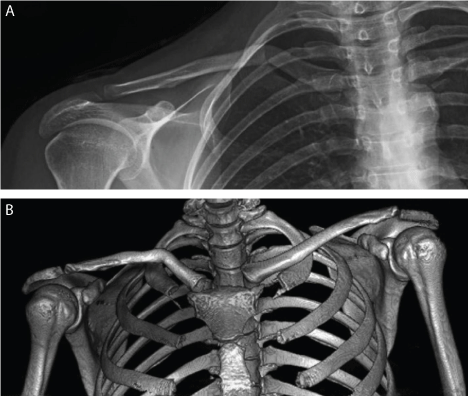
    Figure 1: Anteroposterior radiographs of right clavicle demonstrating
angulated and shortened malunion (A) and preoperative CT of right clavicle
showing 80 degrees of apex postero-superior angulation (B).
After a failed trial of nonoperative measures, to include physical therapy, shoulder injections, and activity modification, the decision was made to proceed with a corrective osteotomy using superior plate fixation and autologous bone graft from the ipsilateral distal radius (Figure 2). Post-operatively, the patient had near anatomic
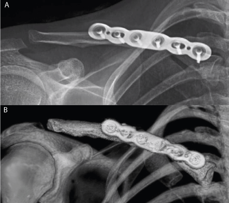
    Figure 2: Radiograph and postoperative CT of right clavicle following
corrective osteotomy with autologous bone grafting and superior plate fixation
of right clavicle.
alignment with restoration of the clavicular length in comparison to the contralateral clavicle with correction of her cosmetic deformity (Figure 3). She followed a progressive physical therapy protocol following surgery begin with 4 weeks of sling immobilization followed by progressive range of motion and strengthening monitored by physical therapy. She reported recovery of full shoulder function at 6 months postoperative and at final follow up at 1 year, she experienced no complications from surgery.
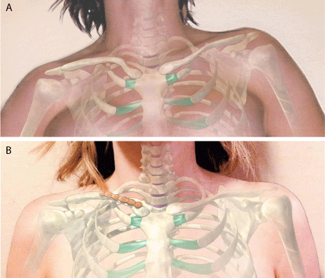
    Figure 3: Cosmetic deformity of patient with asymmetry of shoulder height
(A) compared with shoulder appearance following corrective osteotomy (B).
Discussion & Conclusion
Traditional management of clavicular fractures has been near exclusively nonoperative. Rowe stated, “Fortunately for man, nature has endowed the clavicle with excellent reparative powers” [7]. The classical teaching extrapolated from the inherent ability of the clavicle to unite is that malalignment rarely causes functional problems [8]. However, malunions of the clavicle are more frequently recognized today with a reported incidence of 5%.
Edelson identified distinctive patterns in the anatomy of midshaft clavicle malunions, with shortening, posterior displacement of the lateral fragment, and increasing severity of anterior angulation with more laterally occurring fractures [9]. The result of this deformity is alterations in the biomechanics of the shoulder with decreased moments for shoulder abduction, internal rotation, decreased strength and stability of the shoulder due to loss of muscletendon tension from clavicular shortening, alterations in the glenoid orientation [10-12]. An additionally complication that is often overlooked in the orthopaedic literature is the cosmetic deformity [8], which was the primary complaint in our case, resulting in an inability to use the affected arm.
Potter et al. [13] concluded that although immediate surgical fixation of clavicle fractures has better results and can help prevent the development of the sequela from malunion, corrective osteotomies are an option in patients initially treated with non-operative management and progress to have malunions. Current reports reveal symptomatic clavicular malunions have been successfully treated with a corrective osteotomy using either plate or intramedullary fixation 1-15 years after a clavicle fracture [6,11]. Surgical indications for treating symptomatic clavicular malunions include clavicle shortening greater than1 cm, angulation greater than 30 degrees, translation greater than 1cm, symptoms of thoracic outlet syndrome, rapid fatigability or pain with repetitive activity, difficulty with straps rubbing over the deformity, or substantial disability [14].
Results described in patients with corrective osteotomies include improved patient-based upper-extremity scores and patient satisfaction, however only 46% of patients return to their previous level of activity [15], and complications occur in up to 20% of patients [11], many of these being hardware related.
To our knowledge, this is the first case of a symptomatic malunion of the clavicle, established in childhood, effectively treated with a corrective osteotomy 19 years following the most recent fracture.
References
- Robinson CM. Fractures of the clavicle in the adult. Epidemiology and classification. J Bone Joint Surg Br. 1998; 80: 476-484.
- Hosalkar HS, Parikh G, Bittersohl B. Surgical Fixation of Displaced Clavicle Fractures in Adolescents: A Review of Literature. Orthop Rev (Pavia). 2013; 5: 29.
- Canadian Orthopaedic Trauma Society. Nonoperative treatment compared with plate fixation of displaced midshaft clavicle fractures; a multicenter, randomized clinical trial. J Bone Joint Surg Am. 2007; 89: 1-10.
- LeRoux T, Wasserstein D, Henry P, Khoshbin A, Dwyer T, Ogilvie-Harris D, et al. Rate of and risk factors for reoperations after open reduction and internal fixation of midshaft clavicle fractures. A Population-Based Study in Ontario, Canada. J Bone Joint Surg Am. 2014; 96: 1119-1125.
- Martetschläger F, Gaskill TR, Millett PJ. Management of clavicle nonunion and malunion. J Shoulder Elbow Surg. 2013; 22: 862-868.
- Bae DS, Shah AS, Kalish LA, Kwon JY, Waters PM. Shoulder motion, strength, and functional outcomes in children with established malunion of the clavicle. J Pediatr Orthop. 2013; 33: 544-550.
- Rowe CR. An atlas of anatomy and treatment of midclavicular fractures. Clin Orthop Relat Res. 1968; 58: 29-42.
- McKee MD, Wild LM, Schemitsch EH. Midshaft malunions of the clavicle. J Bone Joint Surg Am. 2003; 85-85: 790-797.
- Edelson JG. The bony anatomy of clavicular malunions. J Shoulder Elbow Surg. 2003; 12: 173-178.
- Patel B, Gustafson PA, Jastifer J. The effect of clavicle malunion on shoulder biomechanics; a computational study. Clin Biomech (Bristol, Avon). 2012; 27: 436-442.
- Hillen RJ, Burger BJ, Pöll RG, de Gast A, Robinson CM. Malunion after midshaft clavicle fractures in adults. Acta Orthop. 2010; 81: 273-279.
- Ledger M, Leeks N, Ackland T, Wang A. Short malunions of the clavicle: an anatomic and functional study. J Shoulder Elbow Surg. 2005; 14: 349-354.
- Potter J, Jones C, Wild L, Schemitsch E, McKee M. Does delay matter? The restoration of objectively measured shoulder strength and patient-oriented outcome after immediate fixation versus delayed reconstruction of displaced midshaft fractures of the clavicle. J Shoulder Elbow Surg. 2007; 16: 514-518.
- McKee MD, Wild LM, Schemitsch EH. Midshaft malunions of the clavicle. Surgical technique. J Bone Joint Surg Am. 2004; 86: 37-43.
- Rosenberg N, Neumann L, Wallace AW. Functional outcome of surgical treatment of symptomatic nonunion and malunion of midshaft clavicle fractures. J Shoulder Elbow Surg. 2007; 16: 510-513.